. 2024 Nov;51(13):3960-3977.
doi: 10.1007/s00259-024-06806-7. Epub 2024 Jul 2.

Efficient characterization of multiple binding sites of small molecule imaging ligands on amyloid-beta, tau and alpha-synuclein

Affiliations

Efficient characterization of multiple binding sites of small molecule imaging ligands on amyloid-beta, tau and alpha-synuclein

Jens Sobek et al. Eur J Nucl Med Mol Imaging. 2024 Nov.

Abstract

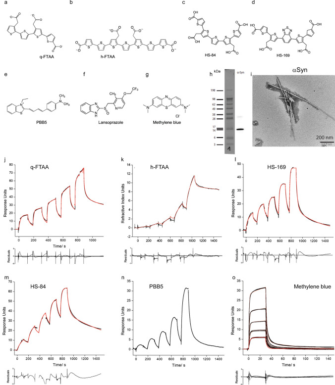
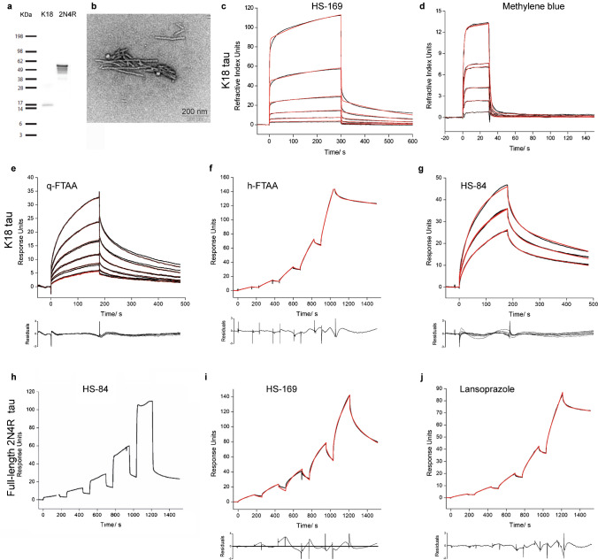
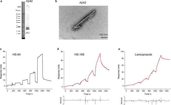
Purpose: There is an unmet need for compounds to detect fibrillar forms of alpha-synuclein (αSyn) and 4-repeat tau, which are critical in many neurodegenerative diseases. Here, we aim to develop an efficient surface plasmon resonance (SPR)-based assay to facilitate the characterization of small molecules that can bind these fibrils.

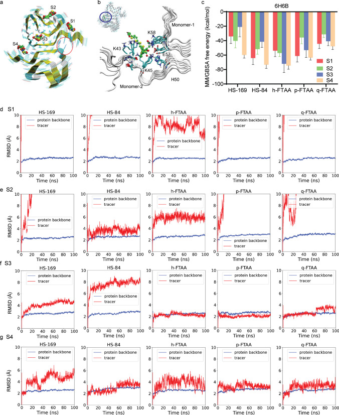
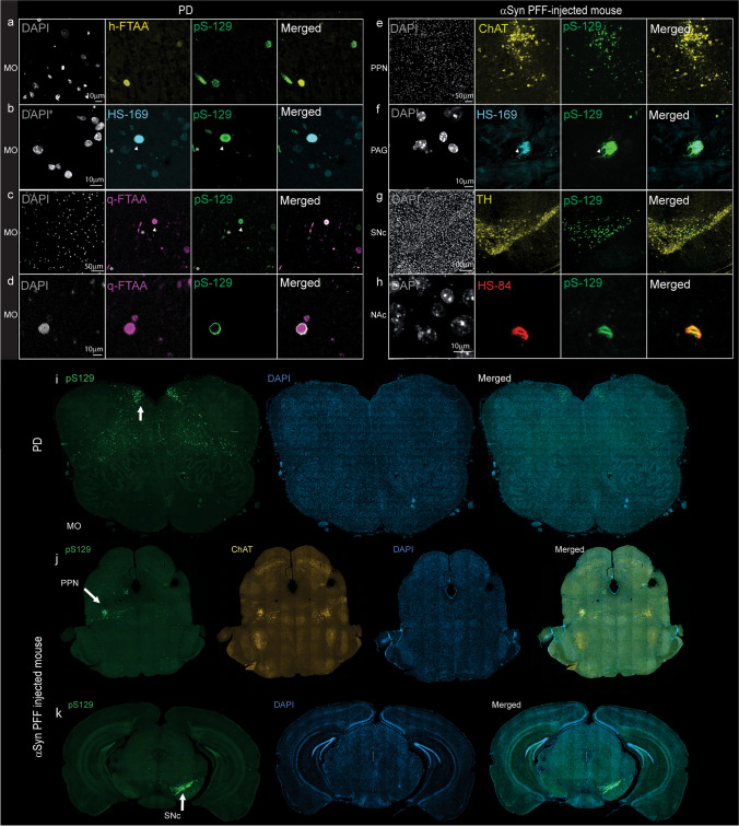
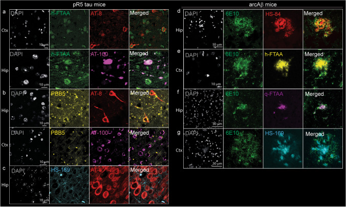
Methods: SPR measurements were conducted to characterize the binding properties of fluorescent ligands/compounds toward recombinant amyloid-beta (Aβ)42, K18-tau, full-length 2N4R-tau and αSyn fibrils. In silico modeling was performed to examine the binding pockets of ligands on αSyn fibrils. Immunofluorescence staining of postmortem brain tissue slices from Parkinson's disease patients and mouse models was performed with fluorescence ligands and specific antibodies.

Results: We optimized the protocol for the immobilization of Aβ42, K18-tau, full-length 2N4R-tau and αSyn fibrils in a controlled aggregation state on SPR-sensor chips and for assessing their binding to ligands. The SPR results from the analysis of binding kinetics suggested the presence of at least two binding sites for all fibrils, including luminescent conjugated oligothiophenes, benzothiazole derivatives, nonfluorescent methylene blue and lansoprazole. In silico modeling studies for αSyn (6H6B) revealed four binding sites with a preference for one site on the surface. Immunofluorescence staining validated the detection of pS129-αSyn positivity in the brains of Parkinson's disease patients and αSyn preformed-fibril injected mice, 6E10-positive Aβ in arcAβ mice, and AT-8/AT-100-positivity in pR5 mice.

Conclusion: SPR measurements of small molecules binding to Aβ42, K18/full-length 2N4R-tau and αSyn fibrils suggested the existence of multiple binding sites. This approach may provide efficient characterization of compounds for neurodegenerative disease-relevant proteinopathies.

Keywords: Alpha-synuclein; Amyloid-beta; Binding sites; In silico; Surface plasmon resonance; Tau.


Conflict of interest statement

KS and RN are associate editors in Eur J Nucl Med Mol Imaging. RMN is an employee and shareholder of Neurimmune AG, Switzerland. The other authors declare no conflicts of interest.

Figures

Fig. 1
Fig. 1
Characterization of ligand binding on recombinant alpha-synuclein fibrils. a-g Chemical structures of q-FTAA, h-FTAA, HS-84, HS-169, PBB5, lansoprazole and methylene blue. h Western blot of αSyn using anti-αSyn antibody, Syn211; i TEM of αSyn fibril; j-o Sensorgrams of q-FTAA(j), h-FTAA(k), HS-169(o), HS-84(m), PBB5(n), and methylene blue(o), binding to αSyn fibrils. The black line represents the experimental data, and the red line represents the fitted curve
Fig. 2
Fig. 2
Characterization of ligand binding on recombinant K18-tau and full-length 2N4R tau fibrils. a Western blot of K18-tau and full-length 2N4R tau using anti-Tau(RD4) antibody, 1E1/A6; b TEM of K18-tau and full-length 2N4R tau fibrils; c-g Sensorgrams of HS-84(c), methylene blue(d), q-FTAA(e), h-FTAA(f), and HS-169(g) binding to K18-tau fibrils. (h-j) Sensorgrams of HS-84 (h), HS-169 (i), and lansoprazole (j) binding to full-length 2N4R tau fibrils. The black line represents the experimental data, and the red line represents the fitted curve
Fig. 3
Fig. 3
Characterization of ligand binding on recombinant Aβ42 fibrils. a Western blot of Aβ42 using the monoclonal anti-β-amyloid antibody BAM-10; b TEM image of Aβ42 fibrils; c-e Sensorgrams of HS-169, HS-84 and lansoprazole binding to Aβ42 fibrils. The black line represents the experimental data, and the red line represents the fitted curve
Fig. 4
Fig. 4
In silico modeling of the binding sites of HS-169, HS-84, h-FTAA, p-FTAA and q-FTAA on the 6H6B αSyn structure. a Four binding sites (S1-S4) on alpha-synuclein fibrils; the red circle indicates the location of site 4; b Zoomed-in view of h-FTAA binding to site 4. c MM/GBSA calculation of free energy indicating that site 4 is preferred by HS-169, HS-84, h-FTAA, p-FTAA and q-FTAA. d-g RMSD analysis of the ligands binding to 4 binding sites (S1-S4)
Fig. 5
Fig. 5
Confocal imaging of alpha-synuclein inclusions in brain tissue sections from PD patients and αSyn PFF-injected mice. a-d Colocalization of the Alexa488-anti-αSYN phosphor S129 antibody pS129 (green) with h-FTAA (yellow), HS-169 (cyan), and q-FTAA (magenta) in the medulla oblongata of PD patients (whole region image in i). αSyn-positive Lewy neurites (*) and Lewy bodies (arrowhead). ef, j pS129-positive inclusions in ChAT-positive neurons (yellow) in the pedunculopontine nucleus (PPN) of αSyn-PFF-injected mice, with whole-brain images in j. (f) Colocalization of pS129-positive inclusions (green) with HS-169 (cyan) in the periaqueductal gray (PAG). g, h pS129-positive inclusions in TH-positive neurons (yellow) in the substantia nigra pars compacta (SNc) of αSyn-PFF-injected mice with whole-brain images in k). (h) Colocalization of pS129-positive inclusions (green) with HS-84 (red) in the nucleus accumbens (NAc)
Fig. 6
Fig. 6
Confocal imaging of ligands with tau and amyloid-beta in mouse models. a-c Colocalization of tau staining by Alexa488-AT8 (red), Alexa488-AT100 (magenta) with h-FTAA (green), PBB5 (yellow), and HS-169 (cyan) in the hippocampus of pR5 tau mice; d-g colocalization of the anti-Aβ1-16 antibody Alexa488-6E10 (green) with HS-84 (red), h-FTAA (yellow), q-FTAA (magenta), and HS-169 (cyan) in the cortex of arcAβ mice. Nuclei were counterstained using DAPI (white)

References

    1. Jucker M, Walker LC. Propagation and spread of pathogenic protein assemblies in neurodegenerative diseases. Nat Neurosci. 2018;21:1341–9. 10.1038/s41593-018-0238-6. - PMC - PubMed
    1. Spillantini MG, Goedert M. Tau pathology and neurodegeneration. Lancet Neurol. 2013;12:609–22. 10.1016/S1474-4422(13)70090-5. - PubMed
    1. Dubois B, Villain N, Frisoni GB, Rabinovici GD, Sabbagh M, Cappa S, et al. Clinical diagnosis of Alzheimer’s disease: recommendations of the International Working Group. Lancet Neurol. 2021;20:484–96. 10.1016/S1474-4422(21)00066-1. - PMC - PubMed
    1. Smith R, Capotosti F, Schain M, Ohlsson T, Touilloux T, Hliva V, et al. Initial clinical scans using [18F]ACI-12589, a novel α-synuclein PET-tracer. Alzheimers Dement. 2022;18:e065394. 10.1002/alz.065394.
    1. Xiang J, Tao Y, Xia Y, Luo S, Zhao Q, Li B, et al. Development of an α-synuclein positron emission tomography tracer for imaging synucleinopathies. Cell. 2023. 10.1016/j.cell.2023.06.004. - PMC - PubMed

MeSH terms