. 2024 Jul 2;73(9):164.
doi: 10.1007/s00262-024-03749-8.

Identification and validation of tumor-specific T cell receptors from tumor infiltrating lymphocytes using tumor organoid co-cultures

Affiliations

Identification and validation of tumor-specific T cell receptors from tumor infiltrating lymphocytes using tumor organoid co-cultures

Zhilang Li et al. Cancer Immunol Immunother. .

Abstract

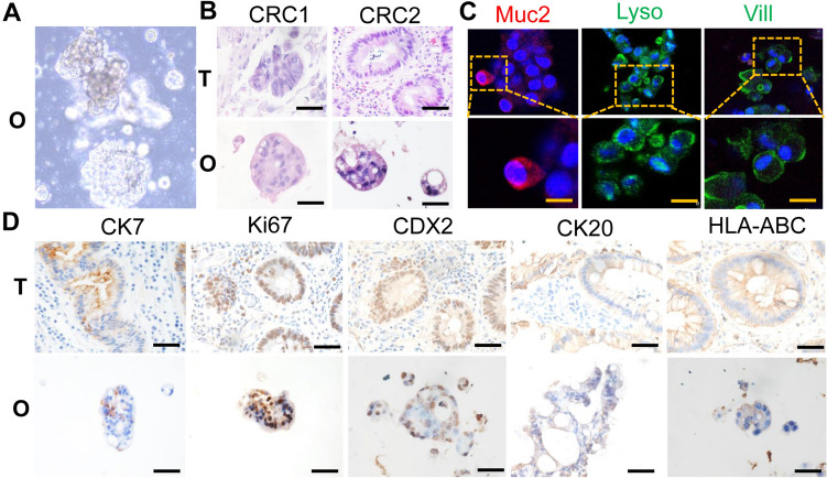
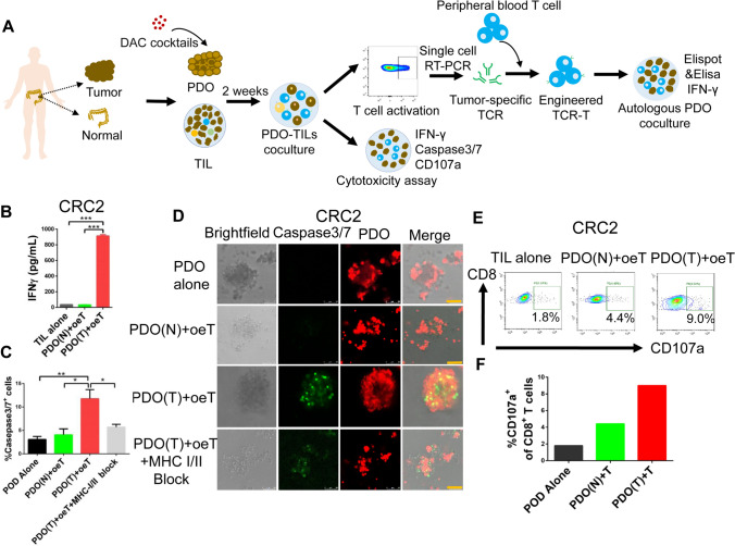
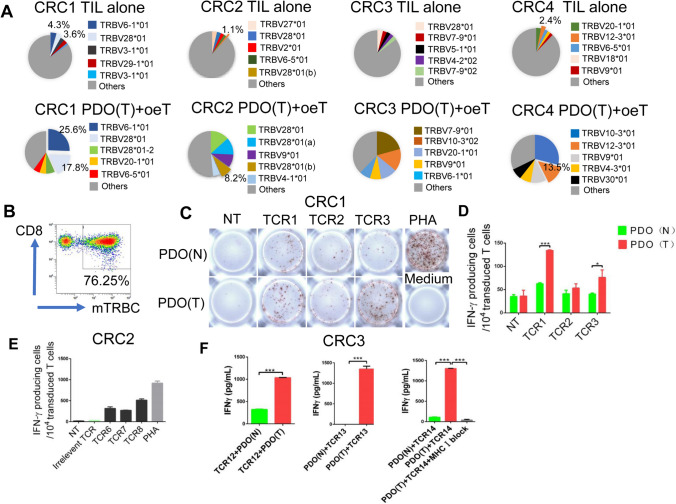
T cell receptor-engineered T cells (TCR-Ts) therapy is promising for cancer immunotherapy. Most studies have focused on identifying tumor-specific T cell receptors (TCRs) through predicted tumor neoantigens. However, current algorithms for predicting tumor neoantigens are unreliable and many neoantigens are derived from non-coding regions. Thus, the technological platform for identifying tumor-specific TCRs using natural antigens expressed on tumor cells is urgently needed. In this study, tumor organoids-enriched tumor infiltrating lymphocytes (oeT) were obtained by repeatedly stimulation of autologous patient-derived organoids (PDO) in vitro. The oeT cells specifically responded to autologous tumor PDO by detecting CD137 expression and the secretion of IFN-γ using enzyme-linked immunospot assay. The measurement of oeT cell-mediated killing of three-dimensional organoids was conducted using a caspase3/7 flow cytometry assay kit. Subsequently, tumor-specific T cells were isolated based on CD137 expression and their TCRs were identified through single-cell RT-PCR analysis. The specificity cytotoxicity of TCRs were confirmed by transferring to primary peripheral blood T cells. The co-culture system proved highly effective in generating CD8+ tumor-specific oeT cells. These oeT cells effectively induced IFN-γ secretion and exhibited specificity in killing autologous tumor organoids, while not eliciting a cytotoxic response against normal organoids. The analysis conducted by TCRs revealed a significant expansion of T cells within a specific subset of TCRs. Subsequently, the TCRs were cloned and transferred to peripheral blood T cells generation engineered TCR-Ts, which adequately recognized and killed tumor cell in a patient-specific manner. The co-culture system provided an approach to generate tumor-specific TCRs from tumor-infiltrating lymphocytes of patients with colorectal cancer, and tumor-specific TCRs can potentially be used for personalized TCR-T therapy.

Keywords: Colorectal cancer; Immunotherapy; Patient derived organoids; T cell receptor-engineered T cells; Tumor-infiltrating lymphocytes.


Conflict of interest statement

The authors declare no conflict of interest.

Figures

Fig. 1
Fig. 1
Histological analysis revealed the morphological and molecular features of PDO. A Representative images of PDO morphology from CRC patients. B H&E staining comparing tumor organoids to their matched patient tumor tissue. Scale bar: 50 μm. C Representative images of immunofluorescent microscopy about cell type compositions in tumor organoids. Mucin2 (Muc2; red) for goblet cells, Lysozyme (Lysz; green) for paneth cells and Villin (Vill; green) for epithelial cells. Scale bar: 20 μm. D Representative images of IHC of CK7, Ki67, CDX2, CK20, HLA-ABC from primary tumor tissue and patient-derived organoids. O: organoids, T: tumor tissue. Scale bar: 50 μm
Fig. 2
Fig. 2
Phenotype analysis of peripheral blood lymphocytes and matched tumor infiltrating lymphocytes form CRC patients. A Typical results of flow cytometry analysis revealed the frequency of CD39+CD103+CD8+ T cells in PBLs and matched TILs of CRC patients. B Statistical analysis of the frequency of CD39+CD103+ T cells in PBLs and matched TILs of CRC patients. C Typical results of the frequency of CD8+PD-1+ T cells in PBL and matched TILs of CRC patients. D Statistical analysis of the frequency of CD8+PD-1+ T cells in PBLs and matched TILs of CRC patients. E Typical results of the frequency of CD8+CXCL13+ T cells in PBLs and matched TILs of CRC patients. F. Statistical analysis of the frequency of CD8+CXCL13+ T cells in PBLs and matched TILs of CRC patients
Fig. 3
Fig. 3
Cytotoxic activity of tumor organoid enriched TILs (oeT). A Schematic representation of the experimental workflow. B ELISA analysis the IFN-γ secretion by oeT cells after co-cultured with autologous tumor organoids or normal organoids for 24 h. C Quantitative summary of the killing efficiency shown by FITC signal (caspase 3/7 probe) in flow cytometry analysis. D oeT cells co-cultured with autologous tumor organoids and normal organoids for 20 h to assess killing efficiency respectively. Organoids (red) were labeled with Cell-Trace FarRed, and apoptotic cells (green) were labeled with caspase-3/7 probe. Scale bars: 50 μm. E Flow cytometry analyzed oeT cell effector marker CD107a after co-cultured with autologous tumor organoids and normal organoids, respectively. F Summary of the frequency of CD107a positive cells after co-cultured with autologous tumor organoids and normal organoids. PDO(N): patient-derived normal organoids, PDO(T): patient-derived tumor organoids
Fig. 4
Fig. 4
Enrichment of Tumor-specific T cell in TILs by co-culture with autologous tumor organoids. A IFN-γ ELISPOT analysis of oeT cells co-cultured with autologous tumor organoids or normal organoids. TILs with no targets are negative controls. PHA well is the positive control. B Quantification of organoid-induced IFN-γ production obtained by oeT cells co-cultured with autologous tumor organoids or normal organoids. C Representative flow cytometry plots of CD8+CD137+T cells after oeT cells stimulated with either tumor organoids and normal organoids. D Quantification of organoids induced CD137 expression of CD8+ T cells directly after oeT cells stimulated with either tumor organoids or normal organoids. PDO (N): patient-derived normal organoids, PDO (T): patient-derived tumor organoids
Fig. 5
Fig. 5
Functional verification of TCR-Ts. A Pie charts showing the distribution of a given TCR beta-chains of CD8+CD137+ T cells stimulated with tumor organoids. B PBLs form healthy donors were transduced with the chimeric TCR and flow sorted by anti-murine TCR-β chain constant region antibody. C IFN-γ ELISPOT analysis on TCR-T cells that were exposed to autologous tumor organoids or normal organoids in CRC1. NT: no transfection. D Quantification of organoid induced IFN-γ production by TCR-T cells co-cultured with autologous tumor organoids or normal organoids in CRC1. E Quantification of organoid induced IFN-γ production by TCR-T cells co-cultured with autologous tumor organoids in CRC2. F ELISA analysis the IFN-γ secretion by TCR-T cells after co-cultured with autologous tumor organoids or normal organoids for 24 h in CRC3. PDO (N): patient-derived normal organoids, PDO (T): patient-derived tumor organoids

References

    1. Parkhurst MR, Yang JC, Langan RC et al (2011) T cells targeting carcinoembryonic antigen can mediate regression of metastatic colorectal cancer but induce severe transient colitis. Mol Ther 19:620–626. 10.1038/mt.2010.272 10.1038/mt.2010.272 - DOI - PMC - PubMed
    1. Robbins PF, Kassim SH, Tran TL et al (2015) A pilot trial using lymphocytes genetically engineered with an NY-ESO-1-reactive T-cell receptor: long-term follow-up and correlates with response. Clin Cancer Res 21:1019–1027. 10.1158/1078-0432.CCR-14-2708 10.1158/1078-0432.CCR-14-2708 - DOI - PMC - PubMed
    1. Meng F, Zhao J, Tan AT et al (2021) Immunotherapy of HBV-related advanced hepatocellular carcinoma with short-term HBV-specific TCR expressed T cells: results of dose escalation, phase I trial. Hep Int 15:1402–1412. 10.1007/s12072-021-10250-2 10.1007/s12072-021-10250-2 - DOI - PMC - PubMed
    1. Leidner R, Sanjuan Silva N, Huang H et al (2022) Neoantigen T-cell receptor gene therapy in pancreatic cancer. N Engl J Med 386:2112–2119. 10.1056/NEJMoa2119662 10.1056/NEJMoa2119662 - DOI - PMC - PubMed
    1. Parkhurst M, Gros A, Pasetto A, Prickett T, Crystal JS, Robbins P, Rosenberg SA (2017) Isolation of T-cell receptors specifically reactive with mutated tumor-associated antigens from tumor-infiltrating lymphocytes based on CD137 expression. Clin Cancer Res 23:2491–2505. 10.1158/1078-0432.CCR-16-2680 10.1158/1078-0432.CCR-16-2680 - DOI - PMC - PubMed

MeSH terms

Substances

LinkOut - more resources