Tracking longitudinal thalamic volume changes during early stages of SCA1 and SCA2
- PMID: 38954239
- PMCID: PMC11322486
- DOI: 10.1007/s11547-024-01839-2
Tracking longitudinal thalamic volume changes during early stages of SCA1 and SCA2
Abstract
Purpose: Spinocerebellar ataxia SCA1 and SCA2 are adult-onset hereditary disorders, due to triplet CAG expansion in their respective causative genes. The pathophysiology of SCA1 and SCA2 suggests alterations of cerebello-thalamo-cortical pathway and its connections to the basal ganglia. In this framework, thalamic integrity is crucial for shaping efficient whole-brain dynamics and functions. The aims of the study are to identify structural changes in thalamic nuclei in presymptomatic and symptomatic SCA1 and SCA2 patients and to assess disease progression within a 1-year interval.
Material and methods: A prospective 1-year clinical and MRI assessment was conducted in 27 presymptomatic and 23 clinically manifest mutation carriers for SCA1 and SCA2 expansions. Cross-sectional and longitudinal changes of thalamic nuclei volume were investigated in SCA1 and SCA2 individuals and in healthy participants (n = 20).
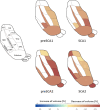
Results: Both SCA1 and SCA2 patients had significant atrophy in the majority of thalamic nuclei, except for the posterior and partly medial nuclei. The 1-year longitudinal evaluation showed a specific pattern of atrophy in ventral and posterior thalamus, detectable even at the presymptomatic stage of the disease.
Conclusion: For the first time in vivo, our exploratory study has shown that different thalamic nuclei are involved at different stages of the degenerative process in both SCA1 and SCA2. It is therefore possible that thalamic alterations might significantly contribute to the progression of the disease years before overt clinical manifestations occur.
Keywords: MRI; Presymptomatic carriers; SCA1; SCA2; Spinocerebellar ataxias; Thalamus.
© 2024. The Author(s).
Conflict of interest statement
The authors have no relevant financial or non-financial interests to disclose.
Figures

Similar articles
-
Conversion of individuals at risk for spinocerebellar ataxia types 1, 2, 3, and 6 to manifest ataxia (RISCA): a longitudinal cohort study.Lancet Neurol. 2020 Sep;19(9):738-747. doi: 10.1016/S1474-4422(20)30235-0. Lancet Neurol. 2020. PMID: 32822634 Clinical Trial.
-
Long-term disease progression in spinocerebellar ataxia types 1, 2, 3, and 6: a longitudinal cohort study.Lancet Neurol. 2015 Nov;14(11):1101-8. doi: 10.1016/S1474-4422(15)00202-1. Epub 2015 Sep 13. Lancet Neurol. 2015. PMID: 26377379
-
Biological and clinical characteristics of individuals at risk for spinocerebellar ataxia types 1, 2, 3, and 6 in the longitudinal RISCA study: analysis of baseline data.Lancet Neurol. 2013 Jul;12(7):650-8. doi: 10.1016/S1474-4422(13)70104-2. Epub 2013 May 22. Lancet Neurol. 2013. PMID: 23707147
-
Thalamic involvement in a spinocerebellar ataxia type 2 (SCA2) and a spinocerebellar ataxia type 3 (SCA3) patient, and its clinical relevance.Brain. 2003 Oct;126(Pt 10):2257-72. doi: 10.1093/brain/awg234. Epub 2003 Jul 7. Brain. 2003. PMID: 12847080 Review.
-
MR Imaging in Spinocerebellar Ataxias: A Systematic Review.AJNR Am J Neuroradiol. 2016 Aug;37(8):1405-12. doi: 10.3174/ajnr.A4760. Epub 2016 May 12. AJNR Am J Neuroradiol. 2016. PMID: 27173364 Free PMC article.
Cited by
-
Radiological Reporting of Brain Atrophy in MRI: Real-Life Comparison Between Narrative Reports, Semiquantitative Scales and Automated Software-Based Volumetry.Diagnostics (Basel). 2025 May 14;15(10):1246. doi: 10.3390/diagnostics15101246. Diagnostics (Basel). 2025. PMID: 40428239 Free PMC article.
References
MeSH terms
Substances
Grants and funding
LinkOut - more resources
Full Text Sources
Medical
Research Materials

