. 2024 Oct 3;147(10):3358-3369.
doi: 10.1093/brain/awae219.

Dopamine and deep brain stimulation accelerate the neural dynamics of volitional action in Parkinson's disease

Affiliations

Dopamine and deep brain stimulation accelerate the neural dynamics of volitional action in Parkinson's disease

Richard M Köhler et al. Brain. .

Abstract

The ability to initiate volitional action is fundamental to human behaviour. Loss of dopaminergic neurons in Parkinson's disease is associated with impaired action initiation, also termed akinesia. Both dopamine and subthalamic deep brain stimulation (DBS) can alleviate akinesia, but the underlying mechanisms are unknown. An important question is whether dopamine and DBS facilitate de novo build-up of neural dynamics for motor execution or accelerate existing cortical movement initiation signals through shared modulatory circuit effects. Answering these questions can provide the foundation for new closed-loop neurotherapies with adaptive DBS, but the objectification of neural processing delays prior to performance of volitional action remains a significant challenge. To overcome this challenge, we studied readiness potentials and trained brain signal decoders on invasive neurophysiology signals in 25 DBS patients (12 female) with Parkinson's disease during performance of self-initiated movements. Combined sensorimotor cortex electrocorticography and subthalamic local field potential recordings were performed OFF therapy (n = 22), ON dopaminergic medication (n = 18) and on subthalamic deep brain stimulation (n = 8). This allowed us to compare their therapeutic effects on neural latencies between the earliest cortical representation of movement intention as decoded by linear discriminant analysis classifiers and onset of muscle activation recorded with electromyography. In the hypodopaminergic OFF state, we observed long latencies between motor intention and motor execution for readiness potentials and machine learning classifications. Both, dopamine and DBS significantly shortened these latencies, hinting towards a shared therapeutic mechanism for alleviation of akinesia. To investigate this further, we analysed directional cortico-subthalamic oscillatory communication with multivariate granger causality. Strikingly, we found that both therapies independently shifted cortico-subthalamic oscillatory information flow from antikinetic beta (13-35 Hz) to prokinetic theta (4-10 Hz) rhythms, which was correlated with latencies in motor execution. Our study reveals a shared brain network modulation pattern of dopamine and DBS that may underlie the acceleration of neural dynamics for augmentation of movement initiation in Parkinson's disease. Instead of producing or increasing preparatory brain signals, both therapies modulate oscillatory communication. These insights provide a link between the pathophysiology of akinesia and its' therapeutic alleviation with oscillatory network changes in other non-motor and motor domains, e.g. related to hyperkinesia or effort and reward perception. In the future, our study may inspire the development of clinical brain computer interfaces based on brain signal decoders to provide temporally precise support for action initiation in patients with brain disorders.

Keywords: Granger causality; bereitschaftspotential; electrocorticography; neuromodulation; oscillations.


Conflict of interest statement

A.A.K. reports personal fees from Medtronic and Boston Scientific. G.H.S. reports personal fees from Medtronic, Boston Scientific and Abbott. W.J.N. serves as consultant to InBrain and reports personal fees from Medtronic.

Figures

Figure 1
Figure 1
Sensorimotor electrocorticography and subthalamic local field potentials during self-initiated movements in Parkinson's disease patients. (A) Twenty-five Parkinson's disease patients were implanted with bilateral deep brain stimulation (DBS) leads and a single electrocorticography (ECoG) strip. (B) ECoG strips were placed on the sensorimotor cortex and DBS electrodes placed into the dorsolateral part of the subthalamic nucleus. Exemplar traces show ECoG activity from the motor cortex and subthalamic local field potentials (STN-LFP) from the hemisphere contralateral to movement during a single movement. (C) Schematic of the rotational handle used by Parkinson's disease patients in the Berlin cohort. (D) Oscillatory activity from the hemisphere contralateral to movement recorded OFF therapy, averaged across patients (n = 22) and electromyography (EMG) of the brachioradial muscle during a self-initiated movement. (E) Unified Parkinson's Disease Rating Scale III (UPDRS-III) scores during experimental sessions of patients recorded both OFF and ON levodopa (29/18 ± 8/8, P = 6 × 10⁻⁵, n = 15; Supplementary Table 1) and UPDRS-III scores at 12 months post-implantation for patients recorded both OFF therapy and on subthalamic deep brain stimulation (STN-DBS; 41/22 ± 14/8, n = 7, unavailable in n = 1; Supplementary Table 2). (F) Readiness potentials of motor cortex (left) and STN-LFP signals (right) contralateral to movement side, averaged across patients. Motor cortex readiness potentials differed significantly from baseline between −1.78 and 1.55 s (OFF therapy), −1.0 and 1.09 s (ON levodopa) and −1.18 and 1.51 s (on STN-DBS). Subthalamic readiness potentials differed significantly from baseline between −1.19 and 1.32 s (OFF therapy), −1.28 and 2.0 s (ON levodopa) and −0.17 and 1.2 s (on STN-DBS; all P ≤ 0.05, cluster corrected). Data are presented as mean ± standard error of the mean. Coloured triangles within subplots and on the lowest x-axis indicate the earliest significant difference.
Figure 2
Figure 2
Dopamine and subthalamic deep brain stimulation reduce motor intention to execution delays in Parkinson's disease. (A) Features of a single motor cortex channel averaged across trials. (B) Classifier outputs averaged across subjects. Classifier outputs of electrocorticography (ECoG) and subthalamic local field potentials (STN-LFP) differed between −2.2 to 1.7 s (OFF therapy), −1.6 to 1.6 s (ON levodopa) and −1.0 to 0.9 s [on subthalamic deep brain stimulation (STN-DBS)]; all P ≤ 0.05, cluster corrected. Data are presented as mean ± standard error of the mean. Coloured triangles within subplots and on the lowest x-axis indicate the earliest significant difference. (C) Time of motor intention of single subjects derived from ECoG classifier outputs. (D) Time of motor intention derived from single-channel ECoG classifier outputs. Left hemispheric channels were flipped onto the right hemisphere. *P ≤ 0.05, **P ≤ 0.01, ***P ≤ 0.001.
Figure 3
Figure 3
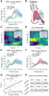
Dopamine and subthalamic deep brain stimulation drive cortico-subthalamic theta coupling that is correlated with shorter intention to execution latencies. (A) At the end of the baseline period (−2 s) motor cortex drives oscillatory cortico-subthalamic coupling in the beta frequency range as measured by time-reverse corrected Granger causality (TRGC). (B) With start of movement intention motor cortex may drive activity through the striatopallidal indirect and monosynaptic hyperdirect pathways to the subthalamic nucleus (STN), that can be modulated by dopaminergic afferences from substantia nigra pars compacta (SNc) and subthalamic deep brain stimulation (STN-DBS). GPe = globus pallidus externus; THAL = thalamus. (C) Comparison of Granger causality OFF versus ON Levodopa reveals stronger coupling in the theta range (dashed cluster, P ≤ 0.05). Peak significance occurred 900 ms before movement onset at 7 Hz (yellow rectangle in inlet), shortly after average decoding time onset (vertical dashed line in blue/green for OFF/ON in inlet). (D) A similar effect was observed when comparing OFF therapy cohorts with the sub-cohort on STN-DBS with a peak significance 900 ms before movement at 6 Hz. Granger causality spectra demonstrate spectrally specific differences for dopamine (E) and STN-DBS (F) in the theta frequency range. (G) Granger causality at peak difference correlated with the time of motor intention in the ON levodopa, but not OFF therapy cohorts. (H) In the hypodopaminergic Parkinson's disease state, intention to execution latency is prolonged and cortico-subthalamic coupling is predominantly observed in the beta frequency range, that could be attributed to excessive indirect pathway (D2) activity. Dopamine and STN-DBS shorten motor intention to execution latencies and shift cortico-subthalamic coupling to the theta frequency band, potentially through suppression and consecutive changes in direct to indirect pathway balance. HD = hyperdirect pathway.

References

    1. Roskies AL. How does neuroscience affect our conception of volition? Annu Rev Neurosci. 2010;33:109–130. - PubMed
    1. Kornhuber HH, Deecke L. Hirnpotentialänderungen bei Willkürbewegungen und passiven Bewegungen des Menschen: Bereitschaftspotential und reafferente Potentiale. Pflügers Arch. 1965;284:1–17. - PubMed
    1. Fried I, Mukamel R, Kreiman G. Internally generated preactivation of single neurons in human medial frontal cortex predicts volition. Neuron. 2011;69:548–562. - PMC - PubMed
    1. Soon CS, Brass M, Heinze H-J, Haynes J-D. Unconscious determinants of free decisions in the human brain. Nat Neurosci. 2008;11:543–545. - PubMed
    1. Deecke L, Kornhuber HH. An electrical sign of participation of the mesial ‘supplementary’ motor cortex in human voluntary finger movement. Brain Res. 1978;159:473–476. - PubMed