Clinical Trial
. 2024 Sep 1;42(25):3033-3046.
doi: 10.1200/JCO.23.02233. Epub 2024 Jul 2.

Final Results From a Phase I Trial and Expansion Cohorts of Cabozantinib and Nivolumab Alone or With Ipilimumab for Advanced/Metastatic Genitourinary Tumors

Affiliations
Clinical Trial

Final Results From a Phase I Trial and Expansion Cohorts of Cabozantinib and Nivolumab Alone or With Ipilimumab for Advanced/Metastatic Genitourinary Tumors

Andrea B Apolo et al. J Clin Oncol. .

Abstract

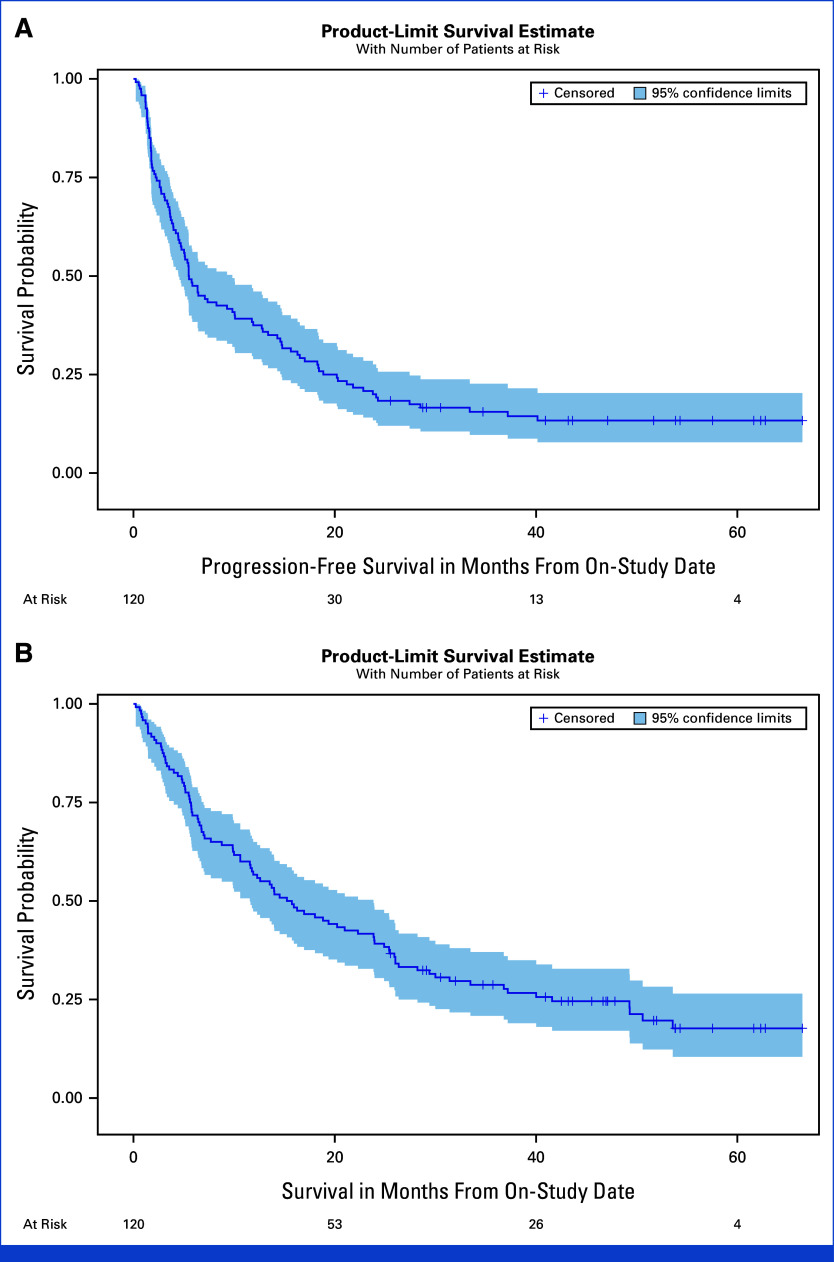
Purpose: Cabozantinib and nivolumab (CaboNivo) alone or with ipilimumab (CaboNivoIpi) have shown promising efficacy and safety in patients with metastatic urothelial carcinoma (mUC), metastatic renal cell carcinoma (mRCC), and rare genitourinary (GU) tumors in a dose-escalation phase I study. We report the final data analysis of the safety, overall response rate (ORR), progression-free survival (PFS), and overall survival (OS) of the phase I patients and seven expansion cohorts.

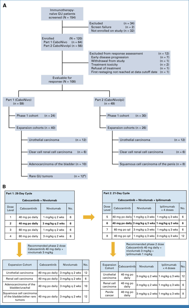
Methods: This is an investigator-initiated, multicenter, phase I trial. CaboNivo doublet expansion cohorts included (1) mUC, (2) mRCC, and (3) adenocarcinoma of the bladder/urachal; CaboNivoIpi triplet expansion cohorts included (1) mUC, (2) mRCC, (3) penile cancer, and (4) squamous cell carcinoma of the bladder and other rare GU tumors (ClinicalTrials.gov identifier: NCT02496208).

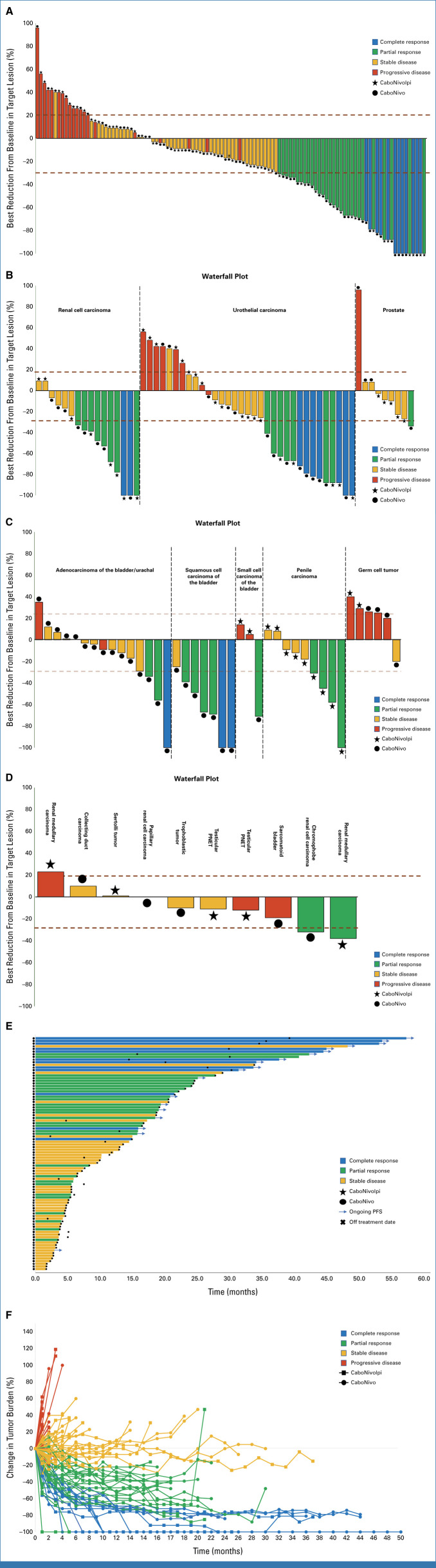
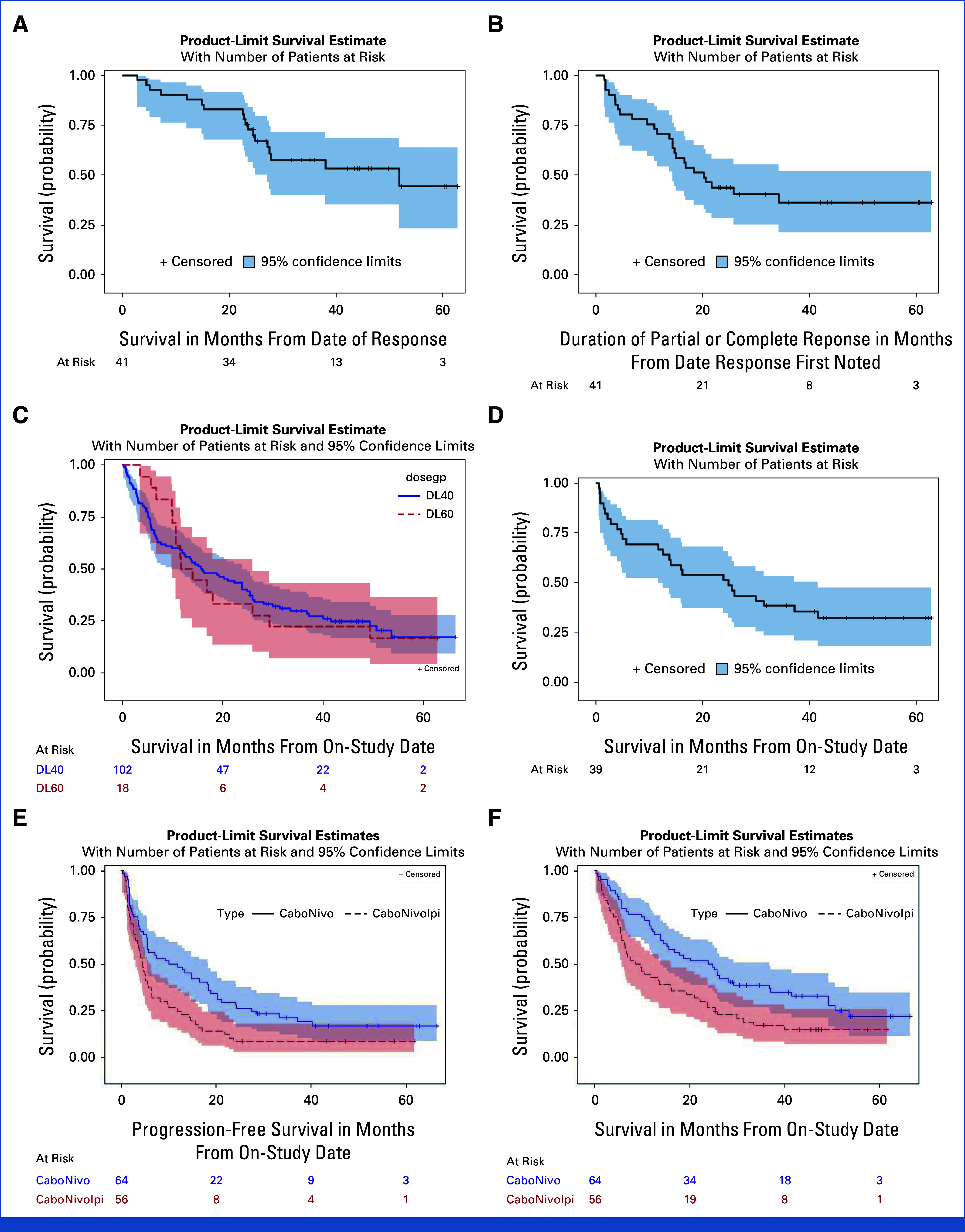
Results: The study enrolled 120 patients treated with CaboNivo (n = 64) or CaboNivoIpi (n = 56), with a median follow-up of 49.2 months. In 108 evaluable patients (CaboNivo n = 59; CaboNivoIpi n = 49), the ORR was 38% (complete response rate 11%) and the median duration of response was 20 months. The ORR was 42.4% for mUC, 62.5% for mRCC (n = 16), 85.7% for squamous cell carcinoma of the bladder (n = 7), 44.4% for penile cancer (n = 9), and 50.0% for renal medullary carcinoma (n = 2). Grade ≥ 3 treatment-related adverse events occurred in 84% of CaboNivo patients and 80% of CaboNivoIpi patients.

Conclusion: CaboNivo and CaboNivoIpi demonstrated clinical activity and safety in patients with multiple GU malignancies, especially clear cell RCC, urothelial carcinoma, and rare GU tumors such as squamous cell carcinoma of the bladder, small cell carcinoma of the bladder, adenocarcinoma of the bladder, renal medullary carcinoma, and penile cancer.


Conflict of interest statement

The following represents disclosure information provided by authors of this manuscript. All relationships are considered compensated unless otherwise noted. Relationships are self-held unless noted. I = Immediate Family Member, Inst = My Institution. Relationships may not relate to the subject matter of this manuscript. For more information about ASCO's conflict of interest policy, please refer to www.asco.org/rwc or ascopubs.org/jco/authors/author-center.

Open Payments is a public database containing information reported by companies about payments made to US-licensed physicians (Open Payments).

Amir Mortazavi

Consulting or Advisory Role: Targeted Oncology

Research Funding: Genentech/Roche (Inst), Merck (Inst), Novartis (Inst), Seagen (Inst), Bristol Myers Squibb (Inst), Astellas Pharma (Inst), GlaxoSmithKline (Inst)

No other potential conflicts of interest were reported.

Figures

FIG 1.
FIG 1.
Antitumor activity and response by measurable disease. (A) Waterfall plot of change in target lesions from baseline. Horizontal upper and lower dashed lines represent RECIST boundaries of 20% increase and 30% decrease in size of target lesions, respectively. (B) Waterfall plots for renal cell carcinoma, urothelial carcinoma, and prostate cohorts. Tumor type for each cohort is listed for each group. (C) Waterfall plots for adenocarcinoma, squamous cell carcinoma of the bladder/urinary tract, small cell carcinoma of the bladder/urinary tract, penile carcinoma, and germ cell tumor cohorts. Tumor type for each cohort is listed for each group. (D) Waterfall plot for rare genitourinary tumor cohort. Tumor type for each individual is listed above each bar. (E) Swimmer's plot showing duration of response, treatment arm, and response. (F) Spider plot depicting response and change in target lesion size (tumor burden) from baseline for individual participants. Red bars/lines represent PD, yellow bars represent SD, green bars represent PR, and blue bars represent CR as best response. CaboNivo, cabozantinib + nivolumab; CaboNivoIpi, cabozantinib + nivolumab + ipilimumab; CR, complete response; PR, partial response; SD, stable disease.
FIG 2.
FIG 2.
Kaplan-Meier estimates for PFS and OS for phase I and expansion cohorts ITT population (n = 120). (A) Median PFS of 5.5 months (95% CI, 4.5 to 9.8 months). (B) Median OS of 15.5 months (95% CI, 11.6 to 23.9). ITT, intention-to-treat; OS, overall survival; PFS, progression-free survival.
FIG 3.
FIG 3.
Summary schema of immune correlative results. This figure summarizes peripheral blood immune subset and functional marker changes by CaboNivo and CaboNivoIpi treatments and association with PFS and OS. CaboNivo and CaboNivoIpi treatments had common and differential impacts on immune subsets. In brief, myeloid cells, including M-MDSC, classical monocytes, and dendritic cells, decreased by both treatments, but association with OS was observed only in CaboNivoIpi treatment group. The activated (Ki67+HLA-DR+, Ki67+ICOS+, or Ki67+GITR+) T cells were robustly increased by CaboNivoIpi treatment; however, association with PFS and OS was observed mostly in the CaboNivo treatment group. The associations with PFS and OS are denoted with (a, b) in the CaboNivo group and with (c, d) in the CaboNivoIpi group. EM, effector memory cells; eTreg, effector regulatory T cells; M-MDSC, monocytic myeloid-derived suppressor cells; mDC, myeloid dendritic cells; OS, overall survival; pDC, plasmacytoid dendritic cells; PFS, progression-free survival.
FIG A1.
FIG A1.
Study design and dose-escalation schematic for CaboNivo and CaboNivoIpi and respective expansion cohorts. (A) CONSORT diagram of patient flow. (B) Dose levels and corresponding doses for cabozantinib plus nivolumab (Part 1—CaboNivo) and cabozantinib, nivolumab, and ipilimumab (Part 2—CaboNivoIpi). Lower panels list the tumor histologies accrued within the expansion cohorts for CaboNivo and CaboNivoIpi. aHistologies enrolled into rare genitourinary tumors cohort included squamous cell carcinoma of the bladder (n = 5), collecting duct carcinoma of the kidney (n = 1), sarcomatoid RCC (n = 1), chromophobe RCC (n = 1), small cell carcinoma of the bladder (n = 1), papillary RCC (n = 1), bladder adenocarcinoma (n = 1), and sarcomatoid bladder (n = 1). GU, genitourinary; po, orally; q 2 wks, once every 2 weeks; q 3 wks, once every 3 weeks; qd, once daily.
FIG A2.
FIG A2.
Exploratory Kaplan-Meier estimate of PFS, OS, and DoR for subgroups. (A) OS for responders (CR + PR) (n = 41) (median OS: 51.8 months [95% CI, 27.1 months to not estimable]; 6 month OS: 92.7% [95% CI, 79.0% to 97.6%]; 12 month OS: 90.2% [95% CI, 76.1% to 96.2%]; 24 month OS: 72.8% [95% CI, 56.3% to 86.6%]). (B) Median DoR for responders (median duration of PR, CR: 20.2 months [95% CI, 14.4 months to not estimable]; 6 month DOR: 80.5% [95% CI, 64.8% to 89.7%]; 12 month DOR: 70.7% [95% CI, 54.3% to 82.2%]; 24 month DOR: 43.9% [95% CI, 28.6% to 58.2%]). (C) OS (N = 120) by cabozantinib dose 40 mg (median OS: 15.8 months [95% CI, 9.9 months to 23.9 months]; 6 month OS: 68.6% [95% CI, 58.7% to 76.7%]; 12 month OS: 57.8% [95% CI, 47.7% to 66.7%]; 24 month OS: 40.2% [95% CI, 30.7% to 49.5%]) and 60 mg (N = 120) (median OS 12.9 months [95% CI, 10.0 months to 26.0 months]; 88.9% [95% CI, 62.4% to 97.1%]; 50.0% [95% CI, 25.9% to 70.1%]; 33.3% [95% CI, 13.7% to 54.5%]). (D) OS for patients with UC (n = 39) (median OS: 24.9 months [95% CI, 11.8 months to 41.6 months]; 6 month OS: 69.2% [95% CI, 52.2% to 81.2%]; 12 month OS: 66.7% [95% CI, 49.6% to 79.1%]; 24 month OS: 51.3% [95% CI, 34.8% to 65.5%]). (E) PFS in patients treated with CaboNivo and CaboNivoIpi (median PFS: CaboNivo: 11.0 months [95% CI, 5.5 months to 18.4 months] CaboNivoIpi: 4.5 months [95% CI, 3.1 months to 5.8 months] P = 0.0069). (F) OS in patients treated with CaboNivo and CaboNivoIpi (median OS CaboNivo: 24.4 months [95% CI, 14.0 months to 36.8 months]; median OS CaboNivoIpi: 9.3 months [95% CI, 5.8 months to 15.9 months] P = 0.0072). CR, complete response; DoR, duration of response; OS, overall survival; PFS, progression-free survival; PR, partial response; UC, urothelial carcinoma.
FIG A3.
FIG A3.
Effect on M-MDSCs and classical monocytes. (A) M-MDSCs decreased by CaboNivoIpi and CaboNivo treatment. (B) Lower M-MDSCs at baseline was associated with better OS with CaboNivoIpi but not with CaboNivo. (C) Classical monocytes significantly decreased by both CaboNivoIpi and CaboNivo. (D) HLA-DR expression on classical monocytes increased after 1 cycle of CaboNivo. (E) Higher HLA-DR expression on classical monocytes at C3D1 was associated with better OS with CaboNivoIpi. (F) HLA-DR expression on intermediate monocytes increased after one cycle of CaboNivo. (G, H) Higher fold change of HLA-DR expression on intermediate monocytes at (G) C2D1 or (H) C3D1 of CaboNivoIpi was associated with better OS and PFS. FC, fold change; IM, intermediate monocytes; MFI, median fluoroscense intensity; OS, overall survival; PFS, progression-free survival.
FIG A4.
FIG A4.
(A) Effect on Ki-67+, activated CD4+ T cells. (A) Percentage of Ki-67+HLA-DR+ cells among total CD4+ T cells markedly increased at C2D1 and C3D1 with CaboNivoIpi. (B, C) Higher fold change in percentage of Ki-67+HLA-DR+ cells at C2D1 was associated with better (B) PFS and (C) OS with CaboNivo. (D) Percentage of Ki-67+ICOS+ cells markedly increased at C2D1 and C3D1 with CaboNivoIpi and increased at C2D1 with CaboNivo. (E, F) Lower-than-median percentage of Ki-67+ICOS+ cells at baseline was associated with poor (E) PFS and (F) OS with CaboNivo. (G) Percentage of Ki-67+GITR+ cells markedly increased at C2D1 and C3D1 with both treatments. (H) Higher fold change in percentage of Ki-67+GITR+ cells at C2D1 with CaboNivo was associated with better PFS. (B) Effect on Ki-67+, activated CD8+ T cells. (A) Percentage of Ki-67+HLA-DR+ cells among total CD8+ T cells greatly increased at C2D1 and C3D1 with CaboNivoIpi. (B) Higher fold change in percentage of Ki-67+HLA-DR+ T cells at C2D1 with CaboNivo was associated with better OS. (C) Percentage of Ki-67+ICOS+ cells among total CD8+ T cells greatly increased at C2D1 and C3D1 with CaboNivoIpi and increased at C2D1 with CaboNivo. (D, E) Higher-than-median percentage of Ki-67+ICOS+ cells at baseline was associated with poor (D) PFS and (E) OS with CaboNivoIpi. (F) Higher-than-median percentage of Ki-67+ICOS+ cells at C3D1 was associated with poor OS with CaboNivoIpi. (G) Percentage of Ki-67+GITR+ cells among total CD8+ T cells greatly increased at C2D1 and C3D1 with CaboNivoIpi. (H, I) Higher fold change in percentage of Ki-67+GITR+ cells at C2D1 with CaboNivo was associated with better (H) PFS and (I) OS. FC, fold change; OS, overall survival; PFS, progression-free survival.
FIG A5.
FIG A5.
DCs decreased by treatment and association with survival in CaboNivoIpi. (A) Percentage of CD141+ mDCs decreased. (B) Higher percentage of CD141+ mDCs at baseline was associated with better OS with CaboNivoIpi. (C) Percentage of CD1c+ mDCs decreased. (D, E) Higher fold change in percentage of CD1c+ mDCs at C2D1 was associated with better (D) PFS and (E) OS with CaboNivoIpi. (F) Percentage of CD303+ pDCs decreased. FC, fold change; mDC, mDC, myeloid dendritic cells; OS, overall survival; PFS, progression-free survival.
FIG A6.
FIG A6.
Effect on eTregs. (A) Percentage of eTregs among CD4+ T cells increased with CaboNivoIpi. (B) Higher percentage of eTregs at C3D1 was associated with better response with CaboNivo. (C) Percent eTreg/CD4 at C3D1 did not associate with OS in CaboNivoIpi treatment. (D) Higher percentage of eTregs at C3D1 was associated with better OS with CaboNivo treatment. OS, overall survival.
FIG A7.
FIG A7.
Effect on naïve T cells and effector memory (EM) T cells. (A) CD8:CD4 ratio increased with treatment. (B) Percentage of naïve cells among CD8+ T cells decreased with CaboNivoIpi. (C) Higher percentage CM cells at C3D1 was associated with better OS with CaboNivo. (D) Percentage of EM1 CD8+ T cells increased with CaboNivoIpi. (E) Percentage of EM1 cells among CD4+ T cells increased with CaboNivoIpi. (F) Percentage of EM2 CD8+ T cells increased with CaboNivoIpi. (G, H) Higher percentage of EM2 CD8+ T cells at C2D1 was associated with better (G) PFS and (H) OS with CaboNivoIpi. OS, overall survival; PFS, progression-free survival.
FIG A8.
FIG A8.
Effect on CTLA-4 and TIM-3 expression. Expression of CTLA-4 on (A) CD4+ T cells and on (B) CD8+ T cells increased with both CaboNivo and CaboNivoIpi. Expression of TIM-3 on (C) CD4+ T cells and on (D) CD8+ T cells increased with both treatments. Higher expression of CTLA-4 on CD4+ T cells at (E, F) C2D1 and at (G, H) C3D1 was associated with improved PFS and OS. OS, overall survival; PFS, progression-free survival.
FIG A9.
FIG A9.
Cytokine changes in response to CaboNivo or CaboNivoIpi at C2D1 and C3D1 relative to baseline. n = 110 for C1D1, n = 96 for C2D1, and n = 91 for C3D1. Black bar represents median; upper red bar represents 75th percentile; lower red bar represents 25th percentile. Table below each plot lists median value at C1D1, C2D1, and C3D1. IFN, interferon; IL, interleukin; PlGF, placental growth factor; TNF, tumor necrosis factor; VEGF, vascular endothelial growth factor.
FIG A10.
FIG A10.
Cytokine levels at baseline and association with PFS and OS with CaboNivo and CaboNivoIpi: n = 110. PFS: (A) VEGF. (B) PIGF. (C) IFN-γ. (D) IL-6. (E) IL-8. (F) IL-10. (G) TNF-α. OS: (H) VEGF. (I) PIGF. (J) IFN-γ. (K) IL-6. (L) IL-8. (M) IL-10. (N) TNF-α. OS, overall survival; PFS, progression-free survival. IFN, interferon; IL, interleukin; PlGF, placental growth factor; TNF, tumor necrosis factor; VEGF, vascular endothelial growth factor.
FIG A11.
FIG A11.
Cytokine changes in response to CaboNivo or CaboNivoIpi at C2D1 and C3D1 relative to baseline. (A) VEGF. (B) PlGF. (C) IFNγ. (D) IL-6. (E) IL-8. (F) IL-10. (G) TNFα. n = 110 for C1D1, n = 96 for C2D1, and n = 91 for C3D1. Violin plots for cytokine level at baseline, separated between responders and nonresponders. Black bar represents median; upper red bar represents 75th percentile; lower red bar represents 25th percentile. IFN, interferon; IL, interleukin; PlGF, placental growth factor; TNF, tumor necrosis factor; VEGF, vascular endothelial growth factor.

References

    1. Siegel RL, Miller KD, Wagle NS, et al. : Cancer statistics, 2023. CA Cancer J Clin 73:17-48, 2023 - PubMed
    1. Nadal R, Bellmunt J: Management of metastatic bladder cancer. Cancer Treat Rev 76:10-21, 2019 - PubMed
    1. de Velasco G, Bex A, Albiges L, et al. : Sequencing and combination of systemic therapy in metastatic renal cell carcinoma. Eur Urol Oncol 2:505-514, 2019 - PubMed
    1. U Gandhy S, Madan RA, Aragon-Ching JB: The immunotherapy revolution in genitourinary malignancies. Immunotherapy 12:819-831, 2020 - PMC - PubMed
    1. Aragon-Ching JB, Pagliaro LC: New developments and challenges in rare genitourinary tumors: Non-urothelial bladder cancers and squamous cell cancers of the penis. Am Soc Clin Oncol Educ Book 37:330-336, 2017 - PubMed

Publication types

MeSH terms

Associated data

LinkOut - more resources