Review
. 2024 Sep:75:103259.
doi: 10.1016/j.redox.2024.103259. Epub 2024 Jun 27.

Protein modification and degradation in ferroptosis

Affiliations
Review

Protein modification and degradation in ferroptosis

Yuan Wang et al. Redox Biol. 2024 Sep.

Abstract

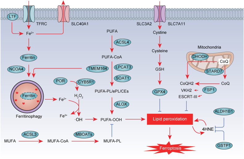
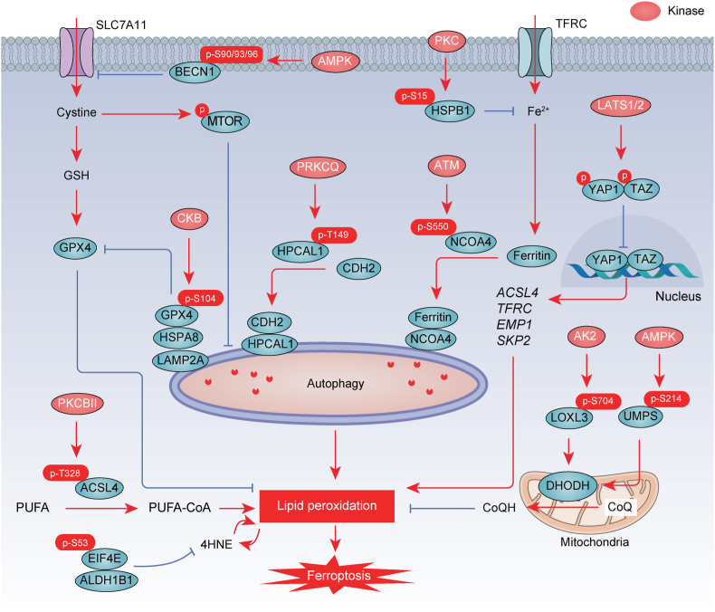
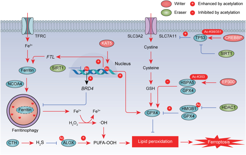
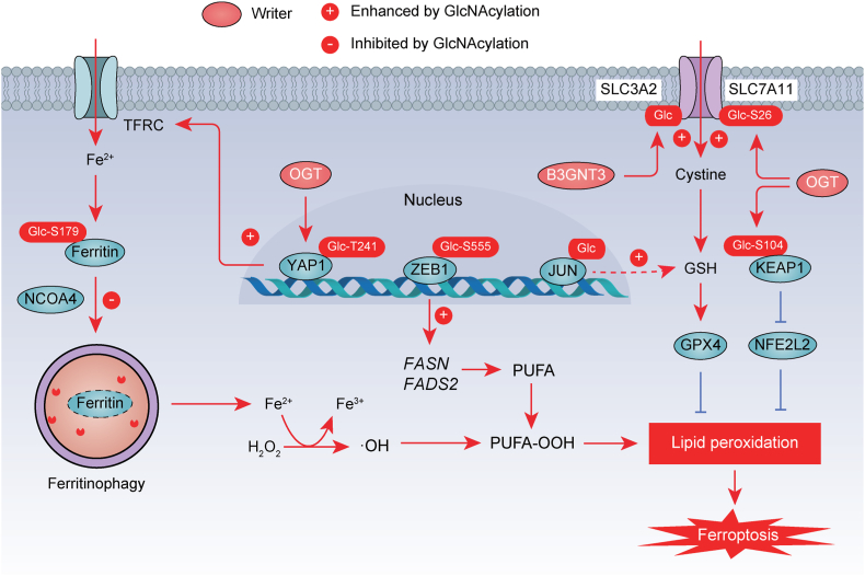
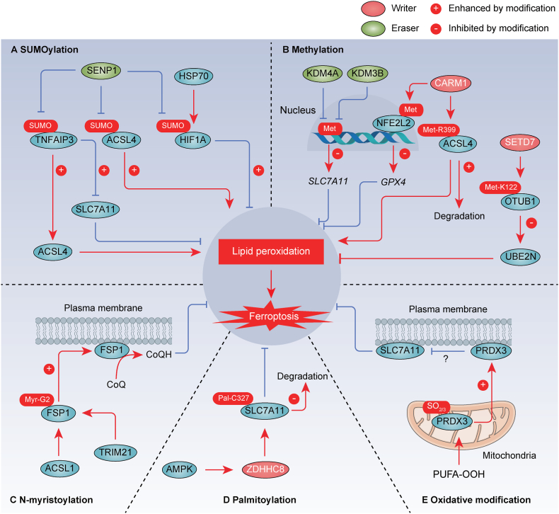
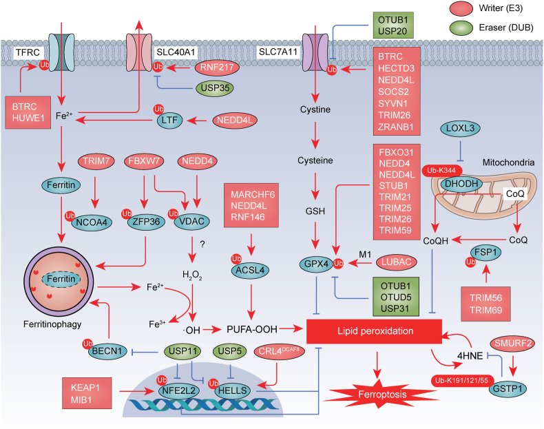
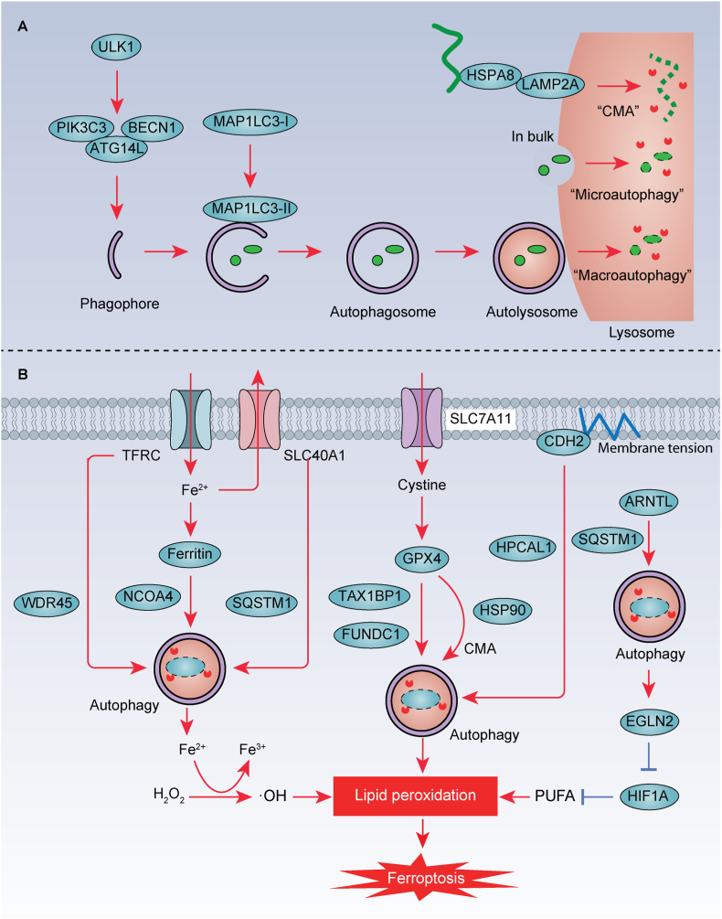
Ferroptosis is a form of iron-related oxidative cell death governed by an integrated redox system, encompassing pro-oxidative proteins and antioxidative proteins. These proteins undergo precise control through diverse post-translational modifications, including ubiquitination, phosphorylation, acetylation, O-GlcNAcylation, SUMOylation, methylation, N-myristoylation, palmitoylation, and oxidative modification. These modifications play pivotal roles in regulating protein stability, activity, localization, and interactions, ultimately influencing both the buildup of iron and lipid peroxidation. In mammalian cells, regulators of ferroptosis typically undergo degradation via two principal pathways: the ubiquitin-proteasome system, which handles the majority of protein degradation, and autophagy, primarily targeting long-lived or aggregated proteins. This comprehensive review aims to summarize recent advances in the post-translational modification and degradation of proteins linked to ferroptosis. It also discusses strategies for modulating ferroptosis through protein modification and degradation systems, providing new insights into potential therapeutic applications for both cancer and non-neoplastic diseases.

Keywords: Autophagy; Degradation; Ferroptosis; Modification; Proteasome.


Conflict of interest statement

Declaration of competing interest The authors declare no competing interests.

Figures

Fig. 1
Fig. 1
The process of ferroptosis. Ferroptosis is a form of nonapoptotic cell death characterized by overwhelming membrane lipid peroxidation and iron accumulation. Ferroptosis involves an imbalance in the oxidation and antioxidant systems. TFRC and LTF mainly promote ferroptosis by transferring Fe2+ into cells, while ferritin and SLC40A1 by storing iron and transporting iron out of cells, respectively. NCOA4-mediated ferritinophagy may play a role in regulating cellular iron levels by targeting ferritin for degradation. Subsequently, Fe2+ generates ROS through Fenton reaction. In particular, ACSL4, LPCAT3, SOAT1, TMEM164, ALOX, and POR pathways mediate the peroxidation of polyunsaturated fatty acids (PUFA), which is necessary for iron-mediated oxidative damage in ferroptosis. To counteract this oxidative stress, antioxidant defense systems have been identified, including SLC7A11-GSH-GPX4 and CoQ. Moreover, phospholipid modifying enzymes ACSL3 and MBOATs inhibit ferroptosis activity by promoting MUFA-PL synthesis. In the intricate network regulating lipid metabolism, ALDH1B1 and GSTP1 emerge as key players in ferroptosis. Their actions restrict the generation of 4-hydroxynonenal (4HNE), effectively acting as suppressors of ferroptosis.
Fig. 2
Fig. 2
Phosphorylation in ferroptosis. Direct phosphorylation of GPX4, ACSL4, BECN1, HPCAL1, PKM2, NCOA4, HSPB1, LOXL3, EIF4E, and UMPS by various kinases is involved in the regulation of ferroptosis. P, phosphorylation.
Fig. 3
Fig. 3
Acetylation in ferroptosis. The acetylation of histones, ALOX, TP53, HSPA5, and HMGB1, orchestrated by acetyltransferases and deacetylases, plays a pivotal role in the regulation of ferroptosis. The promotion or inhibition of protein function by acetylation is indicated by a positive (+) or negative (−) sign, respectively. Ac, acetylation.
Fig. 4
Fig. 4
O-GlcNAcylation in ferroptosis. O-GlcNAcylation mediated by O-linked N-acetylglucosamine transferase (OGT) plays a crucial role in regulating ferroptosis through modifying ferritin, YAP1, ZEB1, JUN, SLC3A2, and SLC7A11. The promotion or inhibition of protein function by acetylation is indicated by a positive (+) or negative (−) sign, respectively. The promotion or inhibition of protein function by O-GlcNAcylation is indicated by a positive (+) or negative (−) sign, respectively. Ac, acetylation.
Fig. 5
Fig. 5
SUMOylation, methylation, N-myristoylation, palmitoylation, and oxidative modification in ferroptosis. A. SUMOylation modifies various regulatory factors (e.g., TNFAIP3/A20, ACSL4, and HIF1A) of ferroptosis. B. Methylation serves as a modifying mechanism for a range of proteins, including histone, NFE2L2/NRF2, ACSL4, and OTUB1, influencing the processes of ferroptosis. C. N-myristoylation of FSP1 at Gly2 recruits it to the plasma membrane, where it functions as an oxidoreductase to generate CoQH2 parallel to GSH system. D. ZDHHC8 catalyzes the S-palmitoylation of SLC7A11 specifically at C327 and diminishes the ubiquitination levels to stabilize SLC7A11. E. PRDX3 yields cysteine oxidation to sulfinic acid (-SO2) and sulfonic acid (-SO3), which induces its translocation from mitochondria to plasma membrane, where it inhibits cystine uptake. The promotion or inhibition of protein function by the modification is indicated by a positive (+) or negative (−) sign, respectively. SUMO, SUMOylation. Met, methylation. Myr, N-myristoylation. Pal, palmitoylation.
Fig. 6
Fig. 6
UPS-mediated protein degradation in ferroptosis. The ubiquitination of proteins is important for the initiation of ferroptosis, which is regulated by various E3 ubiquitin ligases and deubiquitinating enzymes (DUBs). The ubiquitin-proteasome system (UPS) is involved in the degradation and stability of key regulators governing iron metabolism (e.g., TFRC, LTF, and SLC40A1), autophagy (e.g., NCOA4, and ZFP36, and BECN1), antioxidant system (e.g., SLC7A11, GPX4, FSP1, DHODH, and GSTP1), and transcription (e.g., NFE2L2/NRF2 and HELLS/LSH), as well as other proteins, including ACLS4 and VDAC. Ub, ubiquitination.
Fig. 7
Fig. 7
Autophagy-mediated degradation in ferroptosis. A. The process of autophagy, including macroautophagy, microautophagy, and chaperone-mediated autophagy (CMA). B. Selective autophagy is involved in regulating important players in iron metabolism (e.g., TFRC, ferritin, and SLC40A1), antioxidant system (e.g., GPX4), membrane tension (e.g., CDH2), and lipid metabolism (e.g., ARNTL) in the process of ferroptotic cell death. Furthermore, CTSB and cysteine released by lysosome can promote and inhibit ferroptosis, respectively.

References

    1. Galluzzi L., Vitale I., Aaronson S.A., Abrams J.M., Adam D., Agostinis P., Alnemri E.S., Altucci L., Amelio I., Andrews D.W., Annicchiarico-Petruzzelli M., Antonov A.V., Arama E., Baehrecke E.H., Barlev N.A., Bazan N.G., Bernassola F., Bertrand M.J.M., Bianchi K., Blagosklonny M.V., Blomgren K., Borner C., Boya P., Brenner C., Campanella M., Candi E., Carmona-Gutierrez D., Cecconi F., Chan F.K., Chandel N.S., Cheng E.H., Chipuk J.E., Cidlowski J.A., Ciechanover A., Cohen G.M., Conrad M., Cubillos-Ruiz J.R., Czabotar P.E., D'Angiolella V., Dawson T.M., Dawson V.L., De Laurenzi V., De Maria R., Debatin K.M., DeBerardinis R.J., Deshmukh M., Di Daniele N., Di Virgilio F., Dixit V.M., Dixon S.J., Duckett C.S., Dynlacht B.D., El-Deiry W.S., Elrod J.W., Fimia G.M., Fulda S., Garcia-Saez A.J., Garg A.D., Garrido C., Gavathiotis E., Golstein P., Gottlieb E., Green D.R., Greene L.A., Gronemeyer H., Gross A., Hajnoczky G., Hardwick J.M., Harris I.S., Hengartner M.O., Hetz C., Ichijo H., Jaattela M., Joseph B., Jost P.J., Juin P.P., Kaiser W.J., Karin M., Kaufmann T., Kepp O., Kimchi A., Kitsis R.N., Klionsky D.J., Knight R.A., Kumar S., Lee S.W., Lemasters J.J., Levine B., Linkermann A., Lipton S.A., Lockshin R.A., Lopez-Otin C., Lowe S.W., Luedde T., Lugli E., MacFarlane M., Madeo F., Malewicz M., Malorni W., Manic G., Marine J.C., Martin S.J., Martinou J.C., Medema J.P., Mehlen P., Meier P., Melino S., Miao E.A., Molkentin J.D., Moll U.M., Munoz-Pinedo C., Nagata S., Nunez G., Oberst A., Oren M., Overholtzer M., Pagano M., Panaretakis T., Pasparakis M., Penninger J.M., Pereira D.M., Pervaiz S., Peter M.E., Piacentini M., Pinton P., Prehn J.H.M., Puthalakath H., Rabinovich G.A., Rehm M., Rizzuto R., Rodrigues C.M.P., Rubinsztein D.C., Rudel T., Ryan K.M., Sayan E., Scorrano L., Shao F., Shi Y., Silke J., Simon H.U., Sistigu A., Stockwell B.R., Strasser A., Szabadkai G., Tait S.W.G., Tang D., Tavernarakis N., Thorburn A., Tsujimoto Y., Turk B., Vanden Berghe T., Vandenabeele P., Vander Heiden M.G., Villunger A., Virgin H.W., Vousden K.H., Vucic D., Wagner E.F., Walczak H., Wallach D., Wang Y., Wells J.A., Wood W., Yuan J., Zakeri Z., Zhivotovsky B., Zitvogel L., Melino G., Kroemer G. Molecular mechanisms of cell death: recommendations of the nomenclature committee on cell death 2018. Cell Death Differ. 2018;25:486–541. doi: 10.1038/s41418-017-0012-4. - DOI - PMC - PubMed
    1. Tang D., Kang R., Berghe T.V., Vandenabeele P., Kroemer G. The molecular machinery of regulated cell death. Cell Res. 2019;29:347–364. doi: 10.1038/s41422-019-0164-5. - DOI - PMC - PubMed
    1. Yuan J., Ofengeim D. A guide to cell death pathways. Nat. Rev. Mol. Cell Biol. 2023 doi: 10.1038/s41580-023-00689-6. - DOI - PubMed
    1. Dixon S.J., Lemberg K.M., Lamprecht M.R., Skouta R., Zaitsev E.M., Gleason C.E., Patel D.N., Bauer A.J., Cantley A.M., Yang W.S., Morrison B., 3rd, Stockwell B.R. Ferroptosis: an iron-dependent form of nonapoptotic cell death. Cell. 2012;149:1060–1072. doi: 10.1016/j.cell.2012.03.042. - DOI - PMC - PubMed
    1. Dai E., Han L., Liu J., Xie Y., Kroemer G., Klionsky D.J., Zeh H.J., Kang R., Wang J., Tang D. Autophagy-dependent ferroptosis drives tumor-associated macrophage polarization via release and uptake of oncogenic KRAS protein. Autophagy. 2020;16:2069–2083. doi: 10.1080/15548627.2020.1714209. - DOI - PMC - PubMed

LinkOut - more resources