. 2024 Jul 1;12(7):e009243.
doi: 10.1136/jitc-2024-009243.

Mapping the complexity and diversity of tertiary lymphoid structures in primary and peritoneal metastatic gastric cancer

Affiliations

Mapping the complexity and diversity of tertiary lymphoid structures in primary and peritoneal metastatic gastric cancer

Tessa S Groen-van Schooten et al. J Immunother Cancer. .

Abstract

Background: Tertiary lymphoid structures (TLSs) are thought to stimulate antitumor immunity and positively impact prognosis and response to immune checkpoint blockade. In gastric cancers (GCs), however, TLSs are predominantly found in GC with poor prognosis and limited treatment response. We, therefore, hypothesize that immune cell composition and function of TLS depends on tumor location and the tumor immune environment.

Methods: Spatial transcriptomics and immunohistochemistry were used to characterize the phenotype of CD45+ immune cells inside and outside of TLS using archival resection specimens from GC primary tumors and peritoneal metastases.

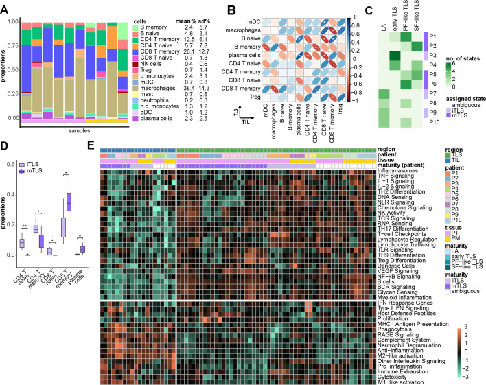
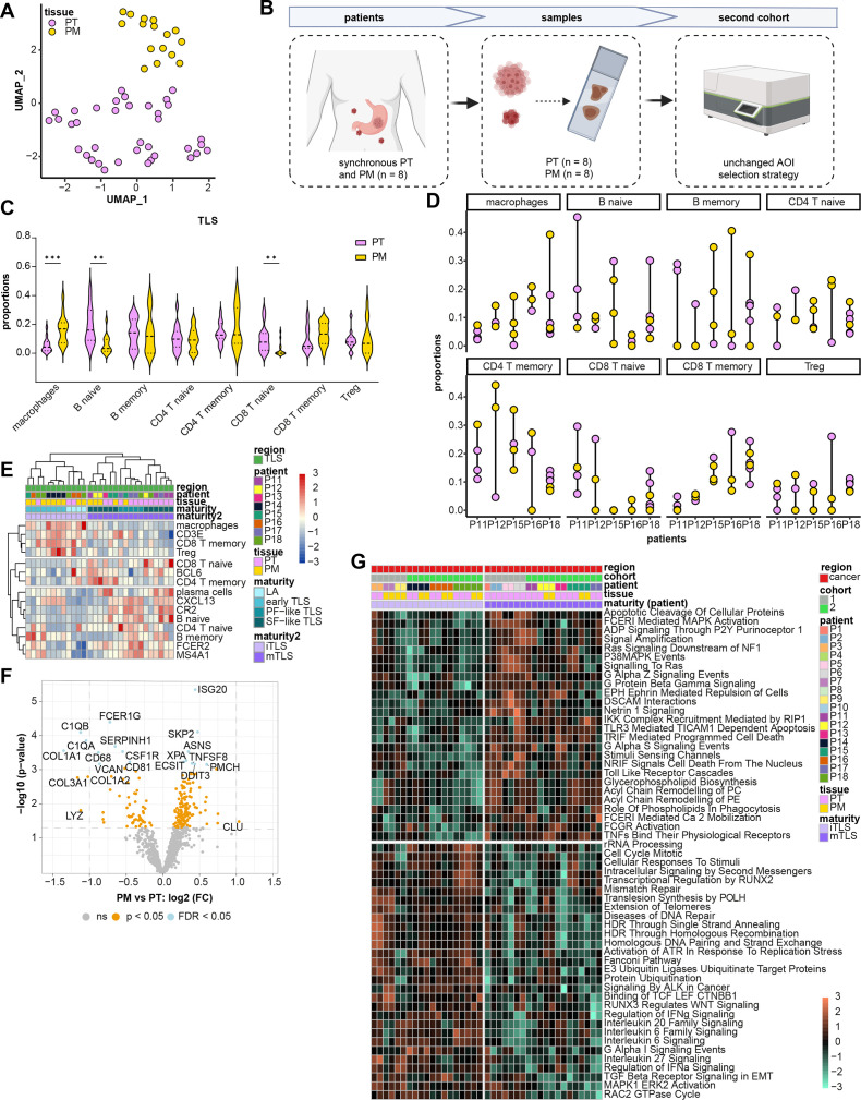
Results: We identified significant intrapatient and interpatient diversity of the cellular composition and maturation status of TLS in GC. Tumor location (primary vs metastatic site) accounted for the majority of differences in TLS maturity, as TLS in peritoneal metastases were predominantly immature. This was associated with higher levels of tumor-infiltrating macrophages and Tregs and less plasma cells compared with tumors with mature TLS. Furthermore, mature TLSs were characterized by overexpression of antitumor immune pathways such as B cell-related pathways, MHC class II antigen presentation while immature TLS were associated with protumor pathways, including T cell exhaustion and enhancement of DNA repair pathways in the corresponding cancer.

Conclusion: The observation that GC-derived peritoneal metastases often contain immature TLS which are associated with immune suppressive regulatory tumor-infiltrating leucocytes, is in keeping with the lack of response to immune checkpoint blockade and the poor prognostic features of peritoneal metastatic GC, which needs to be taken into account when optimizing immunomodulatory strategies for metastatic GC.

Keywords: Adenocarcinoma; B cell; Gastric Cancer; Immunosuppression; Tumor microenvironment - TME.


Conflict of interest statement

Competing interests: None declared.

Figures

Figure 1
Figure 1
Structural and cellular characteristics of TLS in GC PTs and PMs. (A) Cohort of PT (n=6) and unmatched PM (n=4) were characterized using single IHC featuring HE, CD20, PNAd, FDC and DC-LAMP and multiplex IHC using antibodies for CK, CD163, FoxP3, CD3, CD8 and Ki67 (B-C, scaling bar indicates 50 µm). (D) Quantification of number of CD163+, CD3+, CD8+ and FoxP3+ cells within TLS per tumor (n=4–7 TLS analyzed per tumor). FDC, follicular dendritic cells; GC, gastric cancer; IHC, immunohistochemistry; PM, peritoneal metastases; PT, primary tumors; LAMP, lysosomal-associated membrane protein; TLS, tertiary lymphoid structures.
Figure 2
Figure 2
Transcriptomic characterization of TLS in PT and PM. (A) ROI selection of TLS, TIL and tumor using CD45 (pink) and CK (green). (B) TLS subsets, showing mean proportions per patient and annotations for PT (pink) and PM (yellow). (C) Supervised maturation clustering of TLS-associated markers and subset proportions (n=53). (D) UMAP of TLS-ROIs. (E) Differences in proportions of immune subsets across maturity subgroups (pairwise Wilcoxon, FDR correction, *p<0.05, **p<0.01, ***p<0.001). (F) Differential pathway analysis between mTLS and iTLS (p<0.05; those with an * FDR<0.05). (G) Chemokine signature expression among TLS. CK, cytokeratin; FDR, false discovery rate; iTLS, immature TLS; mTLS, mature TLS; n.c., non-classical; PM, peritoneal metastases; PT, primary tumor; ROIs, regions of illumination; TLS, tertiary lymphoid structure.
Figure 3
Figure 3
Relationships between immune features of leukocytes within TLS and outside TLS (tumor-infiltrating leukocytes (TIL)). (A) TIL subsets, showing mean proportions per patient and annotations for PT (pink) and PM (yellow). (B) Spearman correlation between subset proportions in TLS (y-axis) and TIL (x-axis), correlating means per patient (n=10, *p<0.05 and r<−0.6 or >0.6). Red and blue ovals indicate negative and positive correlations, respectively. (C) Maturity states of TLS per patient, assigning the most common state per patient. (D) Comparison of TIL-ROIs in patients with mTLS (n=8) vs iTLS (n=6, Wilcoxon test, *p<0.05, **p<0.01). (E) Differential pathways contrasting TLS vs TIL, FDR<0.05. FDR, false discovery rate; iTLS, immature TLS; mTLS, mature TLS; PM, peritoneal metastases; PT, primary tumor; TIL, tumor-infiltrating leukocyte; TLS, tertiary lymphoid structure.
Figure 4
Figure 4
TLS in GC-derived PT and PM. (A) UMAP of TLS-ROIs from the first cohort (n=53). (B) Set-up of the second cohort containing matched PT and PM samples. (C) Subset proportions from TLS-ROIs from PT (n=15) and PM (n=14). **p<0.01, ***p<0.001. (D) Subset proportions from TLS-ROIs from matched PT (n=5) and PM (n=5). **p<0.01, ***p<0.001. (E) Maturation clustering of TLS-associated markers and subset proportions. (F) Differential gene expression of PT-derived and PM-derived cancer cell ROIs. (G) Differential pathway analysis contrasting cancer cell ROIs from tumors with mTLS vs iTLS. FDR<0.05. FC, fold change; FDR, false discovery rate; PM, peritoneal metastases; PT, primary tumors; ROIs, regions of illumination; TLS, tertiary lymphoid structure; UMAP, uniform manifold approximation and projection.

References

    1. Sung H, Ferlay J, Siegel RL, et al. . Global cancer statistics 2020: GLOBOCAN estimates of incidence and mortality worldwide for 36 cancers in 185 countries. CA A Cancer J Clinicians 2021;71:209–49. 10.3322/caac.21660 - DOI - PubMed
    1. Fridman WH, Meylan M, Pupier G, et al. . Tertiary lymphoid structures and B cells: an intratumoral immunity cycle. Immunity 2023;56:2254–69. 10.1016/j.immuni.2023.08.009 - DOI - PubMed
    1. Sautès-Fridman C, Petitprez F, Calderaro J, et al. . Tertiary lymphoid structures in the era of cancer immunotherapy. Nat Rev Cancer 2019;19:307–25. 10.1038/s41568-019-0144-6 - DOI - PubMed
    1. N J, J T, Sl N, et al. . Tertiary lymphoid structures and B lymphocytes in cancer prognosis and response to immunotherapies. OncoImmunology 2021;10:1900508. 10.1080/2162402X.2021.1900508 - DOI - PMC - PubMed
    1. Engelhard VH, Rodriguez AB, Mauldin IS, et al. . Immune cell infiltration and tertiary lymphoid structures as determinants of antitumor immunity. J Immunol 2018;200:432–42. 10.4049/jimmunol.1701269 - DOI - PMC - PubMed