. 2024 Jul 2;15(1):5530.
doi: 10.1038/s41467-024-48720-4.

Cryo-EM unveils kinesin KIF1A's processivity mechanism and the impact of its pathogenic variant P305L

Affiliations

Cryo-EM unveils kinesin KIF1A's processivity mechanism and the impact of its pathogenic variant P305L

Matthieu P M H Benoit et al. Nat Commun. .

Abstract

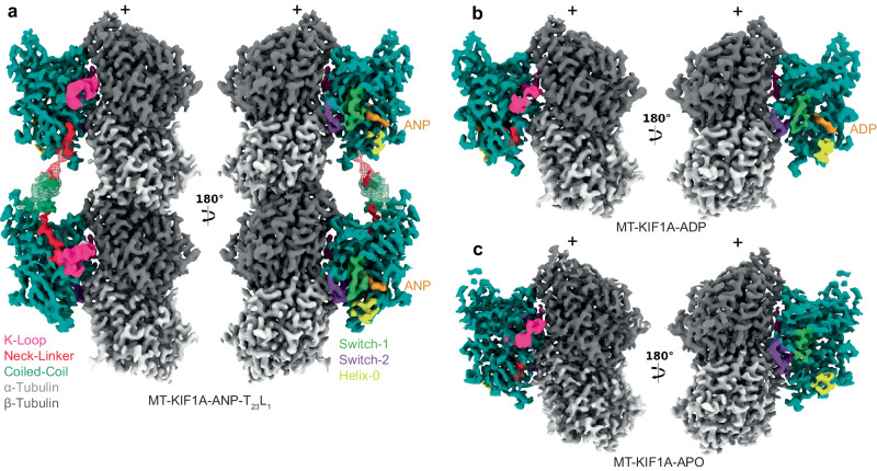
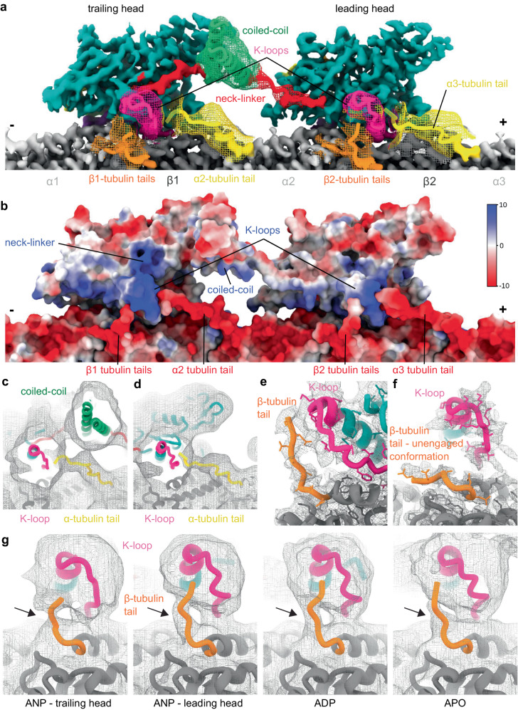
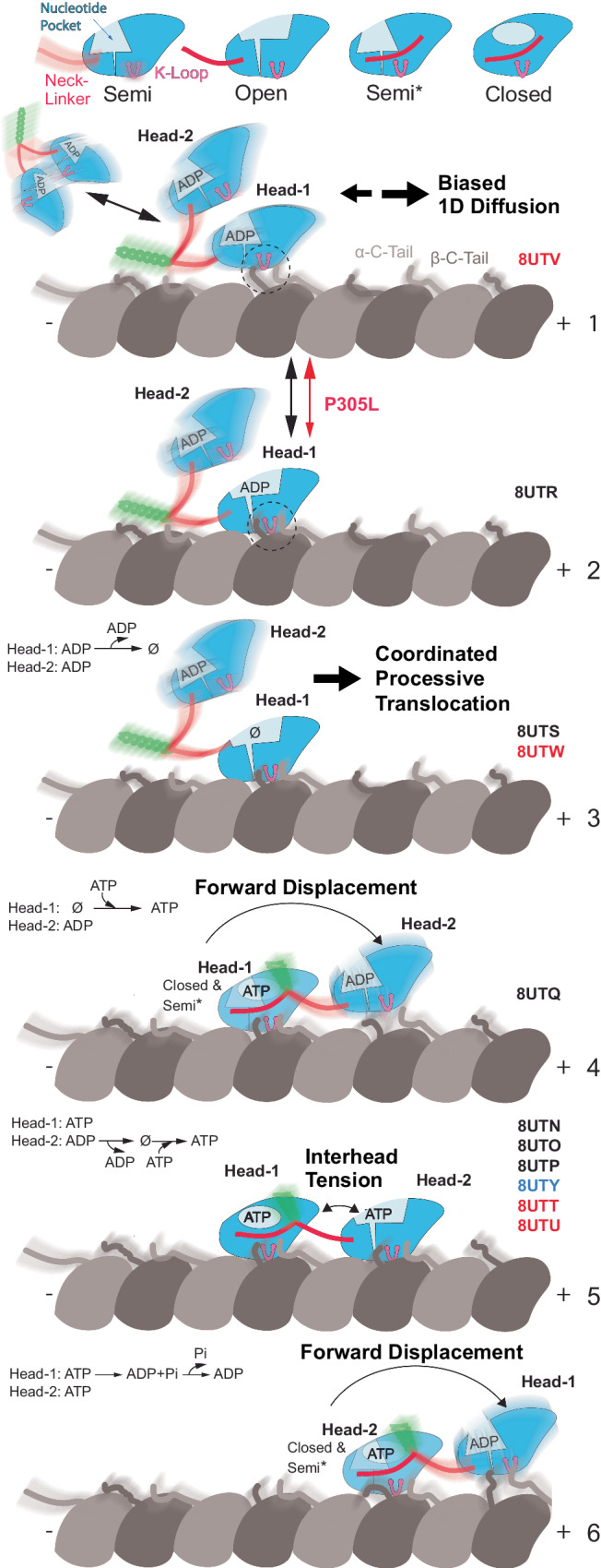
Mutations in the microtubule-associated motor protein KIF1A lead to severe neurological conditions known as KIF1A-associated neurological disorders (KAND). Despite insights into its molecular mechanism, high-resolution structures of KIF1A-microtubule complexes remain undefined. Here, we present 2.7-3.5 Å resolution structures of dimeric microtubule-bound KIF1A, including the pathogenic P305L mutant, across various nucleotide states. Our structures reveal that KIF1A binds microtubules in one- and two-heads-bound configurations, with both heads exhibiting distinct conformations with tight inter-head connection. Notably, KIF1A's class-specific loop 12 (K-loop) forms electrostatic interactions with the C-terminal tails of both α- and β-tubulin. The P305L mutation does not disrupt these interactions but alters loop-12's conformation, impairing strong microtubule-binding. Structure-function analysis reveals the K-loop and head-head coordination as major determinants of KIF1A's superprocessive motility. Our findings advance the understanding of KIF1A's molecular mechanism and provide a basis for developing structure-guided therapeutics against KAND.


Conflict of interest statement

The authors declare no competing interests.

Figures

Fig. 1
Fig. 1. Cryo-EM maps of microtubule-bound KIF1A in different nucleotide states.
Each panel shows two views of the isosurfaces of microtubule-bound KIF1A 3D maps, rotated 180° relative to each other. The surface colors emphasize different structural elements, as indicated in the figure labels. Map densities around the K-loop have been low-passed filtered and displayed at a lower contour level than the rest of the map for enhanced visualization of this mobile part. a Microtubule-bound KIF1A with the ATP analog AMP-PNP (abbreviated as ANP). The map corresponds to the average two-heads-bound configuration (MT-KIF1A-ANP-T23L1). The coiled-coil density and parts of the neck-linker from the leading head have been low-pass filtered and are displayed as a mesh at a lower contour level than that of the main map. Densities from the neck-linkers and coiled-coils connecting the two heads are visible. ANP densities are present in both the leading and trailing heads. Note: One-head-bound configurations were also found in the ANP datasets (Supplementary Figs. 3, 4, Supplementary Table 3). b, c Isosurface representations of KIF1A in the ADP and APO states (datasets MT-KIF1A-ADP and MT-KIF1A-APO). Isosurfaces are colored according to the corresponding structural element in the fitted atomic model, α- and β- tubulin in light and dark gray, respectively, the KIF1A motor domain in cyan with distinct structural elements (the K-loop, the neck-linker, the coiled-coil, the Switch-1, the Switch-2, and the bound nucleotide) colored as indicated in the insert.
Fig. 2
Fig. 2. KIF1A atomic models.
a ANP two-heads-bound configuration (MT-KIF1A-ANP-T23L1). b ADP one-head-bound configuration (MT-KIF1A-ADP). c Apo one-head-bound configuration (MT-KIF1A-APO). d Scatter plot illustrating the average of six distances between Cα carbons of selected residues (R216 and A250 to P14, to S104 and to Y105) across the KIF1A nucleotide-binding pocket (see inset). The average of these distances provides an estimate of the degree of opening of the KIF1A’s nucleotide binding pocket (NP Openness). According to these measurements, the structures can be grouped into three groups, Open, Closed and Semi. The Open group consists of the MT-KIF1A motor domain structures in the ADP and Apo states, and the leading head in the two-heads-bound configuration of the ANP state (three structure classes). The only KIF1A-MT structure in the Semi group corresponds to a class of the ANP state in a single-head-bound configuration (MT-KIF1A-ANP-T1L02*). The cross symbols in the ‘PDB’ column represent KIF1A or KIF1A-MT models deposited in the PDB database, with accession codes: 1I5S, 1I6I, 1IA0, 1VFV, 1VFW, 1VFX, 1VFZ, 2HXF, 2HXH, 2ZFI, 2ZFJ, 2ZFK, 2ZFL, 2ZFM, 4UXO, 4UXP, 4UXR, 4UXS, 7EO9 and 7EOB. Structural elements surrounding the nucleotide pocket are colored as indicated in the inset. All atomic models are colored, as in Fig. 1.
Fig. 3
Fig. 3. Neck-linker structure.
a H6, neck-linker (NL) and coiled-coil (CC) region of the KIF1A ANP two-heads-bound structure (MT-KIF1A-ANP-T23L1). The structure is presented as a ribbon representation with side chain atoms as sticks and color coded as in Fig. 1. The cryo-EM map is shown as an isosurface semitransparent mesh. A composite low-pass filtered version of the map highlights the NL and CC areas, with model side chains omitted. b Comparison of the two-heads-bound configuration structures of KIF1A and KIF14 (gray color, PDB accession code: 6WWL). c Comparison of KIF1A’s trailing head in the two-heads-bound configuration with KIF5B’s docked NL and CC helix crystal structure (shown in gray; PDB accession code: 1MKJ). d Sequence alignment of NL and CC for KIF1A, KIF14 and KIF5B. Residues in the NL or the CC helix, as identified in structures (a) to (c), are colored red and green, respectively. e Kymograph examples showing WT and P364L variant. f Velocities and run-lengths of WT and P364L variant. Green bars represent mean values for velocity or median values for run length with their respective 95% confidence interval (CI). Velocities: WT: 1.91 [1.89, 1.93] µm/s, n = 459; P364L: 1.92 [1.90, 1.94] µm/s n = 514. Run lengths: WT: 14.6 [13.3, 16.1] µm, n = 459; P364L: 10.2 [9.4, 11.0] µm, n = 514. A minimum of three experiments were performed for each construct. Statistical analysis was conducted using an unpaired two-tailed Welch’s test (P = 0.47) for velocities and the Kolmogorov-Smirnov test for run lengths (****P < 0.0001). g Superimposed ANP two-head-bound structure models of WT (MT-KIF1A-ANP-T23L1) and P364L (MT-KIF1AP364L-ANP-TL1), aligned by their tubulin components. The WT structure is colored blue and red, while the P364L variant is represented in gray.
Fig. 4
Fig. 4. K-loop structure.
a Area of the K-loop of the KIF1A-ADP model. The model is shown as a ribbon representation with side chain atoms as stick. Parts colored as in Fig. 1. Cryo-EM density map is represented as a semi-transparent isosurface mesh, with a low-pass filtered version of the map emphasizing the K-loop tip area (left-most section). Model side chains are omitted in this area. The 310-helix, a component of loop-12, is a highly conserved sequence and structural motif within the kinesin superfamily. b Structural comparison of K-loop regions in HsKIF1A, MmKIF14 (PDB accession code: 6WWM) and HsKIF5B (PDB accession code: 1MKJ). Positively-charged residues (K or R) within the K-loop are highlighted in dark blue. A conserved positively-charged residue in kinesin-3’s loop-12 is marked with a * symbol, and a highly conserved R residue in the kinesin superfamily is indicated by an arrowhead. c Sequence alignments of loop-12 across different kinesins, and K-loop-swap mutant (KLS). Positively-charged residues are colored dark blue, with * and arrowhead symbols denoting the same residues marked in the structure (b). d Kymograph examples of KLS, KLS-P364L, and KIF5B. e Velocities and run lengths of KLS, KLS-P364L, and KIF5B. Green bars represent the mean values for velocity or median values for run length with their respective 95% CIs. Velocity: KLS: 2.09 [2.06, 2.11] µm/s n = 398; KLS-P364L: 1.58 [1.56, 1.61] µm/s n = 517; KIF5B: 0.53 [0.52, 0.54] µm/s n = 232. Run-lengths: KLS: 2.7 [2.4, 2.9] µm n = 398; KLS-P364L: 1.7 [1.6, 1.8] µm n = 517; KIF5B: 1.3 [1.2, 1.5] µm n = 232. A minimum of three experiments were performed for each construct. Statistical analysis was conducted using an unpaired two-tailed Welch’s test for velocities and Kolmogorov–Smirnov test for run lengths (****P < 0.0001).
Fig. 5
Fig. 5. Interaction of the K-loop with the C-terminal tubulin tails.
a Composite cryo-EM map of KIF1A microtubule two-heads-bound configuration (MT-KIF1A-ANP-T23L1) interacting with three tubulin dimers. The locally filtered map is displayed as a solid color. The portions with weaker signals were low-pass filtered and displayed as meshes with the underlying model displayed as a ribbon representation. The coiled-coil and β-tubulin tails interacting with the kinesin were low-passed filtered to 6 Å, the α-tubulin tail densities were low-passed filtered to 8 Å and displayed at a low-density threshold. b Representation of the Coulombic electrostatic surface potential of the KIF1A two-heads-bound configuration model. Color scale in kcal·mol−1e−1. c, d 8 Å low-passed filtered cryo-EM map of KIF1A two-heads-bound configuration (MT-KIF1A-ANP-T23L1), showing densities of α-tubulin C-terminal tails reaching the K-loops of the trailing (c) or leading (d) head. e Cryo-EM map of the leading head of the P364L mutant (MT-KIF1AP364L-ANP-TL1) showing a mostly resolved β-tubulin tail conformation interacting with the K-loop. f Cryo-EM map of the trailing head of KIF1A two-heads-bound configuration (MT-KIF1A-ANP-T23L1) showing a portion of a β-tubulin tail conformation lying along the microtubule surface. g 6 Å low-passed filtered map of KIF1A near the K-loop in distinct nucleotide states and motor-domain conformations. The density associated with the β-tubulin C-terminal tail is pointed with an arrow and the threshold was adjusted for each map. Note that in the ANP state the β-tubulin tail density is better resolved in the leading head than in the trailing head.
Fig. 6
Fig. 6. Cryo-EM maps of microtubule-bound KIF1AP305L in different nucleotide states.
a MT-bound KIF1AP305L with ANP. The map represents the two-heads-bound configuration MT-KIF1AP305L-ANP-TL1. The coiled-coil density and part of the neck-linker from the leading head were low-pass filtered and displayed as a mesh. b Isosurface representation of KIF1AP305L in the ADP state (dataset MT-KIF1AP305L-ADP) with the KIF1A switches area displayed as a low-pass filtered mesh. The K-loop area and C-terminal tail of β-tubulin, with connected densities, are low-passed filtered and displayed as a solid surface. c Isosurface representations of KIF1AP305L in the Apo state (dataset MT-KIF1AP305L-APO), illustrated as in (a). d Isosurface representations of the cryo-EM map of WT KIF1A in the ANP state (MT-KIF1A-ANP-T23L1) in the neighborhood of residue P305. The surface is represented as a mesh and colored based on the underlying fitted molecular model (same color convention as in Fig. 1). The model is shown as ribbon representation with displayed side chains. The left and right panels represent two different views of the same area, the left one looking from β-tubulin, on an axis orthogonal to the microtubule axis, and the right one along the microtubule axis towards the microtubule minus end. e Isosurface representations of the cryo-EM map of KIF1AP305L mutant in the ANP state (MT-KIF1AP305L-ANP-TL012*), same view as in (d). f Overlay of the models displayed in (d) and (e) with the KIF1A WT model displayed in gray, the KIF1AP305L mutant model in pink and the residue 305 colored as in (d, e). g Kymograph examples of P305L, F303V/P305L and F303V. h Distribution of velocities and run lengths. The green bars represent the mean for velocity or median values for run length with their respective 95% confidence interval. Velocity: P305L: 0.95 [0.93, 0.96] µm/s, n = 438; F303V/P305L: 0.98 [0.96, 0.99] µm/s n = 441; F303V: 1.58 [1.57, 1.59] µm/s n = 557; WT: 1.91 [1.89, 1.93] µm/s, n = 459. The statistics were performed using an unpaired two-tailed Welch’s test (****P < 0.0001; *P = 0.016). At least 3 experiments were performed for each construct, n is the total number of measurements pooled from all experiments for each construct. Run lengths: P305L: 2.0 [1.9, 2.2] µm n = 438; F303V/P305L: 3.9 [3.5, 4.4] µm n = 441; F303V: 9.5 [8.8, 10.3] µm n = 557; WT: 14.6 [13.3, 16.1] µm n = 459. The statistics were performed using Kolmogorov–Smirnov test (****P < 0.0001).
Fig. 7
Fig. 7. A model for KIF1A’s mechanochemical cycle.
The upper row displays the four primary conformations of the KIF1A motor domain, named based on the degree of openness of the nucleotide-binding pocket. Two types of KIF1A-MT interactions or binding modes can be inferred from the structures, a strong binding mode mediated by stereospecific contacts between the MT and the whole KIF1A-MT interface, and a weak-binding mode mediated by flexible electrostatic interactions between the tubulin C-terminal tails, the KIF1A K-loop and possibly the neck-linker and the start of the coiled-coil domain. Regions of the structures with high mobility are illustrated with a blurred depiction. The PDB accession codes of the structure(s) representing each model step are displayed to the right, with different font colors indicating the KIF1A construct used, black for WT, red for the P305L mutant, and blue for the P364L mutant. The cycle starts with a KIF1A dimer in the ADP state interacting with the MT in a weakly bound mode (state 1). In this state, KIF1A could engage in biased one-dimensional diffusion (Supp. Fig. 11) or in directed motion, where the two motor heads coordinate alternating conformations, nucleotide states, and MT binding modes to move in a hand-over-hand manner in the MT plus-end direction (steps 2 to 6). The P305L mutation alters the structure of the KIF1A MT interface, hindering the transition to the strongly bound configuration (from step 1 to step 2). Several points of the coordination between the two heads can be derived from the structures: In steps 2 and 3 one head is prevented from binding to the MT until the MT-bound head binds ATP (steps 4 and 5). In step 4, the MT-bound head with ATP (head-1) has a reduced probability to reach the fully closed conformation until the partner motor domain binds strongly to the MT in the forward position (step 5). In the two-heads MT-bound configuration (step 5), inter-head tension and the differently oriented neck-linkers maintain the two heads in distinct conformations. In this configuration, nucleotide pocket closure and ATP hydrolysis in the leading head is paused until the trailing head detaches allowing the neck-linker of the leading head to dock. After ATP hydrolysis, product release and detachment of the rear head the neck-linker of the leading head docks moving the detached head forward (step 6).

Update of

References

    1. Carabalona A, Hu DJ, Vallee RB. KIF1A inhibition immortalizes brain stem cells but blocks BDNF-mediated neuronal migration. Nat. Neurosci. 2016;19:253–262. doi: 10.1038/nn.4213. - DOI - PMC - PubMed
    1. Tsai J-W, Lian W-N, Kemal S, Kriegstein AR, Vallee RB. Kinesin 3 and cytoplasmic dynein mediate interkinetic nuclear migration in neural stem cells. Nat. Neurosci. 2010;13:1463–1471. doi: 10.1038/nn.2665. - DOI - PMC - PubMed
    1. Barkus RV, Klyachko O, Horiuchi D, Dickson BJ, Saxton WM. Identification of an axonal kinesin-3 motor for fast anterograde vesicle transport that facilitates retrograde transport of neuropeptides. Mol. Biol. Cell. 2008;19:274–283. doi: 10.1091/mbc.e07-03-0261. - DOI - PMC - PubMed
    1. Hall DH, Hedgecock EM. Kinesin-related gene unc-104 is required for axonal transport of synaptic vesicles in C. elegans. Cell. 1991;65:837–847. doi: 10.1016/0092-8674(91)90391-B. - DOI - PubMed
    1. Lo KY, Kuzmin A, Unger SM, Petersen JD, Silverman MA. KIF1A is the primary anterograde motor protein required for the axonal transport of dense-core vesicles in cultured hippocampal neurons. Neurosci. Lett. 2011;491:168–173. doi: 10.1016/j.neulet.2011.01.018. - DOI - PubMed

LinkOut - more resources