. 2024 Jul 2;15(7):465.
doi: 10.1038/s41419-024-06866-5.

RSL3 enhances ROS-mediated cell apoptosis of myelodysplastic syndrome cells through MYB/Bcl-2 signaling pathway

Affiliations

RSL3 enhances ROS-mediated cell apoptosis of myelodysplastic syndrome cells through MYB/Bcl-2 signaling pathway

Li Liu et al. Cell Death Dis. .

Abstract

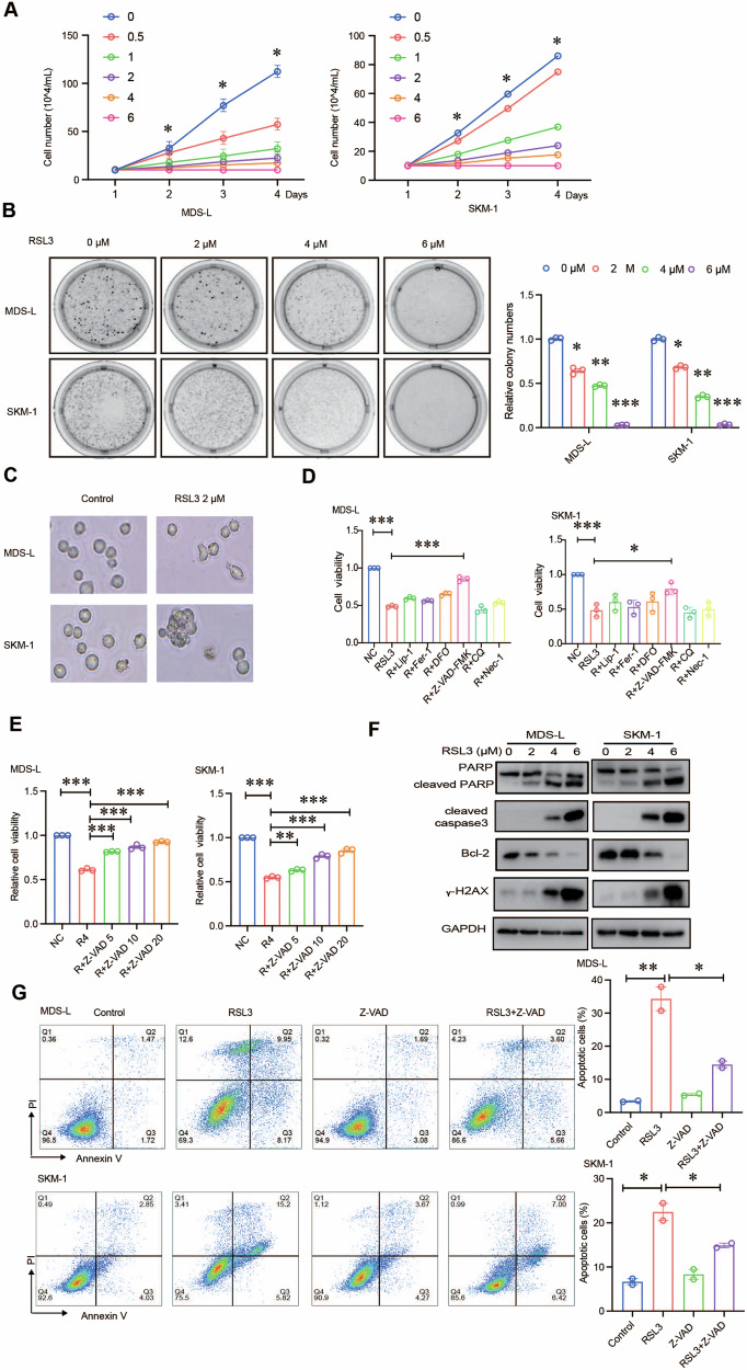
Myelodysplastic syndromes (MDS) are clonal hematopoietic malignancies and seriously threaten people's health. Current therapies include bone marrow transplantation and several hypomethylating agents. However, many elderly patients cannot benefit from bone marrow transplantation and many patients develop drug resistance to hypomethylating agents, making it urgent to explore novel therapy. RSL3 can effectively induce ferroptosis in various tumors and combination of RSL3 and hypomethylating agents is promising to treat many tumors. However, its effect in MDS was unknown. In this study, we found that RSL3 inhibited MDS cell proliferation through inducing ROS-dependent apoptosis. RSL3 inhibited Bcl-2 expression and increased caspase 3 and PARP cleavage. RNA-seq analysis revealed that MYB may be a potential target of RSL3. Rescue experiments showed that overexpression of MYB can rescue MDS cell proliferation inhibition caused by RSL3. Cellular thermal shift assay showed that RSL3 binds to MYB to exert its function. Furthermore, RSL3 inhibited tumor growth and decreased MYB and Bcl-2 expression in vivo. More importantly, RSL3 decreased the viability of bone marrow mononuclear cells (BMMCs) isolated from MDS patients, and RSL3 had a synergistic effect with DAC in MDS cells. Our studies have uncovered RSL3 as a promising compound and MYB/Bcl-2 signaling pathway as a potential target for MDS treatment.


Conflict of interest statement

The authors declare no competing interests.

Figures

Fig. 1
Fig. 1. RSL3 induces apoptosis in MDS cells.
A, B MDS cell proliferation was evaluated by manual counting and soft agar clonogenic assay. RSL3 significantly suppressed MDS-L and SKM-1 cell proliferation in a concentration-dependent manner. C Morphological changes associated with cell death after RSL3 treatment were visualized by optical microscopy. Scale bar, 100 μm. D, E MDS cells were treated with RSL3 (4 μM) in combination with Lip-1 (2 μM), Fer-1 (2 μM), DFO (100 μM), Z-VAD-FMK (10 μM), CQ (10 μM), Nec-1 (10 μM), respectively. The CCK-8 rescue assay results showed that Z-VAD-FMK notably reversed the inhibition of proliferation induced by RSL3. F Western blotting of the levels of apoptotic proteins, including cleaved PARP, cleaved caspase3, γ-H2AX, and Bcl-2. G Flow cytometric analysis showed that RSL3 increased the percentage of apoptotic cells and that Z-VAD-FMK reversed this effect after 24 h. ***p < 0.001, **p < 0.01, *p < 0.05.
Fig. 2
Fig. 2. RSL3-mediated apoptosis is dependent on ROS.
A, B Flow cytometric analysis after treatment with RSL3 alone or in combination with NAC. RSL3 increased ROS levels in MDS cells, and NAC (5 mM) partially reversed RSL3-triggered ROS accumulation. C Fluorescence microscopy analysis showed that RSL3 treatment intensified ROS fluorescence in MDS cells. NAC treatment reduced ROS fluorescence. D The effect of NAC on reversing the RSL3-mediated decrease in MDS cell viability was evaluated by a CCK-8 assay. E Cleaved PARP, Bcl-2, and cleaved caspase 3 levels were measured by western blotting after treatment with RSL3 alone or in combination with NAC. F Flow cytometric analysis showed that NAC reduced the RSL3-mediated apoptosis rates in MDS-L and SKM-1 cells. ***p < 0.001, **p < 0.01, *p < 0.05. NS not significant.
Fig. 3
Fig. 3. MYB is an efficacious target of RSL3-mediated apoptosis.
A PCA plot analysis between RSL3-treated group and the control. B Volcano plot indicated the differential genes between the RSL3-treated group and control group. The up‐regulated genes (red), and the down‐regulated genes (green) with P adjust < 0.05 and | log2 (FoldChange)| ≥ 1. C GSEA enrichment analysis showed that the differentially expressed genes were significantly enriched in apoptosis pathway. D Venn analysis showed that MYB was involved in RSL3-mediated apoptosis in MDS after RSL3 treatment and the results were shown in the heatmap. E qRT-PCR showed that RSL3 downregulated expression of MYB. F The transfection efficiency of the MYB overexpression plasmid was determined in MDS cells. G The CCK-8 rescue assay showed that cell viability, which was decreased by RSL3 treatment, could be enhanced by overexpression of MYB in MDS cells. H Flow cytometric analysis showed that MYB overexpression rescued ROS production caused by RSL3. I Western blotting showed that MYB overexpression increased the Bcl-2 protein level and decreased the level of cleaved caspase 3 and γ-H2AX and reversed the effect of apoptotic protein expression caused by RSL3. J Flow cytometric analysis showed that MYB overexpression blocked RSL3-mediated MDS cell apoptosis after 48 h. ***p < 0.001, **p < 0.01, *p < 0.05, NS not significant.
Fig. 4
Fig. 4. RSL3 reduces MYB expression via binding MYB.
A, B Western blotting showed that RSL3 inhibited MYB expression in a concentration-dependent and time-dependent manner. C Western blotting indicated that RSL3-mediated MYB degradation could be rescued by MG132 after 6 h. D MDS cells were treated by DMSO or RSL3 at 10 μM. 1 h later, they were put into the water bath at 37 °C, 42 °C, and 47 °C. As the temperature increased, the degradation of MYB protein was reduced in the RSL3-treated group.
Fig. 5
Fig. 5. RSL3 inhibits tumor growth in vivo.
A Schematic of the anticancer effect of RSL3 (20 mg/kg) in the SKM-1 cell subcutaneous xenograft model. B The body weight of mice in each group was determined every 2 days. C The tumor volume of mice in each group was calculated every 2 days. D Excised tumors on day 10. E Tumor weight of mice in each group. F, G Immunofluorescence staining showed that RSL3 treatment decreased MYB and Bcl-2 expression in the tumor tissue. Scale bar, 50 μm. H Western blotting showed that RSL3 decreased MYB and Bcl-2 expression in the tumor tissue. ***p < 0.001, **p < 0.01, *p < 0.05.
Fig. 6
Fig. 6. RSL3 treatment had therapeutic efficiency in MDS patients’ derived samples.
AD The CCK-8 assay results showed that RSL3 decreased cell viability in MDS patients but not healthy individuals. E, F Combination index (CI) values of RSL3 and DAC in MDS-L cells and SKM-1 cells. Both were treated with different concentrations of RSL3, DAC, or RSL3 plus DAC for 48 h. Cell viability was assessed using the CCK-8 assay. ***p < 0.001, **p < 0.01, *p < 0.05, NS not significant.
Fig. 7
Fig. 7. A schematic diagram of the proposed anti-MDS activity of RSL3.
RSL3 directly targets MYB protein leads its degradation through proteasome, then leads to decreased Bcl-2 expression and increased ROS production further to promote MDS cell apoptosis.

References

    1. Li H, Hu F, Gale RP, Sekeres MA, Liang Y. Myelodysplastic syndromes. Nat Rev Dis Prim. 2022;8:74. doi: 10.1038/s41572-022-00402-5. - DOI - PubMed
    1. DeRosa PA, Roche KC, Nava VE, Singh S, Liu ML, Agarwal A. Concurrent Waldenstrom’s macroglobulinemia and myelodysplastic syndrome with a sequent t(10;13)(p13;q22) translocation. Curr Oncol. 2022;29:4587–92. doi: 10.3390/curroncol29070363. - DOI - PMC - PubMed
    1. Volpe VO, Garcia-Manero G, Komrokji RS. Myelodysplastic syndromes: a new decade. Clin Lymphoma Myeloma Leuk. 2022;22:1–16. doi: 10.1016/j.clml.2021.07.031. - DOI - PubMed
    1. Liu Z, Wang P, Yuan S, Wang Y, Cao P, Wen F, et al. LncRNA BC200/miR-150-5p/MYB positive feedback loop promotes the malignant proliferation of myelodysplastic syndrome. Cell Death Dis. 2022;13:126. doi: 10.1038/s41419-022-04578-2. - DOI - PMC - PubMed
    1. Šimoničová K, Janotka Ľ, Kavcová H, Sulová Z, Breier A, Messingerova L. Different mechanisms of drug resistance to hypomethylating agents in the treatment of myelodysplastic syndromes and acute myeloid leukemia. Drug Resist Updat. 2022;61:100805. doi: 10.1016/j.drup.2022.100805. - DOI - PubMed