. 2024 Jul 2;15(1):5379.
doi: 10.1038/s41467-024-49519-z.

Intrinsic signaling pathways modulate targeted protein degradation

Affiliations

Intrinsic signaling pathways modulate targeted protein degradation

Yuki Mori et al. Nat Commun. .

Abstract

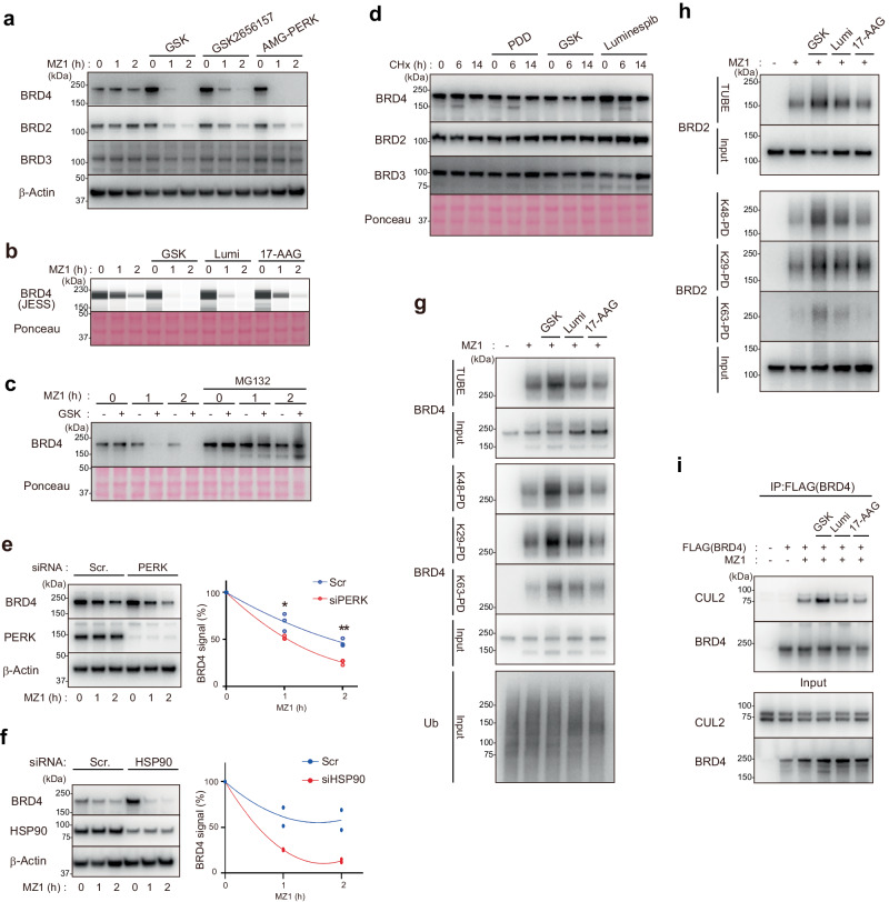
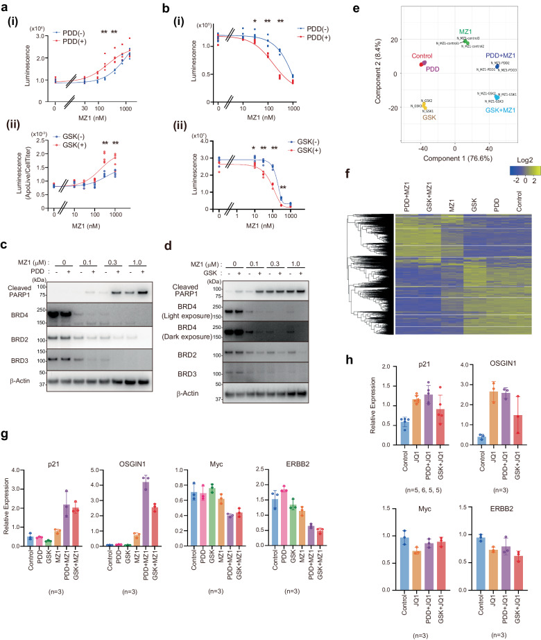
Targeted protein degradation is a groundbreaking modality in drug discovery; however, the regulatory mechanisms are still not fully understood. Here, we identify cellular signaling pathways that modulate the targeted degradation of the anticancer target BRD4 and related neosubstrates BRD2/3 and CDK9 induced by CRL2VHL- or CRL4CRBN -based PROTACs. The chemicals identified as degradation enhancers include inhibitors of cellular signaling pathways such as poly-ADP ribosylation (PARG inhibitor PDD00017273), unfolded protein response (PERK inhibitor GSK2606414), and protein stabilization (HSP90 inhibitor luminespib). Mechanistically, PARG inhibition promotes TRIP12-mediated K29/K48-linked branched ubiquitylation of BRD4 by facilitating chromatin dissociation of BRD4 and formation of the BRD4-PROTAC-CRL2VHL ternary complex; by contrast, HSP90 inhibition promotes BRD4 degradation after the ubiquitylation step. Consequently, these signal inhibitors sensitize cells to the PROTAC-induced apoptosis. These results suggest that various cell-intrinsic signaling pathways spontaneously counteract chemically induced target degradation at multiple steps, which could be liberated by specific inhibitors.


Conflict of interest statement

The authors declare no competing interests.

Figures

Fig. 1
Fig. 1. Screening of pathways modulating targeted degradation of BRD4.
a Scheme of screening using HiBiT-BRD4–expressing cells in (d, e). b, c MZ1-dependent decrease of HiBiT luminescence in a HCT116 cell line expressing HiBiT-BRD4. HiBiT-BRD4 cells were treated with the indicated concentration (nM) of MZ1 for 2 h (b) or for the indicated number of hours (c) before HiBiT luminescence analysis (n = 5 (b) or 4 (c), biological replicates). d Heatmap presentation of chemicals that enhance or repress BRD4 degradation (n = 3, biological replicates). HiBiT-BRD4 cells were treated with candidate chemicals for 6 h and with 30 nM MZ1 for 2 h. e Chemicals that enhance HiBiT-BRD4 degradation in the presence of MZ1. The data in (d) are presented to analyze chemicals that enhance/repress the BRD4 degradation in the presence (y-axis) or absence (x-axis) of MZ1 (n = 3, biological replicates). f, g HiBiT-BRD4 cells were treated with PDD, GSK, or luminespib (1, 3, or 10 μM) for 4 h and with 30 nM MZ1 (f) or 50 nM dBET6 (g) for 2 h (n = 4 (f) or 3 (g), biological replicates).
Fig. 2
Fig. 2. PARG inhibition promotes targeted protein degradation of BRD4.
a PDD promotes MZ1-induced degradation of BRD4 and BRD2. HeLa cells were treated with 3 μM PDD and/or 100 nM MZ1 for the indicated number of hours. The lower panel shows the band intensities of the blots from three biological replicates. P values in ANOVA are shown. b PDD promotes BRD4 degradation at a lower concentration than the concentration at which it exhibits cytotoxicity. (i) HiBiT-BRD4 cells were treated with the indicated concentration of PDD (6 h) together with MZ1 (2 h), and BRD4 degradation was quantified (n = 6, biological replicates). Data were normalized to the MZ1 treatment alone. Asterisk: * P = 0.0003 or ** P < 0.0001 in ANOVA. (ii) The parental HCT116 cells were treated with the indicated concentration of PDD for 3 days, and cell viability was quantified (n = 5, biological replicates). Asterisk: *P = 0.0003 or **P < 0.0001 in ANOVA. c HCT116 cells were treated with 3 μM PDD for 6 h, and total RNA was isolated and subjected to RNA-sequencing analysis (n = 3, biological replicates). d Knockdown of PARG promotes BRD4 degradation. HT1080 cells were transfected with the indicated siRNAs for 3 days, then treated with MZ1 as indicated. e, f PDD promotes BRD4 degradation induced by different PROTACs. HeLa (e) or HCT116 (f) cells were treated with PDD and/or ARV771 (e) or with the CRL4CRBN-based dBET6 (f), as indicated. g, h Targeted degradation of ERα or MEK1 is not affected by PDD. MCF7 (g) or HCT116 (h) cells were treated with the indicated chemicals. i HiBiT-BRD4 cells were pre-treated with either PDD (4 h) and/or the indicated inhibitors (0.5 h) and then with 30 nM MZ1 for an additional 2 h (n = 4, biological replicates).
Fig. 3
Fig. 3. PARG inhibition facilitates BRD4–MZ1–CRL2VHL ternary complex formation and K29/K48-branched ubiquitylation.
a PDD promotes ubiquitylation of BRD4 and BRD2. HeLa cells were treated with the indicated concentration of MZ1 for 1 h. MG132 was added 5 min prior to the treatment with MZ1. The ubiquitylated proteins were purified from the cell lysates using TUBE-conjugated agarose. Input or TUBE-pulldown samples were subjected to western blotting, as indicated. b TUBE pulldown using either K29-, K48-, or K63-specific TUBEs. c BRD4 was modified with K48- and K29-linked ubiquitin chains. Endogenous BRD4 was immunopurified from HCT116 cells treated with PDD (3 μM, 6 h) and MZ1 (100 nM, 1 h) together with MG132 (20 μM, 1 h) and subsequently subjected to PRM-based ubiquitin linkage quantification (n = 2, biological replicates). d HCT116 cells were treated with PDD (3 μM, 6 h) and/or MZ1 (100 nM, 1 h), and BRD4-modified ubiquitin chains were analyzed using PRM. The data show abundance (normalized to the vehicle) of signature peptides for K29 ubiquitin linkages and total ubiquitin (n = 2, biological replicates). e HT1080 cells transfected with the indicated siRNAs were treated as in (a, b) to pulldown K29-linked ubiquitin chains. f Knockdown of TRIP12 partially canceled PDD-dependent promotion of BRD4 degradation. HeLa cells were transfected with the indicated siRNAs and treated with PDD (3 μM, 6 h) and/or MZ1 (100 nM, 1 or 2 h). g BRD4–MZ1–CRL2VHL ternary complex assembly. 293T cells were transfected with FLAG-BRD4 (lanes 2–5) and/or HA-VHL (lanes 1–5), and cell lysates were subjected to immunoprecipitation using anti-FLAG antibody. h The samples in (g) were subjected to LC-MS and label-free quantification (n = 3, biological replicates, ANOVA). i Heatmaps of ChIP-seq signals of BRD4 in the cells treated either with control, PDD17273, or MZ1. BRD4-binding regions in control cells (n = 3562) were subtracted for plotting, and peaks were divided into three clusters. j Percentage of BRD4-binding regions consisting of the three clusters. k Metaplots of ChIP-seq signals of BRD4 over the center of peaks. l Metaplots depicting H3K27ac ChIP-seq signals in HCT116 cells over BRD4-binding regions. m HCT116 cells were treated with PDD (5 μM, 4 h), and cell fractionation was performed. PARylation of chromatin fractions was analyzed.
Fig. 4
Fig. 4. Proteostatic pathways promote BRD4 degradation through multiple steps.
a PERK inhibitors promote BRD4 degradation. HCT116 cells were treated with either GSK, GSK2656157, or AMG-PERK (10 μM, 6 h) and/or MZ1 (100 nM, 1 or 2 h). b HSP90 inhibitors promote BRD4 degradation. HCT116 cells were treated with either GSK, luminespib, or 17-AAG (10 μM, 6 h) and/or MZ1 (100 nM, 1 or 2 h). c BRD4 degradation in the presence of GSK is proteasome-dependent. HCT116 cells were treated with either GSK (10 μM, 6 h), MG132 (20 μM, 2 h), or MZ1 (100 nM, 1 or 2 h). d HCT116 cells were treated with either PDD, GSK, or luminespib (10 μM, 14 h) and/or 50 ng/mL CHX for the indicated number of hours. Total cell lysates were subjected to Western blotting. e, f HCT116 cells were transfected with the indicated siRNAs and treated with MZ1 (100 nM, 1 or 2 h). (Right) BRD4 band intensities were quantified (n = 3 (d) or 2 (e), biological replicates). Asterisk: *P = 0.0014 or **P = 0.0002 in ANOVA. g, h HCT116 cells were treated with either GSK, luminespib, or 17-AAG (10 μM, 6 h) and/or MZ1 (100 nM, 1 h). Cell lysates were subjected to pulldown using the indicated TUBEs. i 293T cells were transfected with FLAG-BRD4 (lanes 2–6) and/or HA-VHL (lanes 1–6) and then treated with the indicated chemicals; co-immunoprecipitation was subsequently performed.
Fig. 5
Fig. 5. Consequence of enhanced BRD4 degradation by inhibiting signaling pathways.
a HeLa (i) or HCT116 (ii) cells were treated with PDD or GSK together with the indicated concentration of MZ1 for 24 h, and caspase 3/7 activity was measured. Asterisk: **P < 0.0001 in ANOVA (n = 5, biological replicates). b HeLa (i) or HCT116 (ii) cells were treated with PDD or GSK together with the indicated concentration of MZ1 for 3 days, and cell viability was measured. Asterisk: (i) *P = 0.0023 or **P < 0.0001 in ANOVA (n = 5, biological replicates). (ii) *P = 0.0001 or **P < 0.0001 in ANOVA (n = 5, biological replicates). c, d HeLa (c) or HCT116 (d) cells were treated as in (a), and cell lysates were subjected to western blotting. e, f RNA-sequencing analysis. HCT116 cells were treated with either PDD, GSK, or MZ1 for 24 h, and total RNA was isolated. Multiple dimension analysis (e) and clustering analysis (f) are shown (n = 3, biological replicates). g, h HCT116 cells were treated with either vehicle, PDD, GSK, or MZ1 (g) or JQ1 (h) for 24 h, and total RNA was isolated. Quantitative RT-PCR was performed (n is indicated in each panel; biological replicates). Error bars show SD.
Fig. 6
Fig. 6. Application of enhancing targeted protein degradation.
a, b PDD or GSK promotes BRD4 degradation induced by SIM1. HeLa (a) or HCT116 (b) cells were treated with PDD, GSK, and/or MZ1 as indicated, and cell lysates were subjected to western blotting. c HiBiT-BRD4 cells were treated as indicated, and luminescence was measured (n = 3, biological replicates). d, e PDD or GSK promotes cell death induced by SIM1. HeLa cells were treated with PDD, GSK, and/or MZ1 for 3 days, as indicated. Asterisk: (d) *P = 0.0006 or **P < 0.0001 in ANOVA (n = 5, biological replicates). e **P < 0.0001 in ANOVA (n = 5, biological replicates). f, hj HeLa (f), HCT116 (h), MCF7 (i), or MDA-MB231 (j) cells were treated with the indicated chemicals for 6 h or for the indicated number of hours (100 nM ThalSNS, 30 nM ARV471, 0.5 μM SJF8240). Total cell lysates were subjected to Western blotting. The lower panel shows the band intensities of the blots from two biological replicates. g HeLa cells were treated with the indicated chemicals for 3 days, and cell viability was quantified. Asterisk: *P = 0.026 or **P < 0.0001 in ANOVA (n = 5, biological replicates). k Schematic model. Various cell-intrinsic pathways spontaneously counteract target degradation at multiple steps. Inhibitors to PARG, PERK, or HSP90 robustly enhance the targeted degradation of BRD4 as well as BRD2/3 and sensitize cells to PROTAC-induced apoptosis. PARG inhibition promotes TRIP12-mediated K29/K48-branched ubiquitylation of BRD4 by facilitating the BRD4-PROTAC-CRL2VHL ternary complex, while HSP90 inhibition promotes BRD4 degradation after the ubiquitylation step.

References

    1. Burslem GM, Crews CM. Proteolysis-targeting chimeras as therapeutics and tools for biological discovery. Cell. 2020;181:102–114. doi: 10.1016/j.cell.2019.11.031. - DOI - PMC - PubMed
    1. Verma R, Mohl D, Deshaies RJ. Harnessing the power of proteolysis for targeted protein inactivation. Mol. Cell. 2020;77:446–460. doi: 10.1016/j.molcel.2020.01.010. - DOI - PubMed
    1. Cowan AD, Ciulli A. Driving E3 ligase substrate specificity for targeted protein degradation: lessons from nature and the laboratory. Annu. Rev. Biochem. 2022;91:295–319. doi: 10.1146/annurev-biochem-032620-104421. - DOI - PubMed
    1. Winter GE, et al. DRUG DEVELOPMENT. Phthalimide conjugation as a strategy for in vivo target protein degradation. Science. 2015;348:1376–1381. doi: 10.1126/science.aab1433. - DOI - PMC - PubMed
    1. Bekes M, Langley DR, Crews CM. PROTAC targeted protein degraders: the past is prologue. Nat. Rev. Drug Discov. 2022;21:181–200. doi: 10.1038/s41573-021-00371-6. - DOI - PMC - PubMed

MeSH terms