Review
. 2024 Jul 2;10(1):71.
doi: 10.1038/s41421-024-00689-6.

Cell signaling and transcriptional regulation of osteoblast lineage commitment, differentiation, bone formation, and homeostasis

Affiliations
Review

Cell signaling and transcriptional regulation of osteoblast lineage commitment, differentiation, bone formation, and homeostasis

Siyu Zhu et al. Cell Discov. .

Abstract

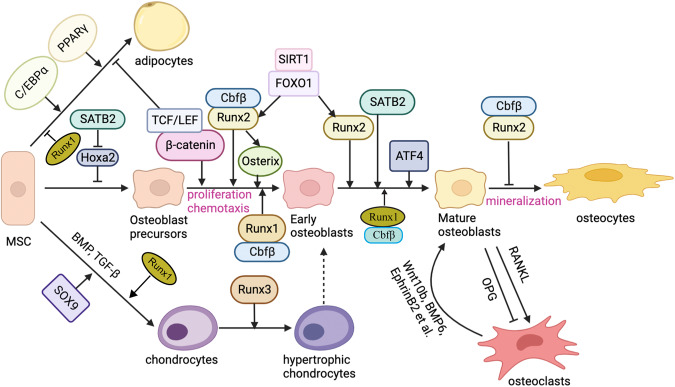
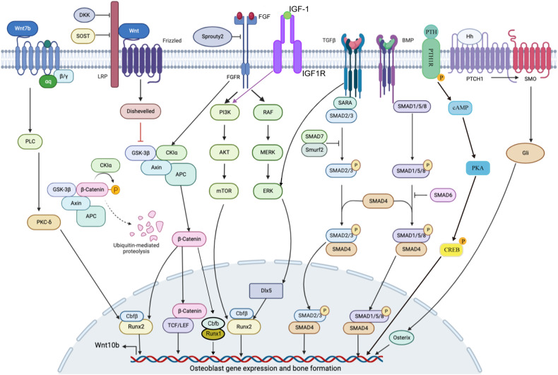
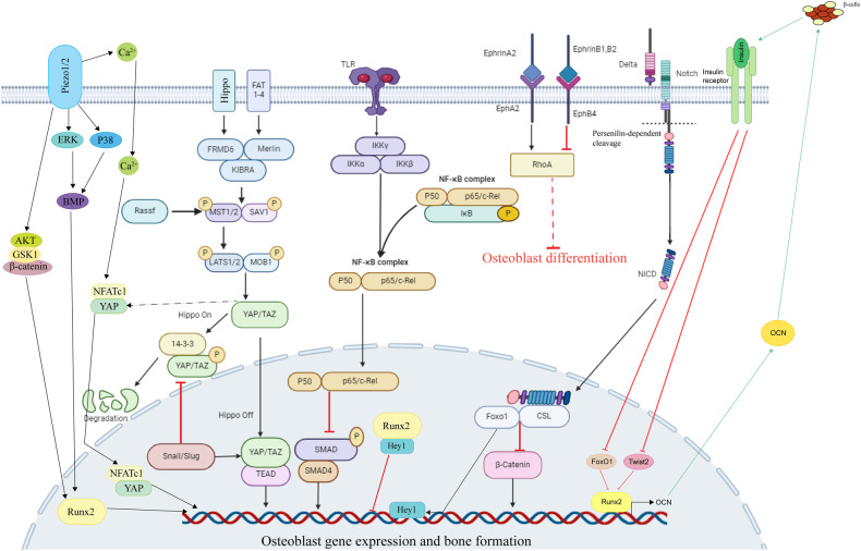
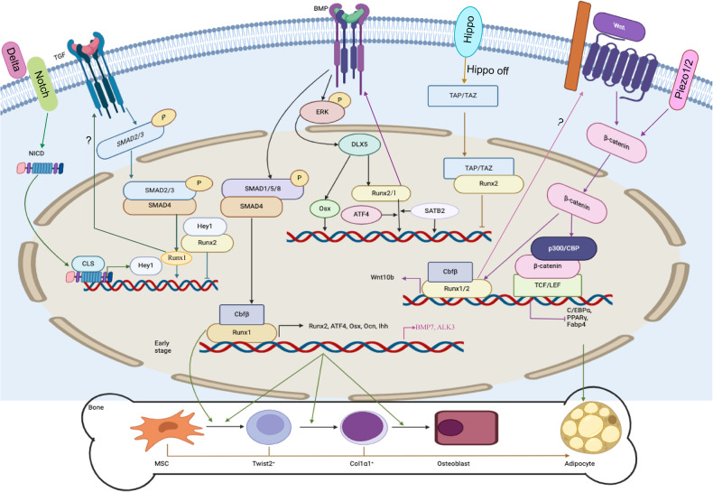
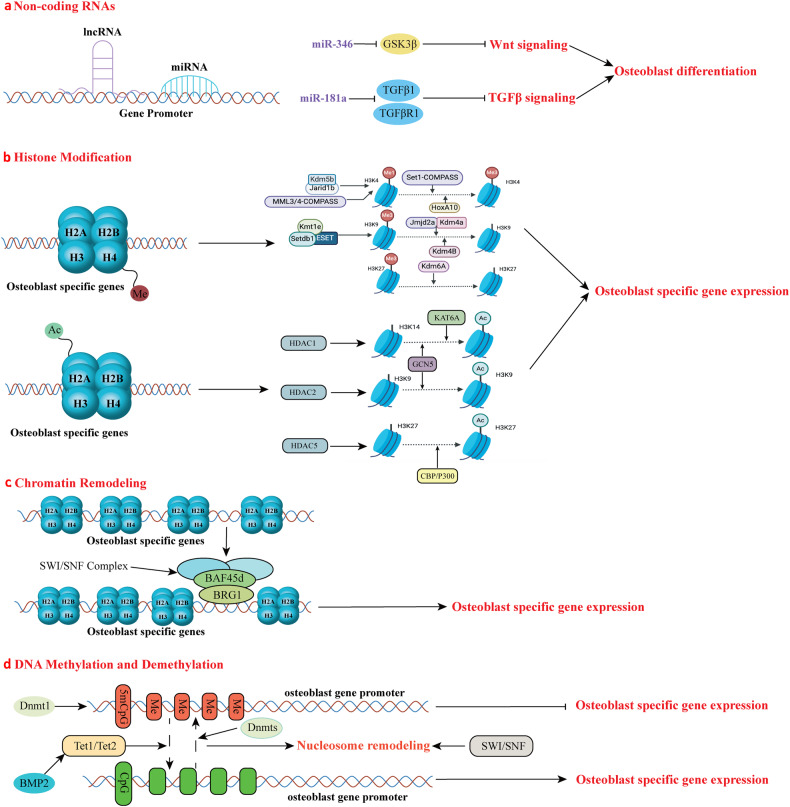
The initiation of osteogenesis primarily occurs as mesenchymal stem cells undergo differentiation into osteoblasts. This differentiation process plays a crucial role in bone formation and homeostasis and is regulated by two intricate processes: cell signal transduction and transcriptional gene expression. Various essential cell signaling pathways, including Wnt, BMP, TGF-β, Hedgehog, PTH, FGF, Ephrin, Notch, Hippo, and Piezo1/2, play a critical role in facilitating osteoblast differentiation, bone formation, and bone homeostasis. Key transcriptional factors in this differentiation process include Runx2, Cbfβ, Runx1, Osterix, ATF4, SATB2, and TAZ/YAP. Furthermore, a diverse array of epigenetic factors also plays critical roles in osteoblast differentiation, bone formation, and homeostasis at the transcriptional level. This review provides an overview of the latest developments and current comprehension concerning the pathways of cell signaling, regulation of hormones, and transcriptional regulation of genes involved in the commitment and differentiation of osteoblast lineage, as well as in bone formation and maintenance of homeostasis. The paper also reviews epigenetic regulation of osteoblast differentiation via mechanisms, such as histone and DNA modifications. Additionally, we summarize the latest developments in osteoblast biology spurred by recent advancements in various modern technologies and bioinformatics. By synthesizing these insights into a comprehensive understanding of osteoblast differentiation, this review provides further clarification of the mechanisms underlying osteoblast lineage commitment, differentiation, and bone formation, and highlights potential new therapeutic applications for the treatment of bone diseases.


Conflict of interest statement

The authors declare no competing interests.

Figures

Fig. 1
Fig. 1. Different transcription factors regulate three different differentiation fates — adipocytes, osteoblasts, and chondrocytes from MSCs.
MSCs have three different differentiation fates — adipocytes, osteoblasts, and chondrocytes — which are regulated by different genes. In the differentiation process, some cells in the three differentiation pathways also have a reciprocal transformation relationship through the regulation of related genes, such as interactions between mature osteoblasts and mature osteoclasts or hypertrophic chondrocytes and early osteoblasts. Transcription factors have different functions in different stages of osteoblast differentiation. Runx2 is a vital factor in all osteoblast differentiation stages; Runx2 promotes osteoblast differentiation in the early stage while inhibiting mature osteoblast differentiation into osteocytes. Cbfβ is the major co-factor of Runx2 and Runx1. Runx3 can promote chondrocytes into hypertrophic chondrocytes. SIRT1 and FOXO1 can promote Runx2 expression. Osx and β-catenin also have important functions in the early stage of osteoblast differentiation. SATB2 and ATF4 are important in promoting the terminal differentiation stage of osteoblast. SATB2 inhibits Hoxa2 activity in the early stage of osteoblast differentiation. Runx1 plays an important role in inhibiting adipocyte differentiation and promoting chondrocyte differentiation. The interaction between osteoblasts and osteoclasts is also very important. Osteoblasts regulate osteoclast differentiation via RANKL signaling and inhibit osteoclast differentiation through OPG. Similarly, osteoclasts can regulate osteoblast differentiation through the Wnt10b, BMP6, or Ephrin signaling pathway.
Fig. 2
Fig. 2. Canonical signaling pathways in osteoblast differentiation.
Several canonical signaling pathways control the activity of key transcription factors to mediate osteoblast differentiation. Wnt, TGF-β, BMP, FGF, and Hedgehog pathways are the most classic pathways that have been studied during osteoblast differentiation. Wnt binds with FZD receptors, causing the β-catenin accumulation. β-catenin then moves to the nucleus, in which it causes target genes to be transcribed. Wnt signaling also regulates Runx1 and Runx2 functions. TGF-β and BMP signaling regulate osteoblast-specific gene expression through multiple Smad proteins. TGF-β signaling mainly activates Smad2/3, while BMP signaling activates Smad1/5/8. FGF and FGFR can also regulate osteoblast differentiation and osteoblast-specific gene expression via downstream pathways such as PI3K-AKT and ERK pathways. Runx2, Osterix, and several other transcription factors are also necessary for osteoblast differentiation, and these transcription factors are regulated by these classic pathways. Hedgehog signaling is activated through Hh ligand binding to the 12-transmembrane receptor Patched 1 (PTCH1), which relieves inhibition of the seven-pass transmembrane G protein-coupled receptor Smoothened (SMO). Actived SMO can initiate the intracellular cascade leading to the activation of three Gli transcription factors, and Gli can then translocate into the nucleus and regulate Osx activation, thereby modulating osteoblast-specific gene expression. PTH regulates osteoblast differentiation by binding with PTH1R, subsequently activating cAMP and PKA, leading to the phosphorylation of CREB to regulate osteoblast-specific gene expression. IGF-1 binds to its receptor IGF1R and activates PI3K-Akt pathway, resulting in the activation of mTOR and promotion of osteoblast differentiation.
Fig. 3
Fig. 3. Noncanonical signaling pathways in osteoblast differentiation.
Notch, NF-κB, Hippo, Ephrin, and Piezo1/2 signaling pathways also play important roles in osteoblast differentiation. Osteoblast differentiation is inhibited by the Notch and NF-κB signaling pathways, with the Notch signaling pathway’s downstream factors NICD binding to CLS and inhibiting β-catenin. NICD/CLS/Foxo1 complex can promote Hey1 expression, then Hey1 can bind with Runx2 to inhibit Runx2 activity. When NF-κB signaling is stimulated, the P50 and P65 complex translocates into the nucleus and inhibits Smad protein activity. Hippo signaling is a crucial pathway in cell growth and development. The important downstream factors YAP/TAZ in Hippo signaling can regulate osteoblast differentiation-related gene expression. When Hippo signaling is in an “off” state, the YAP/TAZ complex will not be degraded and will translocate into the nucleus to regulate osteoblast-specific gene expression. The snail/slug complex can promote YAP/TAZ activity in nuclear and inhibit YAP/TAZ degradation in the cytoplasm. Ephrin signaling has different regulatory effects on osteoblast differentiation through different pathways and plays a specific role in the regulation of osteoblasts and osteoclasts. The EphrinA2 and EphrinB2 expressed on osteoclast membranes can interact with EphA2 and EphB4 expressed on osteoblast membranes to regulate osteoblast differentiation. Ephrin signaling regulated osteoblast-specific gene expression mainly through RhoA. EphrinA2 binds with EphA2 and promotes RhoA activity, while EphrinB1/2 binds with EphB4 and inhibits RhoA activity. When RhoA is activated, it will inhibit osteoblast differentiation. Piezo1/2 is a recently discovered pathway functioning in osteoblast differentiation. Piezo1/2 can regulate osteoblast-specific gene expression by regulating downstream pathways such as ERK and P38 and interacting with Hippo signaling. Piezo1/2 can regulate β-catenin activation to active Runx2 and regulate osteoblast gene expression. Piezo1/2 can also activate NFATc1 and YAP via calcium signal, and then NFATc1 and YAP can translocate into the nucleus and regulate osteoblast differentiation. Insulin signaling inhibits the production of FoxO1 and Twist2, which can inhibit the expression of Runx2 and Ocn. Ocn improves insulin sensitivity and energy expenditure through multiple mechanisms like activating to β-cells. The direct effect of osteocalcin as an insulin-sensitizing hormone is speculative and remains to be determined.
Fig. 4
Fig. 4. Signaling and transcriptional regulation of osteoblast cell lineage commitment, differentiation, and bone formation.
Among the many transcription factors that participate in osteoblast differentiation, the Runx family, Osx, ATF4, Cbfβ, and SATB2 are significant. Cbfβ binds to Runx family proteins to form heterodimers, improving Runx2’s and Runx1’s stability and subsequently facilitating Runx2 or Runx1 binding to target DNA sequences. Runx1 positively regulates osteoblast lineage gene expression at various stages of differentiation. Runx1 plays a significant role in postnatal bone homeostasis by binding to ATF4, Ocn, and Runx2 promoters to activate the corresponding genes and promote osteoblast early differentiation. Runx1 promotes BMP7 and Alk3 expression to regulate BMP signaling. How Runx1 can regulate Wnt10b and TGF-β signaling remains unclear. Runx1 can also regulate osteoblast-adipocyte lineage via inducing Wnt/β-catenin signaling, TGF-β signaling, and restraining adipogenic gene transcription. Cbfβ is also crucial in stimulating osteogenesis by inhibiting the expression of the adipogenesis regulatory gene C/EBPα and activating Wnt10b/β-catenin signaling. Wnt binds with FZD receptors, causing the ß-catenin accumulation. Pizeo1/2 can also regulate ß-catenin. ß-catenin then moves to the nucleus, in which it causes target genes to be transcribed by interacting with P300/CBP and TCF/LEF. TGF-β and BMP regulate transcription factors through SMAD proteins to activate Runx1 and Runx2 activity, and the SMAD itself can also regulate osteoblast-specific gene expression. BMP signaling can also activate Dlx5 through ERK and promote Runx2, ATF4, and Osx expression. When the Hippo signaling is at the “off” state, YAP/TAZ can translocate into the nucleus and bind with Runx2 to inhibit its activity. NICD1 in Notch signaling can translocate into the nucleus and promote the expression Hey1, which in turn inhibits Runx2 function by binding with Runx2. The activation of β-catenin and Runx2 will also inhibit the expression of C/EBPα, PPAR-γ, and Fabp4; these genes are important in adipocyte differentiation.
Fig. 5
Fig. 5. Epigenetic modification of transcriptional genes regulation in osteoblasts.
a The non-coding RNAs have a regulatory effect on osteoblast differentiation; miRNA-346 can inhibit GSK3β to promote Wnt signaling activation, and miRNA-181a can inhibit TGFβ1 and TGFβR1 to promote TGFβ signaling activation to promote osteoblast differentiation. b The methylation and acetylation of histone H3 control the promoter of the transcriptional genes. The enrichment of H3K4me1, H3K9me3, and H3K27me3 leads to transcriptional silencing of gene promoters, while the enrichment of H3K4me3 promotes transcriptional activation. Demethylation of H3K9me3 and H3K27me3 also induces transcriptional activation of gene promoters. This group of proteins regulates the enrichment of different methylation groups and their methylation degree. Jarid1/Kdm5 can convert H3K4me3 and H3K4me2 to H3K4me1, while MLL3/4-COMPASS-like can catalyze the deposition of H3K4me1 on the enhancer elements. Set1-COMPASS and HoxA10 promote H3K4 methylation. Jmjd2a, Kdm4a and Kdm4B promote H3K9 demethylation. Kmt1e/Setdb1/ESET can mediate H3K9 trimethylation. Osteoblast differentiation is facilitated by the acetylation of H3K9, H3K14, and H3K27. HDAC1/2/5 promotes deacetylation of H3K9, H3K14, and H3K27, while GCN5 promotes H3K9 and H3K14 histone acetylation, KATA5 promotes H3K14 acetylation and CBP/P300 promotes H3K27 acetylation. c SWI/SNF regulates osteoblast differentiation through chromatin remodeling. BRG1 is one of the subunits of the SWI/SNF complex. Baf45d also belongs to SWI/SNF complex. When activated, it can regulate osteoblast early differentiation. SWI/SNF chromatin remodeling complex is required for Runx2-dependent initiation of skeletal gene expression. d The degree of DNA methylation affects the gene promoters. The Dnmts family, such as Dnmt1, can regulate DNA methylation while Tet1/2 can demethylate 5mCpG to promote Runx2 and Osx gene transcription, thereby promoting osteoblast differentiation. BMP2 can induce Tet1/2 activity to convert 5mCpG to 5hmCpG on Osx promoter.

References

    1. Murshed M, Harmey D, Millán JL, McKee MD, Karsenty G. Unique coexpression in osteoblasts of broadly expressed genes accounts for the spatial restriction of ECM mineralization to bone. Genes Dev. 2005;19:1093–1104. doi: 10.1101/gad.1276205. - DOI - PMC - PubMed
    1. Ferron M, et al. Insulin signaling in osteoblasts integrates bone remodeling and energy metabolism. Cell. 2010;142:296–308. doi: 10.1016/j.cell.2010.06.003. - DOI - PMC - PubMed
    1. Karsenty G, Wagner EF. Reaching a genetic and molecular understanding of skeletal development. Dev. Cell. 2002;2:389–406. doi: 10.1016/S1534-5807(02)00157-0. - DOI - PubMed
    1. Bar-Shavit Z. The osteoclast: a multinucleated, hematopoietic-origin, bone-resorbing osteoimmune cell. J. Cell Biochem. 2007;102:1130–1139. doi: 10.1002/jcb.21553. - DOI - PubMed
    1. Soltanoff CS, Yang S, Chen W, Li YP. Signaling networks that control the lineage commitment and differentiation of bone cells. Crit. Rev. Eukaryot. Gene Expr. 2009;19:1–46. doi: 10.1615/CritRevEukarGeneExpr.v19.i1.10. - DOI - PMC - PubMed

LinkOut - more resources