. 2024 Jul 1;28(1):212.
doi: 10.1186/s13054-024-05001-2.

Vitamin K: a potential missing link in critical illness-a scoping review

Affiliations

Vitamin K: a potential missing link in critical illness-a scoping review

Michelle Carmen Paulus et al. Crit Care. .

Abstract

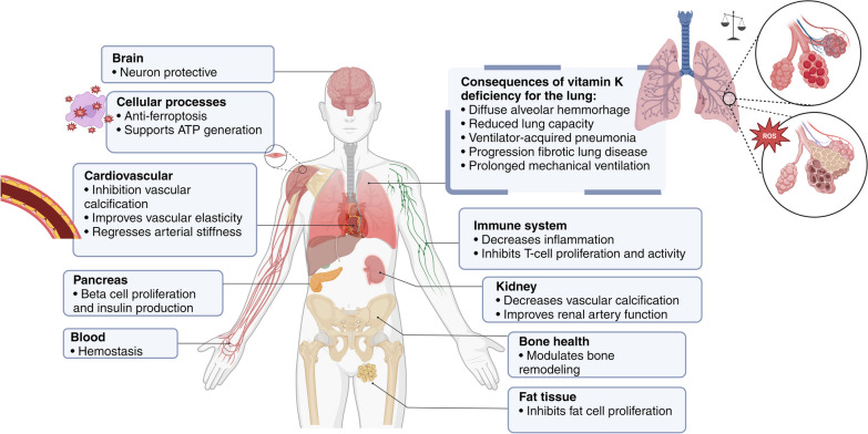
Background: Vitamin K is essential for numerous physiological processes, including coagulation, bone metabolism, tissue calcification, and antioxidant activity. Deficiency, prevalent in critically ill ICU patients, impacts coagulation and increases the risk of bleeding and other complications. This review aims to elucidate the metabolism of vitamin K in the context of critical illness and identify a potential therapeutic approach.

Methods: In December 2023, a scoping review was conducted using the PRISMA Extension for Scoping Reviews. Literature was searched in PubMed, Embase, and Cochrane databases without restrictions. Inclusion criteria were studies on adult ICU patients discussing vitamin K deficiency and/or supplementation.

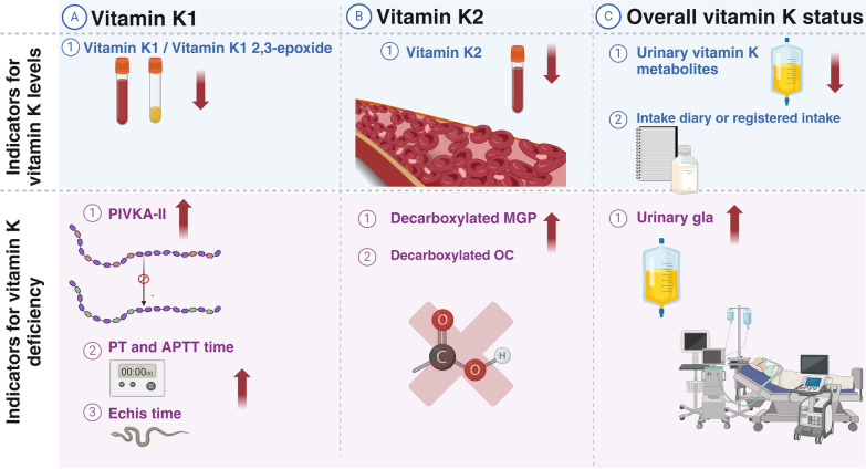
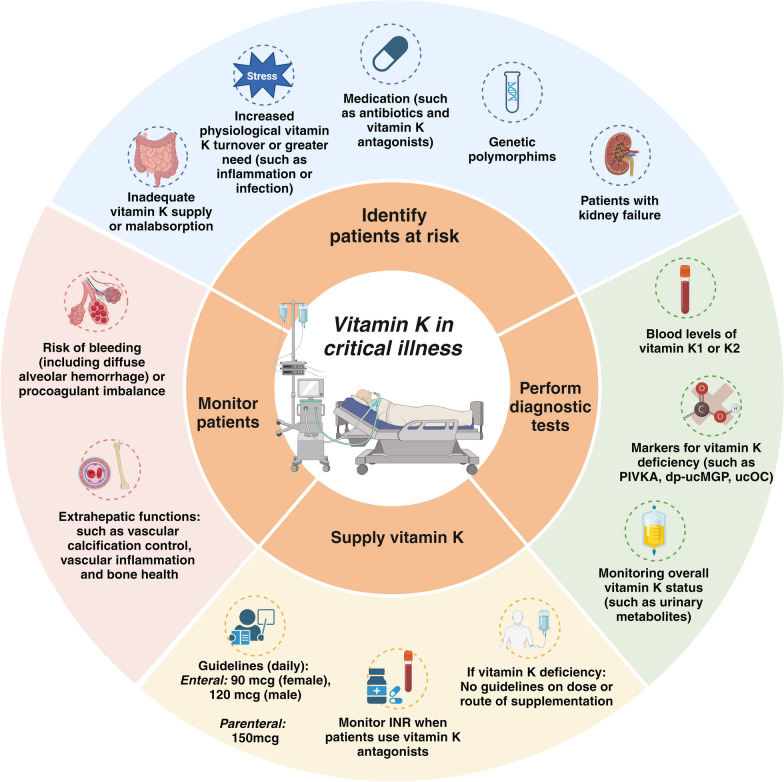
Results: A total of 1712 articles were screened, and 13 met the inclusion criteria. Vitamin K deficiency in ICU patients is linked to malnutrition, impaired absorption, antibiotic use, increased turnover, and genetic factors. Observational studies show higher PIVKA-II levels in ICU patients, indicating reduced vitamin K status. Risk factors include inadequate intake, disrupted absorption, and increased physiological demands. Supplementation studies suggest vitamin K can improve status but not normalize it completely. Vitamin K deficiency may correlate with prolonged ICU stays, mechanical ventilation, and increased mortality. Factors such as genetic polymorphisms and disrupted microbiomes also contribute to deficiency, underscoring the need for individualized nutritional strategies and further research on optimal supplementation dosages and administration routes.

Conclusions: Addressing vitamin K deficiency in ICU patients is crucial for mitigating risks associated with critical illness, yet optimal management strategies require further investigation.

Impact research: To the best of our knowledge, this review is the first to address the prevalence and progression of vitamin K deficiency in critically ill patients. It guides clinicians in diagnosing and managing vitamin K deficiency in intensive care and suggests practical strategies for supplementing vitamin K in critically ill patients. This review provides a comprehensive overview of the existing literature, and serves as a valuable resource for clinicians, researchers, and policymakers in critical care medicine.

Keywords: Gla protein; ICU; Micronutrients; PIVKA-II; Vitamin K.


Conflict of interest statement

Prof. Dr. Van Zanten reported receiving honoraria for advisory board meetings, lectures, research, and travel expenses from AOP Pharma, Abbott, Baxter, Cardinal Health, Danone-Nutricia, DIM3, Fresenius Kabi, GE Healthcare, InBody, Mermaid, Rousselot, and Lyric. The other authors have nothing to declare.

Figures

Fig. 1
Fig. 1
Physiological functions of vitamin K in the body. The diagram demonstrates the numerous roles vitamin K plays in the human body. From facilitating blood clotting in the liver to promoting bone health and cardiovascular function, vitamin K is essential to maintaining overall health. In critical care, vitamin K deficiency can significantly impact patients. Low vitamin K levels impair blood clotting and increase the risk of uncontrolled bleeding, especially when clot formation is critical to the patient's recovery. There is also an increased risk of microbleeding in the lungs, which can lead to diffuse alveolar haemorrhage. This process may also contribute to the development of lung fibrosis by inducing oxidative stress and inflammation. Created with BioRender.com
Fig. 2
Fig. 2
Absorption of vitamin K. Phylloquinone (vitamin K1) is mainly found in green leafy vegetables, while bacteria synthesize menaquinone (vitamin K2), mainly from the fat fraction of dairy products. Vitamin K2 exists in several forms, called MK-n, depending on the side chain. MK-4 can be formed by the conversion of phylloquinone in the intestinal mucosa during absorption or by tissue-specific conversion in the body. Medium- and long-chain MK-n (MK-6 or higher) are synthesized by bacteria and anaerobes in the human colonic microbiota [8]. Vitamin K1 is absorbed in the upper small intestine, particularly in the jejunum and ileum. Vitamin K absorption is facilitated by bile acids and specific transport proteins such as Niemann-Pick C1-like 1 (NPC1L1) and scavenger receptor class B-type I [3]. Following absorption in the small intestine, vitamin K is incorporated into chylomicron remnants and transported through the lymphatic capillaries to the liver [9]. Created with BioRender.com
Fig. 3
Fig. 3
Vitamin K metabolism in the liver. In the liver, vitamin K uptake is regulated by receptor-mediated endocytosis via lipoprotein receptors. Some of it is utilized to synthesize clotting factors, while the remaining amount re-enters the systemic circulation through very low-density lipids. These lipids undergo conversion into low-density lipoproteins (LDL), which serve as carriers for transporting vitamin K to extrahepatic tissues [9]. Initially, vitamin K epoxide (VKO) is converted to vitamin K quinone through vitamin K epoxide reductase (VKOR). Subsequently, vitamin K reductase (VKR) and vitamin K quinone reductases 1 and 2 (VKQR, DT diaphorase) further convert it into VKH. Vitamin K antagonists exert their effect by inhibiting the enzymatic activity of VKOR and VKR, thereby impeding the conversion of vitamin K to its active form. CYP4F2 has been found to play a minor role in the metabolism of vitamin K in its inactive form [11]. This inhibition has implications for both the hepatic and extrahepatic actions of vitamin K [12]. Different cytochrome P450 enzymes are involved in metabolizing coumarins into inactive metabolites. Vitamin K is excreted in the feces via bile and urine. In the absence of warfarin, bile excretion is the predominant route. However, a higher proportion of vitamin K is excreted in the urine when warfarin is used [9]. The different forms of vitamin K have different half-lives. Vitamin K1 and MK-4 have short half-lives of hours, whereas long-chain MK has a much longer half-life of several days [13]. Created with BioRender.com
Fig. 4
Fig. 4
Assessment of Vitamin K status. Figure 4 Red arrows indicate changes in measurements due to vitamin K deficiency. Quantifying vitamin K status is challenging due to various dietary intakes and the complexity of detecting vitamin K2 without supplementation [84]. Measurement accuracy may require adjustments and fasting samples, as vitamin K circulates with triglyceride-rich lipoproteins [84, 114]. Assessment of hepatic vitamin K status commonly relies on prothrombin time (PT) and PT-internal normalized ratio (PT-INR), but PT lacks sensitivity, particularly in the presence of liver dysfunction or hematological diseases. Furthermore, PT offers restricted insights as it exclusively concentrates on procoagulants while neglecting anticoagulants and extrahepatic functions. Consequently, it provides an incomplete reflection of overall vitamin K status [117]. Uncarboxylated gla proteins, such as uncarboxylated factor II (PIVKA-II), desphospho-uncarboxylated MGP (dp-uc MGP), and uncarboxylated osteocalcin (ucOC) are gaining attention for assessing extrahepatic vitamin K use. However, elevated dp-uc MGP and ucOC levels do not always indicate suboptimal carboxylation of hepatic proteins [106, 118]. In critical illness, PIVKA-II levels rise, possibly due to the acute phase response, complicating interpretation [15]. Echis time, using viper venom, provides an alternative method to assess vitamin K status. Echis time uses viper venom (Echis carinatus) to activate normal prothrombin and PIVKA-II to form thrombin. Consequently, the Echis time remains within the normal range in the presence of vitamin K deficiency and is only prolonged in the presence of inadequate clotting factor production. However, its applicability in other critically ill patients requires further validation [119]. Urinary biomarkers such as y-carboxyglutamic acid (gla) reflect overall vitamin K-dependent protein status but have limitations, such as the need for 24-h urine samples, lack of correlation with dietary intake, and dependence on lean body mass. Created with BioRender.com
Fig. 5
Fig. 5
Practical tool to manage Vitamin K for critically ill patients in daily clinical practice. This figure provides an overview of the management of vitamin K during critical illness. Critically ill patients are known to have a high prevalence of vitamin K deficiency, which can worsen during ICU admission. Patient categories at risk are listed. The complexity of assessing vitamin K levels is compounded by the availability of various diagnostic tests. So far, no consensus on the optimal method to diagnose vitamin K deficiency in critically ill patients exists. International guidelines have been developed for the minimum daily intake of vitamin K in critically ill patients. However, uncertainty remains regarding the optimal dosage and route of administration to correct vitamin K deficiency. Furthermore, the figure emphasises the importance of monitoring patients at risk of vitamin K deficiency. Abbreviations: PIVKA: proteins induced by vitamin K absence or antagonist, dp-ucMGP: dephospho-uncarboxylated matrix Gla protein, ucOC: undercarboxylated osteocalcin. Created with BioRender.com

References

    1. Berkner KL. The vitamin K-dependent carboxylase. Annu Rev Nutr. 2005;25:127–149. doi: 10.1146/annurev.nutr.25.050304.092713. - DOI - PubMed
    1. Suttie JW. Vitamin K-dependent carboxylase. Ann Rev Biochem. 1985;54(1):459–477. doi: 10.1146/annurev.bi.54.070185.002331. - DOI - PubMed
    1. Bus K, Szterk A. Relationship between structure and biological activity of various vitamin K forms. Foods. 2021;10(12):3136. doi: 10.3390/foods10123136. - DOI - PMC - PubMed
    1. Stanley TB, Jin DY, Lin PJ, Stafford DW. The propeptides of the vitamin K-dependent proteins possess different affinities for the vitamin K-dependent carboxylase. J Biol Chem. 1999;274(24):16940–16944. doi: 10.1074/jbc.274.24.16940. - DOI - PubMed
    1. Chatrou ML, Winckers K, Hackeng TM, Reutelingsperger CP, Schurgers LJ. Vascular calcification: the price to pay for anticoagulation therapy with vitamin K-antagonists. Blood Rev. 2012;26(4):155–166. doi: 10.1016/j.blre.2012.03.002. - DOI - PubMed

Publication types

MeSH terms

LinkOut - more resources