

Dynamics of DNA methylation during osteogenic differentiation of porcine synovial membrane mesenchymal stem cells from two metabolically distinct breeds
- PMID: 38956836
- PMCID: PMC11225923
- DOI: 10.1080/15592294.2024.2375011
Dynamics of DNA methylation during osteogenic differentiation of porcine synovial membrane mesenchymal stem cells from two metabolically distinct breeds
Abstract
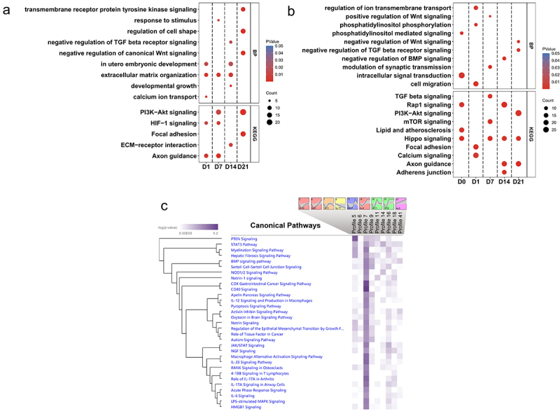
Mesenchymal stem cells (MSCs), with the ability to differentiate into osteoblasts, adipocytes, or chondrocytes, show evidence that the donor cell's metabolic type influences the osteogenic process. Limited knowledge exists on DNA methylation changes during osteogenic differentiation and the impact of diverse donor genetic backgrounds on MSC differentiation. In this study, synovial membrane mesenchymal stem cells (SMSCs) from two pig breeds (Angeln Saddleback, AS; German Landrace, DL) with distinct metabolic phenotypes were isolated, and the methylation pattern of SMSCs during osteogenic induction was investigated. Results showed that most differentially methylated regions (DMRs) were hypomethylated in osteogenic-induced SMSC group. These DMRs were enriched with genes of different osteogenic signalling pathways at different time points including Wnt, ECM, TGFB and BMP signalling pathways. AS pigs consistently exhibited a higher number of hypermethylated DMRs than DL pigs, particularly during the peak of osteogenesis (day 21). Predicting transcription factor motifs in regions of DMRs linked to osteogenic processes and donor breeds revealed influential motifs, including KLF1, NFATC3, ZNF148, ASCL1, FOXI1, and KLF5. These findings contribute to understanding the pattern of methylation changes promoting osteogenic differentiation, emphasizing the substantial role of donor the metabolic type and epigenetic memory of different donors on SMSC differentiation.
Keywords: DNA methylation; Epigenetic pattern; Mesenchymal stem cells; Osteogenic differentiation; Pig breeds.
Conflict of interest statement
No potential conflict of interest was reported by the author(s).
Figures


Similar articles
-
DNA methylation in adipocyte differentiation of porcine mesenchymal stem cells and the impact of the donor metabolic type.Genomics. 2025 May;117(3):111050. doi: 10.1016/j.ygeno.2025.111050. Epub 2025 Apr 28. Genomics. 2025. PMID: 40306557
-
Effect of metabolically divergent pig breeds and tissues on mesenchymal stem cell expression patterns during adipogenesis.BMC Genomics. 2024 Apr 25;25(1):407. doi: 10.1186/s12864-024-10308-z. BMC Genomics. 2024. PMID: 38664635 Free PMC article.
-
Transcriptome changes during osteogenesis of porcine mesenchymal stem cells derived from different types of synovial membranes and genetic background.Sci Rep. 2023 Jun 21;13(1):10048. doi: 10.1038/s41598-023-37260-4. Sci Rep. 2023. PMID: 37344635 Free PMC article.
-
Epigenetic Regulation of Osteogenic Differentiation of Mesenchymal Stem Cells.Curr Stem Cell Res Ther. 2016;11(3):235-46. doi: 10.2174/1574888x10666150528153313. Curr Stem Cell Res Ther. 2016. PMID: 26018226 Review.
-
The role of epigenetic modifications in the osteogenic differentiation of adipose-derived stem cells.Connect Tissue Res. 2019 Nov;60(6):507-520. doi: 10.1080/03008207.2019.1593395. Epub 2019 Jun 17. Connect Tissue Res. 2019. PMID: 31203665 Review.
References
-
- Mochizuki T, Muneta T, Sakaguchi Y, et al. Higher chondrogenic potential of fibrous synovium– and adipose synovium–derived cells compared with subcutaneous fat–derived cells: Distinguishing properties of mesenchymal stem cells in humans. Arthritis Rheum. 2006;54(3):843–15. doi: 10.1002/art.21651 - DOI - PubMed
MeSH terms
LinkOut - more resources
Full Text Sources
Other Literature Sources
Molecular Biology Databases
Research Materials