. 2024 Aug 7;32(8):2624-2640.
doi: 10.1016/j.ymthe.2024.06.030. Epub 2024 Jul 2.

Optimized rAAV8 targeting acinar KLF4 ameliorates fibrosis in chronic pancreatitis via exosomes-enriched let-7s suppressing pancreatic stellate cells activation

Affiliations

Optimized rAAV8 targeting acinar KLF4 ameliorates fibrosis in chronic pancreatitis via exosomes-enriched let-7s suppressing pancreatic stellate cells activation

Yating Zhao et al. Mol Ther. .

Abstract

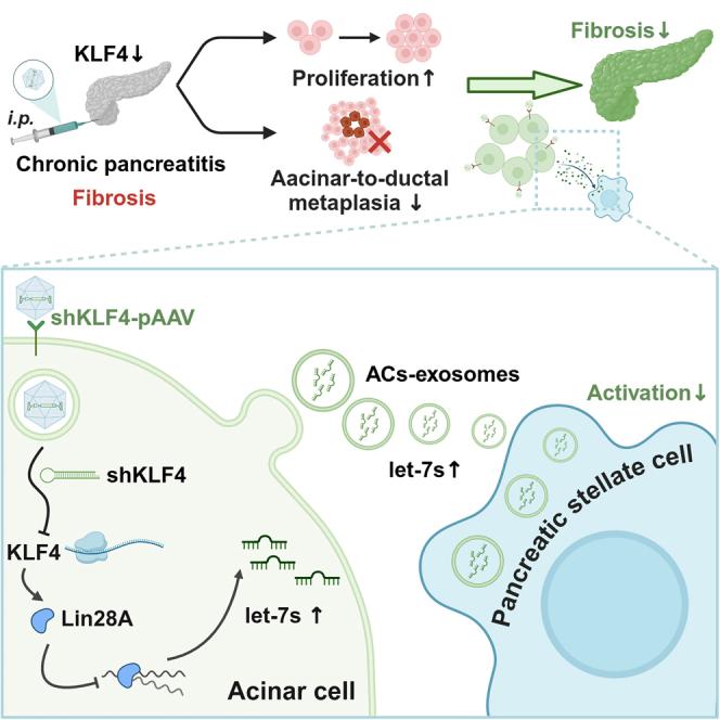
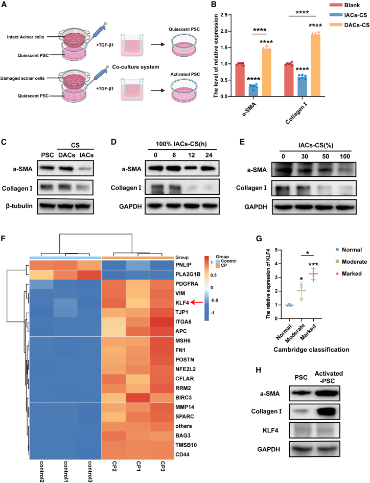
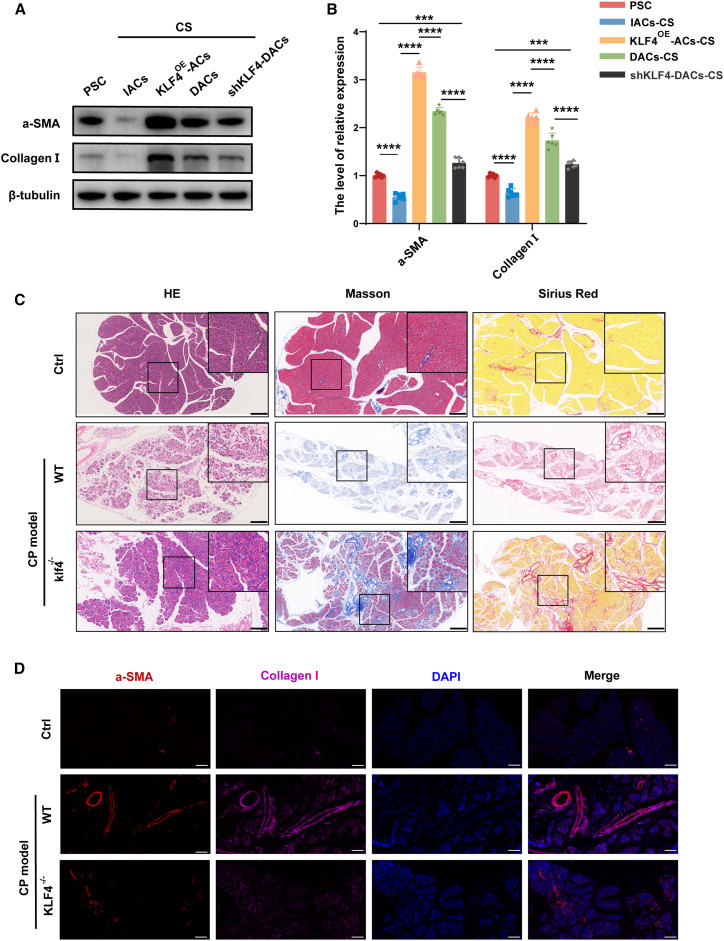
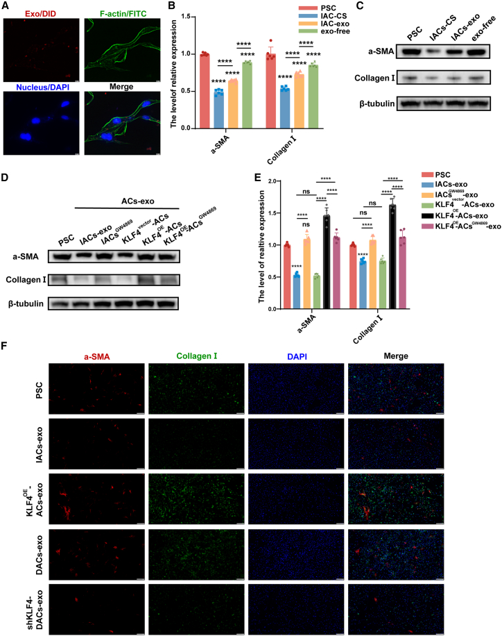
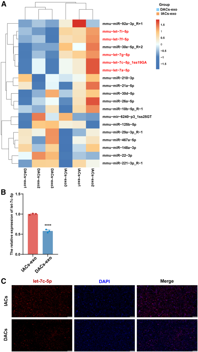
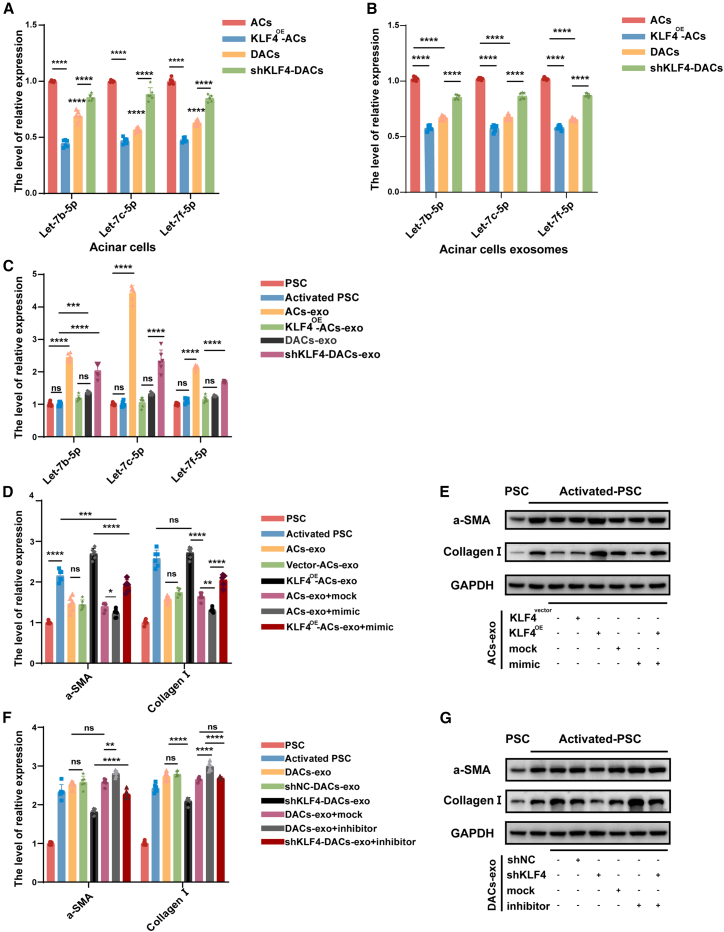
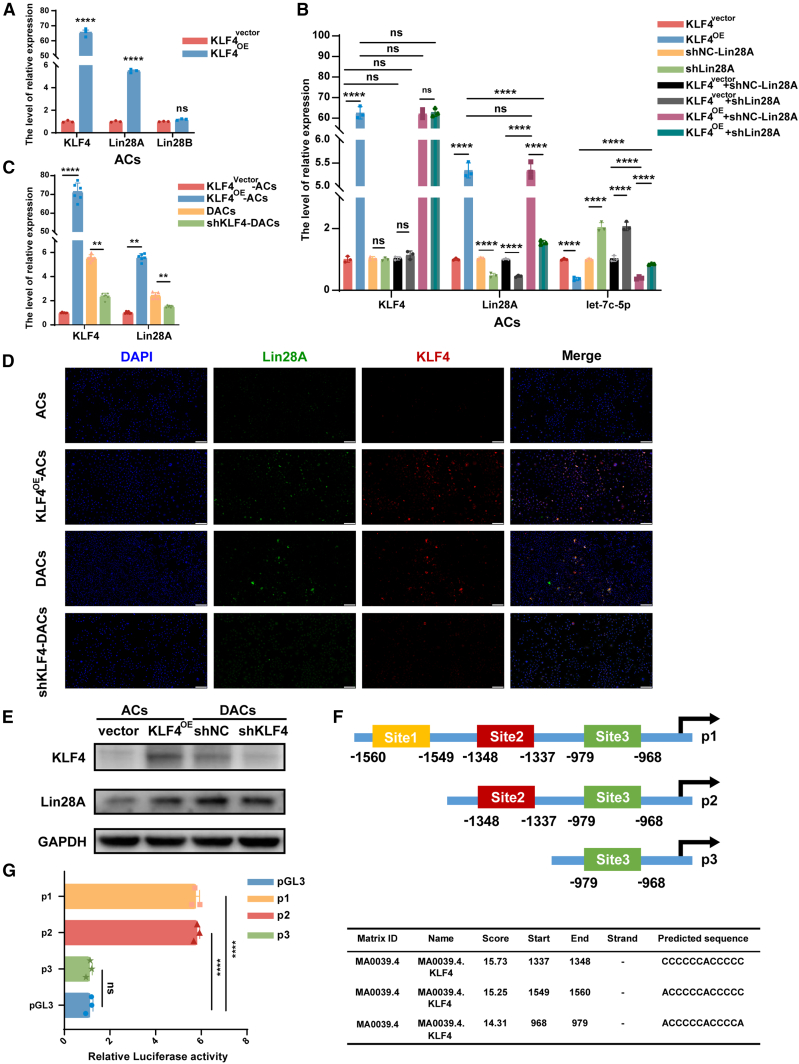
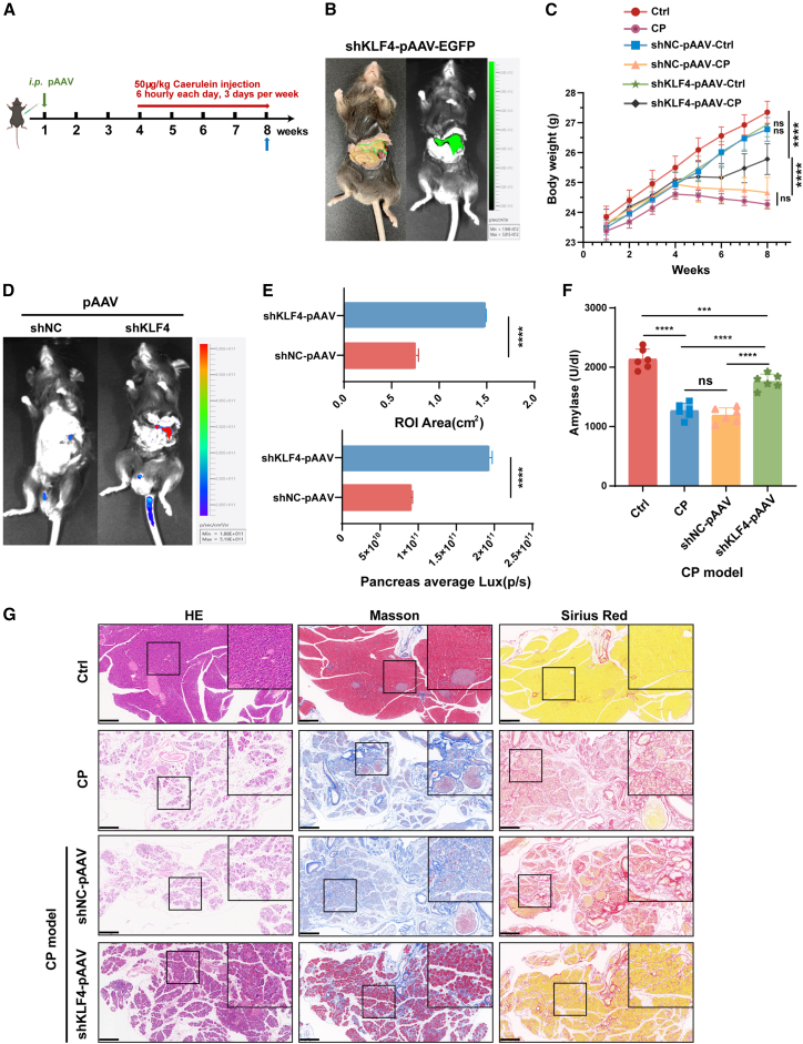
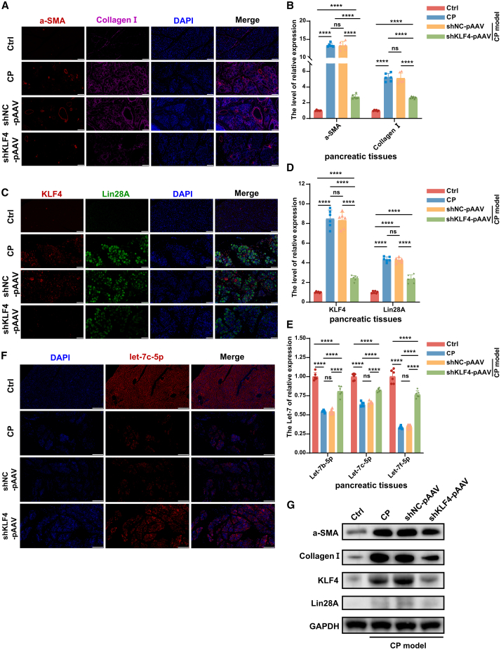
Chronic pancreatitis (CP) is marked by progressive fibrosis and the activation of pancreatic stellate cells (PSCs), accompanied by the destruction of pancreatic parenchyma, leading to the loss of acinar cells (ACs). Few research studies have explored the mechanism by which damaged ACs (DACs) contribute to PSCs activation and pancreatic fibrosis. Currently, there are no effective drugs for curing CP or limiting the progression of pancreatic fibrosis. In this research, co-culture with intact acinar cells (IACs) suppressed PSC activation, while co-culture with DACs did the opposite. Krüppel-like factor 4 (KLF4) was significantly upregulated in DACs and was established as the key molecule that switches ACs from PSCs-suppressor to PSCs-activator. We revealed the exosomes of IACs contributed to the anti-activated function of IACs-CS on PSCs. MiRNome profiling showed that let-7 family is significantly enriched in IAC-derived exosomes (>30% miRNome), which partially mediates IACs' suppressive impacts on PSCs. Furthermore, it has been observed that the enrichment of let-7 in exosomes was influenced by the expression level of KLF4. Mechanistic studies demonstrated that KLF4 in ACs upregulated Lin28A, thereby decreasing let-7 levels in AC-derived exosomes, and thus promoting PSCs activation. We utilized an adeno-associated virus specifically targeting KLF4 in ACs (shKLF4-pAAV) to suppress PSCs activation in CP, resulting in reduced pancreatic fibrosis. IAC-derived exosomes hold potential as potent weapons against PSCs activation via let-7s, while activated KLF4/Lin28A signaling in DACs diminished such functions. ShKLF4-pAAV holds promise as a novel therapeutic approach for CP.

Keywords: AAV; KLF4; Lin28A; MiRNome; chronic pancreatitis; exosome; fibrosis; let-7.


Conflict of interest statement

Declaration of interests The authors declare no competing interests.

Figures

None
Graphical abstract
Figure 1
Figure 1
Two distinct states of acinar cells exert different effects on PSCs due to KLF4 (A) The experimental model of cocultured intact acinar cells (IACs) and pancreatic stellate cells (PSCs). (B) Activation levels of PSCs were quantified by qPCR analysis after 12 h treatment with culture supernatant from IACs (IACs-CS) and damaged acinar cells (DACs, injured with CCK 24 h). (C) The protein levels of α-SMA and Collagen Ι in PSCs were assessed using western blot analysis after 12-h treatment with culture medium (Blank), culture supernatant from IACs (IACs-CS), and DACs (DACs-CS). (D) Western blot analysis of PSCs activation after treatment with conditioned media from IACs at different time points (0, 6 h, 12 h, 24 h). (E) Western blot analysis of PSCs activation after 12 h treatment with different concentrations of culture supernatant from IACs (0%, 30%, 50%, 100%). (F) The heatmap presents top 20 differential gene expression (KLF4, red arrowhead) in CP mouse tissues (n = 3) by analyzing available public databases GEO and GeneCards. (G) qPCR was utilized to assess the relative mRNA expression levels of KLF4 in pancreatic tissues obtained from patients categorized into distinct Cambridge classification of CP. (Normal, n = 3; Moderate, n = 3; Marked, n = 4; ∗p < 0.05, ∗∗p < 0.01, ∗∗∗p < 0.001, ∗∗∗∗p < 0.0001, ns, not significant). (H) The protein levels of KLF4 in PSCs and activated PSCs were examined using western blot analysis.
Figure 2
Figure 2
KLF4 acts as a key molecular switch between IACs and DACs regarding their impacts on PSCs (A and B) The relative expression of α-SMA and Collagen Ι in activated PSCs after 12 h treatment with culture supernatants from ACs overexpressing KLF4 and from DACs with downregulated KLF4 were detected by western blot (A) and qPCR analysis (B). (n = 6, ∗p < 0.05, ∗∗p < 0.01, ∗∗∗p < 0.001, ∗∗∗∗p < 0.0001, ns, not significant.) (C) Representative pancreatic images of H&E and Masson staining for Ctrl, WT, and klf4−/− after induction of the chronic pancreatitis model. 100×: scale bar, 300 μm. (D)The pancreatic immunofluorescence image of α-SMA and Collagen Ι in Ctrl, WT, and klf4−/− after induction of CP model was presented. (200×: scale bar, 150 μm) (Ctrl, wild-type mice injected with saline during the model; CP model: WT, wild-type mice; klf4−/−, klf4 conditional knockout mice).
Figure 3
Figure 3
Exosomes contribute to mediating the suppressive effects of IACs against PSCs activation (A) The immunofluorescence image of IACs-exo taken by PSCs. DiD was used to label exosome phospholipid bilayers. Green fluorescence represents F-actin, and blue fluorescence represents the PSCs nucleus. (1000×: scale bar, 10 μm). (B and C) The qPCR (B) and western blot (C) detected the expression of α-SMA, Collagen Ι in PSCs after being treated for 12 h by IACs-CS, IACs-exo, and exo-free medium. (D and E) After manipulating ACs with different levels of KLF4 expression and treating them with the exosome secretion inhibitor GW4869, the exosomes were extracted from ACs-CS and used to intervene PSCs 12 h. Subsequently, the protein and mRNA expression levels of α-SMA and Collagen Ι in PSCs were assessed through western blot (D) and qPCR (E). (n = 6, ∗p < 0.05, ∗∗p < 0.01, ∗∗∗p < 0.001, ∗∗∗∗p < 0.0001, ns, not significant). (F) The immunofluorescence detected the expression levels of α-SMA and Collagen Ι in PSC after 12-h treatment with exosomes from acinar cells expressing different levels of KLF4 (200×: scale bar, 150 μm).
Figure 4
Figure 4
Lowered levels of let-7s in exosomes contribute to loss of inhibitory effects of DACs against PSCs activation (A) The miRNome sequencing analysis of IACs- and DACs-exo. (B) The relative expression of let-7c-5p in exosomes of IACs and DACs using qPCR. (n = 3, ∗p < .05, ∗∗p < .01, ∗∗∗p < .001, ∗∗∗∗p < .0001, ns, not significant.) (C) The FISH images present the relative expression levels of let-7c-5p in IACs and DACs. FISH, fluorescence in situ hybridization. 200×: scale bar, 150 μm.
Figure 5
Figure 5
Let-7s serve as a major effector in mediating the regulatory impacts of ACs-derived exosomes by KLF4 (A) The expression of let-7s family (let-7b-5p, let-7c-5p, let-7f-5p) in ACs overexpressed KLF4 (KLF4OE-ACs) and DACs knocked down KLF4 (shKLF4-DACs) was analyzed by qPCR. (B) The expression of let-7s family (let-7b-5p, let-7c-5p, let-7f-5p) in exosomes of KLF4OE-ACs and shKLF4-DACs was analyzed by qPCR. (C) The expression of let-7s family (let-7b-5p, let-7c-5p, let-7f-5p) in PSC treated with KLF4OE-ACs-exo and shKLF4-DACs-exo for 12 h detected by qPCR. (D) The expression of α-SMA and Collagen Ι in PSC after being treated for 12 h by exosomes of KLF4OE-ACs transfected with let-7c-5p mimic detected by the qPCR. (E) Western blot was performed to assess the protein expression levels of α-SMA and Collagen Ι in PSCs following 12 h treatment with exosomes secreted by ACs that were transfected with let-7c-5p mimic and the KLF4 overexpression plasmid. Vector is an empty control plasmid of KLF4. Mock is the control of the mimic. (F) The expression of α-SMA and Collagen Ι in PSCs after being treated 12 h by exosomes of shKLF4-DACs transfected with let-7c-5p inhibitor detected by the qPCR. (G) Western blot was performed to assess the protein expression levels of α-SMA and Collagen I in PSCs, following 12 h treatment with exosomes secreted by DACs that were transfected with let-7c-5p inhibitor and shKLF4. shKLF4 is a short hairpin RNA of KLF4. shNC is the negative control of shKLF4. Mock is the control of the inhibitor. (n = 6, ∗p < 0.05, ∗∗p < 0.01, ∗∗∗p < 0.001, ∗∗∗∗p < 0.0001, ns, not significant.).
Figure 6
Figure 6
Acinar KLF4 suppresses the generation of let-7 family by activating Lin28A (A) The relative expression of Lin28A and Lin28B after overexpressed KLF4 in ACs with qPCR analysis (n = 3). (B) The relative expression of KLF4, Lin28A, and let-7c-5p after treated by KLF4 plasmid and shLin28A in ACs with qPCR analysis. The shLin28A is the short hairpin RNA of Lin28A (n = 3). (C–E) The relative expression of KLF4 and Lin28A in KLF4OE-ACs and shKLF4-DACs with qPCR analysis(C) (n = 6), immunofluorescence (D) and western blot (E). 200×: scale bar, 150 μm. Vector is an empty control of the plasmid. (F) Schematic structure of the full-length Lin28A promoter-reporter and its plasmids of deletion mutants (p1, p2, p3) (up). The three predicted binding sites (site1, site2, site3) between KLF4 with promoter of Lin28A in JASPAR database analysis (down). (G) The plasmids of full-length Lin28A promoter (p1) and deletion mutants of it (p2, p3) and pGL3 were transfected into 293T cells with pcDNA3.1-KLF4 (pKLF4) for 48h. The pGL3 is the empty vector of Lin28A promoter plasmids. The relative luciferase activity was examined using a dual-luciferase assay kit (n = 3, ∗p < 0.05, ∗∗p < 0.01, ∗∗∗p < 0.001, ∗∗∗∗p < 0.0001, ns, not significant).
Figure 7
Figure 7
Knockdown of KLF4 via pancreatic-specific AAV exhibits excellent targeting specificity (A) Experimental flowchart of CP mouse model and AAV intraperitoneal injection. (B) In vivo optical imaging in mice pancreas after pAAV treatment. (C) The body weight records of different groups during the pAAV treatment and CP model. (D and E) After pAAV injection and CP model, the mice representative images are shot by in vivo optical imaging system (D). The pancreatic average luciferase intensity qualified by Lux (fluorescence intensity) and ROI (fluorescence area) (E). (F)The mice serum amylase levels in Ctrl, CP, shNC-AAV, and shKLF4-AAV groups. (n = 6, ∗p < 0.05, ∗∗p < 0.01, ∗∗∗p < 0.001, ∗∗∗∗p < 0.0001, ns, not significant.) (G) Representative images of pancreatic histology with H&E, Masson, and Sirius Red staining in four groups mice. 50×: scale bar, 400 μm.
Figure 8
Figure 8
shKLF4-pAAV attenuates pancreatic fibrosis via let-7 in vivo (A and B) The relative expressions of α-SMA and Collagen Ι in pancreas tissues of four groups of mice were demonstrated by immunofluorescence (IF) (A) and qPCR (B). (IF: 400×: scale bar, 100 μm). (C and D) The relative expressions of KLF4 and Lin28A in pancreas tissues of four groups of mice were demonstrated by IF (C) and qPCR (D) (IF: 400×: scale bar = 100 μm). (E and F) The expressions of let-7c-5p in pancreas tissues of four groups of mice were analyzed by qPCR (E) and FISH (F). FISH, fluorescence in situ hybridization. 200×: scale bar, 100 μm. (n = 6, ∗p < 0.05, ∗∗p < 0.01, ∗∗∗p < 0.001, ∗∗∗∗p < 0.0001, ns, not significant.) (G) The protein expressions of α-SMA, Collagen Ι, KLF4, Lin28A, and GAPDH in pancreatic tissues by western blot.

Similar articles

Cited by

References

    1. Beyer G., Habtezion A., Werner J., Lerch M.M., Mayerle J. Chronic pancreatitis. Lancet. 2020;396:499–512. doi: 10.1016/S0140-6736(20)31318-0. - DOI - PubMed
    1. Bynigeri R.R., Jakkampudi A., Jangala R., Subramanyam C., Sasikala M., Rao G.V., Reddy D.N., Talukdar R. Pancreatic stellate cell: Pandora’s box for pancreatic disease biology. World J. Gastroenterol. 2017;23:382–405. doi: 10.3748/wjg.v23.i3.382. - DOI - PMC - PubMed
    1. Masamune A., Watanabe T., Kikuta K., Shimosegawa T. Roles of pancreatic stellate cells in pancreatic inflammation and fibrosis. Clin. Gastroenterol. Hepatol. 2009;7:S48–S54. doi: 10.1016/j.cgh.2009.07.038. - DOI - PubMed
    1. Wang Q., Wang H., Jing Q., Yang Y., Xue D., Hao C., Zhang W. Regulation of Pancreatic Fibrosis by Acinar Cell-Derived Exosomal miR-130a-3p via Targeting of Stellate Cell PPAR-γ. J. Inflamm. Res. 2021;14:461–477. doi: 10.2147/JIR.S299298. - DOI - PMC - PubMed
    1. Kalluri R., LeBleu V.S. The biology, function, and biomedical applications of exosomes. Science. 2020;367 doi: 10.1126/science.aau6977. - DOI - PMC - PubMed

MeSH terms

LinkOut - more resources