Review
. 2024 Jun 24:2024:4421589.
doi: 10.1155/2024/4421589. eCollection 2024.

Renal Manifestations of IgG4-Related Disease: A Concise Review

Affiliations
Review

Renal Manifestations of IgG4-Related Disease: A Concise Review

Shahrukh T Towheed et al. Int J Nephrol. .

Abstract

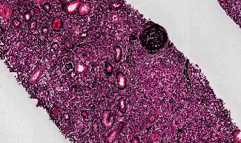
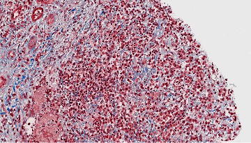
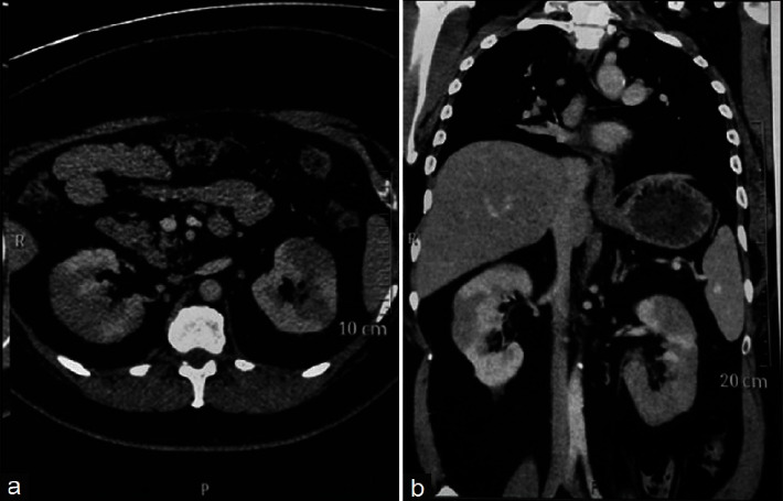
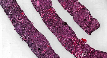
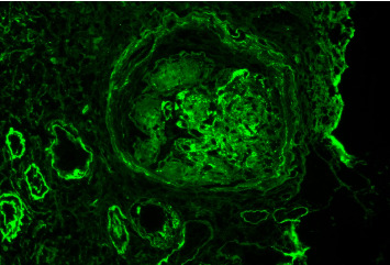
IgG4-related disease (IgG4-RD) is an immune-mediated disorder marked by fibro-inflammatory masses that can infiltrate multiple organ systems. Due to its relatively recent discovery and limited understanding of its pathophysiology, IgG4-related disease may be difficult to recognize and is consequently potentially underdiagnosed. Renal involvement is becoming regarded as one of the key features of this disease. To date, the most well-recognized renal complication of IgG4-related disease is tubulointerstitial nephritis, but membranous glomerulonephritis, renal masses, and retroperitoneal fibrosis have also been reported. This concise review has two objectives. First, it will briefly encapsulate the history, epidemiology, and presentation of IgG4-related disease. Second, it will examine the reported renal manifestations of IgG4-related disease, exploring the relevant histology, imaging, clinical features, and treatment considerations. This synthesis will be highly relevant for nephrologists, rheumatologists, general internists, and renal pathologists to raise awareness and help improve early recognition of IgG4-related kidney disease (IgG4-RKD).


Conflict of interest statement

The authors declare that they have no conflicts of interest.

Figures

Figure 1
Figure 1
Storiform fibrosis highlighted on silver stain. Reproduced with permission from PathologyOutlines.com.
Figure 2
Figure 2
Storiform fibrosis highlighted on trichrome stain. Reproduced with permission from PathologyOutlines.com.
Figure 3
Figure 3
Contrast-enhanced computed tomography scan of the abdomen highlighting IgG4-related heterogenous enhancement of the kidneys with hypodensities at the lower pole. Adapted from Korivi et al. [62] as published in the Indian Journal of Nephrology. Reproduced under the Creative Commons Attribution-Noncommercial-Share Alike 3.0 Unported License.
Figure 4
Figure 4
Marked tubulointerstitial effacement highlighted on silver stain in IgG4-RD. Reproduced with permission from PathologyOutlines.com.
Figure 5
Figure 5
Granular IgG deposits within glomerular mesangial regions and capillary loops, as well as within tubular basement membranes and Bowman capsule. Background glomerulus shows features suggestive of diabetes. Reproduced with permission from PathologyOutlines.com.

References

    1. Stone J. H., Zen Y., Deshpande V. IgG4-related disease. New England Journal of Medicine . 2012;366(6):539–551. doi: 10.1056/nejmra1104650. - DOI - PubMed
    1. Kamisawa T., Funata N., Hayashi Y., et al. A new clinicopathological entity of IgG4-related autoimmune disease. Journal of Gastroenterology . 2003;38(10):982–984. doi: 10.1007/s00535-003-1175-y. - DOI - PubMed
    1. Hamano H., Kawa S., Horiuchi A., et al. High serum IgG4 concentrations in patients with sclerosing pancreatitis. New England Journal of Medicine . 2001;344(10):732–738. doi: 10.1056/nejm200103083441005. - DOI - PubMed
    1. Yamamoto M., Takahashi H., Ohara M., et al. A new conceptualization for Mikulicz’s disease as an IgG4-related plasmacytic disease. Modern Rheumatology . 2006;16(6):335–340. doi: 10.3109/s10165-006-0518-y. - DOI - PMC - PubMed
    1. Himi T., Takano K., Yamamoto M., Naishiro Y., Takahashi H. A novel concept of Mikulicz’s disease as IgG4-related disease. Auris Nasus Larynx . 2012;39(1):9–17. doi: 10.1016/j.anl.2011.01.023. - DOI - PubMed

LinkOut - more resources