. 2024 Jul 1;121(6):e20230675.
doi: 10.36660/abc.20230675. eCollection 2024.

Protective Effect of Long Noncoding RNA OXCT1-AS1 on Doxorubicin-Induced Apoptosis of Human Myocardial Cells by the Competitive Endogenous RNA Pattern

[Article in Portuguese, English]
Affiliations

Protective Effect of Long Noncoding RNA OXCT1-AS1 on Doxorubicin-Induced Apoptosis of Human Myocardial Cells by the Competitive Endogenous RNA Pattern

[Article in Portuguese, English]
Zhen Chen et al. Arq Bras Cardiol. .

Abstract

Background: The anthracycline chemotherapeutic antibiotic doxorubicin (DOX) can induce cumulative cardiotoxicity and lead to cardiac dysfunction. Long non-coding RNAs (lncRNAs) can function as important regulators in DOX-induced myocardial injury.

Objective: This study aims to investigate the functional role and molecular mechanism of lncRNA OXCT1 antisense RNA 1 (OXCT1-AS1) in DOX-induced myocardial cell injury in vitro.

Methods: Human cardiomyocytes (AC16) were stimulated with DOX to induce a myocardial cell injury model. OXCT1-AS1, miR-874-3p, and BDH1 expression in AC16 cells were determined by RT-qPCR. AC16 cell viability was measured by XTT assay. Flow cytometry was employed to assess the apoptosis of AC16 cells. Western blotting was used to evaluate protein levels of apoptosis-related markers. Dual-luciferase reporter assay was conducted to verify the binding ability between miR-874-3p and OXCT1-AS1 and between miR-874-3p and BDH1. The value of p<0.05 indicated statistical significance.

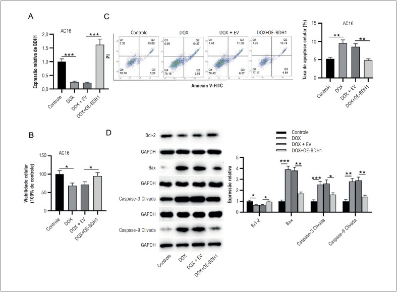
Results: OXCT1-AS1 expression was decreased in DOX-treated AC16 cells. Overexpression of OXCT1-AS1 reversed the reduction of cell viability and promotion of cell apoptosis caused by DOX. OXCT1-AS1 is competitively bound to miR-874-3p to upregulate BDH1. BDH1 overexpression restored AC16 cell viability and suppressed cell apoptosis under DOX stimulation. Knocking down BDH1 reversed OXCT1-AS1-mediated attenuation of AC16 cell apoptosis under DOX treatment.

Conclusion: LncRNA OXCT1-AS1 protects human myocardial cells AC16 from DOX-induced apoptosis via the miR-874-3p/BDH1 axis.

Fundamento: O antibiótico quimioterápico antraciclina doxorrubicina (DOX) pode induzir cardiotoxicidade cumulativa e levar à disfunção cardíaca. RNAs não codificantes longos (lncRNAs) podem funcionar como importantes reguladores na lesão miocárdica induzida por DOX.

Objetivo: Este estudo tem como objetivo investigar o papel funcional e o mecanismo molecular do RNA antisense lncRNA OXCT1 1 (OXCT1-AS1) na lesão celular miocárdica induzida por DOX in vitro.

Métodos: Cardiomiócitos humanos (AC16) foram estimulados com DOX para induzir um modelo de lesão celular miocárdica. A expressão de OXCT1-AS1, miR-874-3p e BDH1 em células AC16 foi determinada por RT-qPCR. A viabilidade das células AC16 foi medida pelo ensaio XTT. A citometria de fluxo foi empregada para avaliar a apoptose de células AC16. Western blotting foi utilizado para avaliar os níveis proteicos de marcadores relacionados à apoptose. O ensaio repórter de luciferase dupla foi conduzido para verificar a capacidade de ligação entre miR-874-3p e OXCT1-AS1 e entre miR-874-3p e BDH1. O valor de p<0,05 indicou significância estatística.

Resultados: A expressão de OXCT1-AS1 foi diminuída em células AC16 tratadas com DOX. A superexpressão de OXCT1-AS1 reverteu a redução da viabilidade celular e a promoção da apoptose celular causada pela DOX. OXCT1-AS1 está ligado competitivamente ao miR-874-3p para regular positivamente o BDH1. A superexpressão de BDH1 restaurou a viabilidade das células AC16 e suprimiu a apoptose celular sob estimulação com DOX. A derrubada do BDH1 reverteu a atenuação da apoptose de células AC16 mediada por OXCT1-AS1 sob tratamento com DOX.

Conclusão: LncRNA OXCT1-AS1 protege células miocárdicas humanas AC16 da apoptose induzida por DOX através do eixo miR-874-3p/BDH1.


Conflict of interest statement

Potencial conflito de interesse

Não há conflito com o presente artigo.

Figures

None
Graphical abstract
Figura 1
Figura 1. A superexpressão de OXCT1-AS1 inibe a apoptose de células miocárdicas humanas desencadeada por DOX. A) Células AC16 foram tratadas com DOX por 0,5, 2, 6, 12, 24 e 48 h, e a expressão relativa de OXCT1-AS1 em células miocárdicas humanas AC16 foi medida por RT-qPCR. B) A viabilidade das células AC16 foi determinada pelo ensaio XTT. C) A citometria de fluxo foi realizada para detectar apoptose celular. D) Os níveis de proteína dos genes relacionados à apoptose foram analisados por western blotting. n=3.*p< 0,05, **p< 0,01, ***p< 0,001. DOX: doxorrubicina; EV: vetor vazio; OE: superexpressão.
Figura 2
Figura 2. OXCT1-AS1 liga-se ao miR-874-3p. A-C) Os níveis de expressão de possíveis miRNAs a jusante de OXCT1-AS1 em AC16 tratado com DOX foram examinados por RT-qPCR. D) A expressão de miR-874-3p foi mostrada por RT-qPCR após superexpressão de OXCT1-AS1. E) O sítio de ligação entre OXCT1-AS1 e miR-874-3p foi previsto pelo banco de dados ENCORI. F) O ensaio repórter da luciferase foi conduzido para verificar a capacidade de ligação entre miR-874-3p e OXCT1-AS1 em células AC16, n=3. *p< 0,05, **p< 0,01 vs. 0 h ou EV.DOX, doxorrubicina; EV: vetor vazio; OE: superexpressão; NC: controle negativo; WT: tipo largo; MUT: mutante.
Figura 3
Figura 3. miR-874-3p tem como alvo BDH1. A)Os níveis de expressão de dez mRNAs que possuem potenciais sítios de ligação ao miR-874-3p foram avaliados por RT-qPCR. (B) RT-qPCR foi realizado para avaliar a expressão de RGS4, BDH1 e HEG1 em células AC16 com ou sem inibição de miR-874-3p. (C) RT-qPCR mostrou o nível de expressão de BDH1 após superexpressão de OXCT1-AS1. (D) O sítio de ligação entre miR-874-3p e OXCT1-AS1 foi previsto pelo TargetScan. (E). O ensaio repórter da luciferase foi realizado para validar a relação de ligação entre miR-874-3p e BDH1 em células AC16. n=3.**p< 0,01, ***p< 0,001. DOX: doxorrubicina; EV: vetor vazio; OE: superexpressão; NC: controle negativo; WT: tipo largo; MUT: mutante.
Figura 4
Figura 4. BDH1 suprime a apoptose de células miocárdicas humanas estimuladas por DOX. A) A expressão de BDH1 em células AC16 de diferentes grupos (controle, DOX, DOX-EV e DOX+OE-OXCT1-AS1) foi examinada por RT-qPCR. (B) A viabilidade das células AC16 após os tratamentos indicados foi detectada pelo ensaio XTT. (C) A apoptose das células AC16 em cada grupo foi examinada por citometria de fluxo. (D) Os efeitos da superexpressão de BDH1 nos níveis de proteína de genes relacionados à apoptose (Bax, caspase-3 clivada, caspase-9 clivada e Bcl-2) em células AC16 tratadas com DOX foram analisados por western blotting. n=3.*p< 0,05, **p< 0,01, ***p< 0,001. DOX: doxorrubicina; EV: vetor vazio; OE: superexpressão.
Figura 5
Figura 5. O silenciamento de BDH1 resgata o efeito supressor de OXCT1-AS1 na apoptose de células AC16 tratadas com DOX. Células AC16 foram transfectadas com EV, OE-OXCT1-AS1 ou OE-OXCT1-AS1 + sh-BDH1, seguido de estimulação com DOX. (A) A viabilidade das células AC16 foi avaliada pelo ensaio XTT. (B) A apoptose de células AC16 foi demonstrada por citometria de fluxo. (C) Western blotting foi utilizado para avaliar os níveis de proteína relacionados à apoptose em células AC16. n=3.*p< 0,05, **p< 0,01. DOX: doxorrubicina; EV: vetor vazio; OE: superexpressão; sh: RNA em gancho curto.
None
Graphical abstract
Figure 1
Figure 1. Overexpression of OXCT1-AS1 inhibits DOX-triggered apoptosis of human myocardial cells. A) AC16 cells were treated with DOX for 0.5, 2, 6, 12, 24, and 48 h, and relative expression of OXCT1-AS1 in human myocardial cells AC16 was measured by RT-qPCR. B) The viability of AC16 cells was determined by XTT assay. C) Flow cytometry was conducted to detect cell apoptosis. D) Protein levels of apoptosis-related genes were analyzed by western blotting. n=3. *p< 0.05, **p< 0.01, ***p< 0.001. DOX: doxorubicin; EV: empty vector; OE: overexpression.
Figure 2
Figure 2. OXCT1-AS1 binds to miR-874-3p. A-C) Expression levels of possible downstream miRNAs of OXCT1-AS1 in DOX-treated AC16 were examined by RT-qPCR. D) miR-874-3p expression was shown by RT-qPCR after overexpressing OXCT1-AS1. E) The binding site between OXCT1-AS1 and miR-874-3p was predicted by the ENCORI database. F) Luciferase reporter assay was conducted to verify the binding ability between miR-874-3p and OXCT1-AS1 in AC16 cells. n=3. *p< 0.05, **p< 0.01 vs. 0 h or EV. DOX, doxorubicin; EV, empty vector; OE, overexpression; NC, negative control; WT, wide type; MUT, mutant.
Figure 3
Figure 3. miR-874-3p targets BDH1. A) The expression levels of ten mRNAs that have potential binding sites to miR-874-3p were evaluated by RT-qPCR. (B) RT-qPCR was performed to evaluate the expression of RGS4, BDH1, and HEG1 in AC16 cells with or without miR-874-3p inhibition. (C) RT-qPCR showed the expression level of BDH1 after OXCT1-AS1 overexpression. (D) The binding site between miR-874-3p and OXCT1-AS1 was predicted by TargetScan. (E). Luciferase reporter assay was performed to validate the binding relation between miR-874-3p and BDH1 in AC16 cells. n=3. **p< 0.01, ***p< 0.001. DOX, doxorubicin; EV, empty vector; OE, overexpression; NC, negative control. WT, wide type; MUT, mutant.
Figure 4
Figure 4. BDH1 suppresses the apoptosis of DOX-stimulated human myocardial cells. A) BDH1 expression in AC16 cells of different groups (control, DOX, DOX-EV, and DOX+OE-OXCT1-AS1) was examined by RT-qPCR. B) The viability of AC16 cells after indicated treatments was detected by XTT assay. C) The apoptosis of AC16 cells in each group was examined by flow cytometry. D) Effects of BDH1 overexpression on the protein levels of apoptosis-related genes (Bax, Cleaved caspase-3, Cleaved caspase-9, and Bcl-2) in DOX-treated AC16 cells were analyzed by western blotting. n=3. *p< 0.05, **p< 0.01, ***p< 0.001. DOX, doxorubicin; EV, empty vector; OE, overexpression.
Figure 5
Figure 5. Silencing of BDH1 rescues the suppressive effect of OXCT1-AS1 on the apoptosis of DOX-treated AC16 cells. AC16 cells were transfected with EV, OE-OXCT1-AS1, or OE-OXCT1-AS1 + sh-BDH1, followed by DOX stimulation. A) The viability of AC16 cells was evaluated by XTT assay. B) AC16 cells apoptosis was shown by flow cytometry. C) Western blotting was used to assess apoptosis-related protein levels in AC16 cells. n=3. *p< 0.05, **p< 0.01. DOX, doxorubicin; EV: empty vector; OE: overexpression; sh: short hairpin RNA.

References

    1. Tan L, Xiong D, Zhang H, Xiao S, Yi R, Wu J. ETS2 Promotes Cardiomyocyte Apoptosis and Autophagy in Heart Failure by Regulating lncRNA TUG1/miR-129-5p/ATG7 Axis. FASEB J. 2023;37(6):e22937. doi: 10.1096/fj.202202148RR. - DOI - PubMed
    1. Sun M, Mao S, Wu C, Zhao X, Guo C, Hu J, et al. Piezo1-Mediated Neurogenic Inflammatory Cascade Exacerbates Ventricular Remodeling After Myocardial Infarction. Circulation. 2024;0:1–8. doi: 10.1161/CIRCULATIONAHA.123.065390. - DOI - PubMed
    1. Mi S, Huang F, Jiao M, Qian Z, Han M, Miao Z, et al. Inhibition of MEG3 Ameliorates Cardiomyocyte Apoptosis and Autophagy by Regulating the Expression of miRNA-129-5p in a Mouse Model of Heart Failure. Redox Rep. 2023;28(1):2224607–2224607. doi: 10.1080/13510002.2023.2224607. - DOI - PMC - PubMed
    1. Hu L, Xu Y, Wang Q, Liu M, Meng L, Yan D, et al. Yiqi Huoxue Recipe Inhibits Cardiomyocyte Apoptosis Caused by Heart Failure Through Keap1/Nrf2/HIF-1α Signaling Pathway. Bioengineered. 2021;12(1):969–78. doi: 10.1080/21655979.2021.1900634. - DOI - PMC - PubMed
    1. Zhao H, Yu J, Zhang R, Chen P, Jiang H, Yu W. Doxorubicin Prodrug-based Nanomedicines for the Treatment of Cancer. Eur J Med Chem. 2023;258:115612–115612. doi: 10.1016/j.ejmech.2023.115612. - DOI - PubMed

LinkOut - more resources