. 2024 Jul:103:320-334.
doi: 10.1016/j.neuro.2024.06.017. Epub 2024 Jul 1.

Neuroprotective efficacy of the glucocorticoid receptor modulator PT150 in the rotenone mouse model of Parkinson's disease

Affiliations

Neuroprotective efficacy of the glucocorticoid receptor modulator PT150 in the rotenone mouse model of Parkinson's disease

Amanda S Latham et al. Neurotoxicology. 2024 Jul.

Abstract

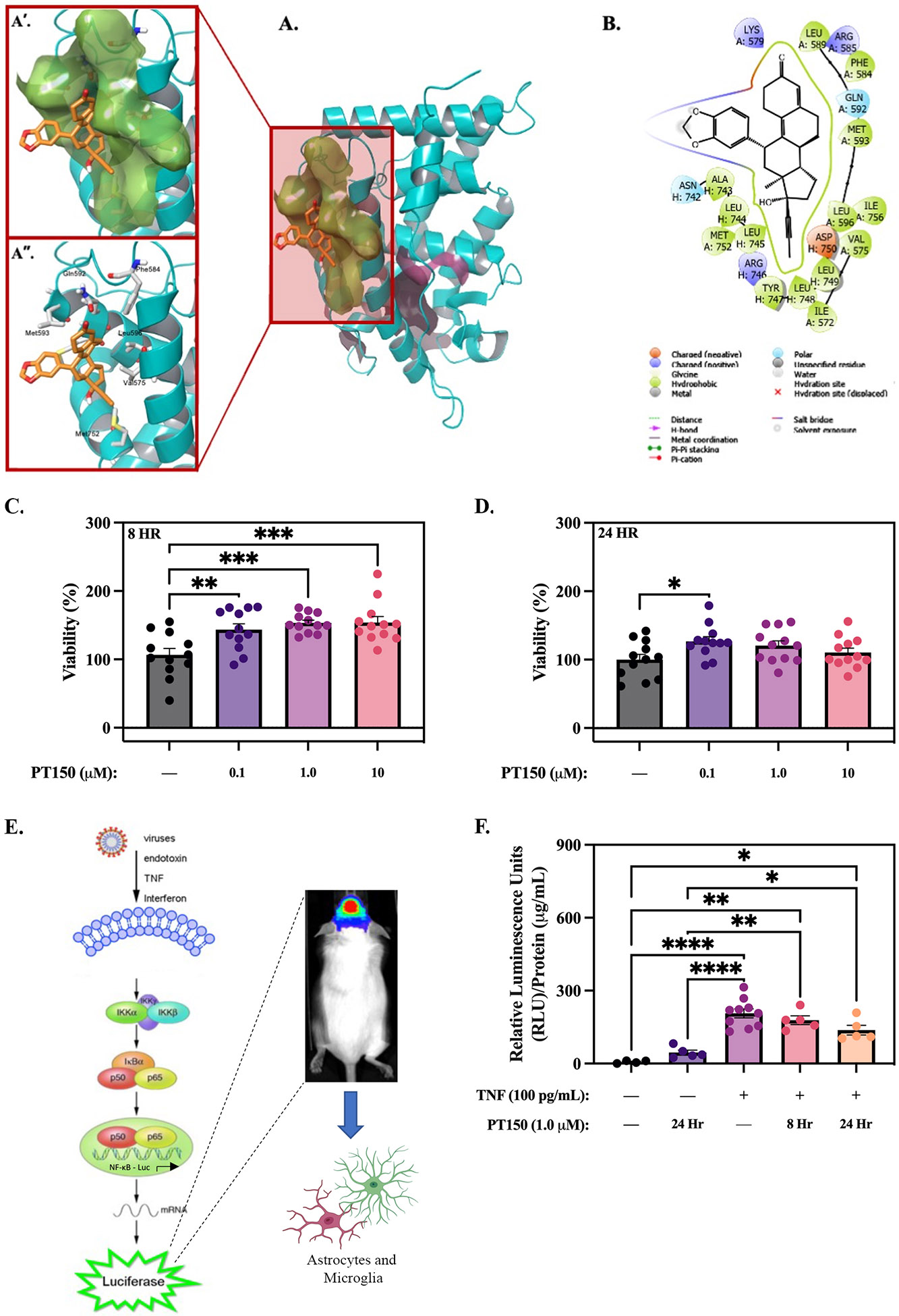
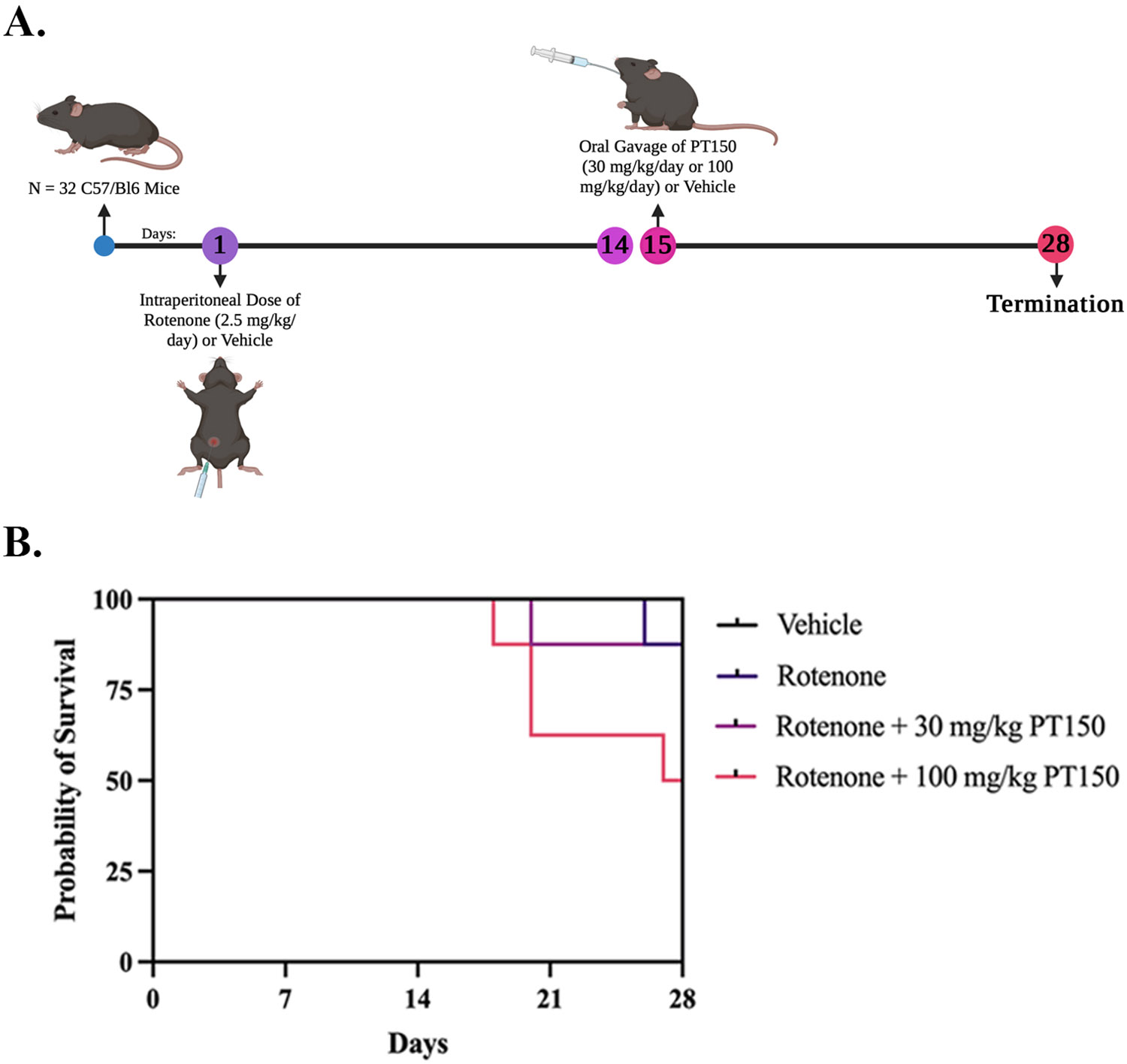
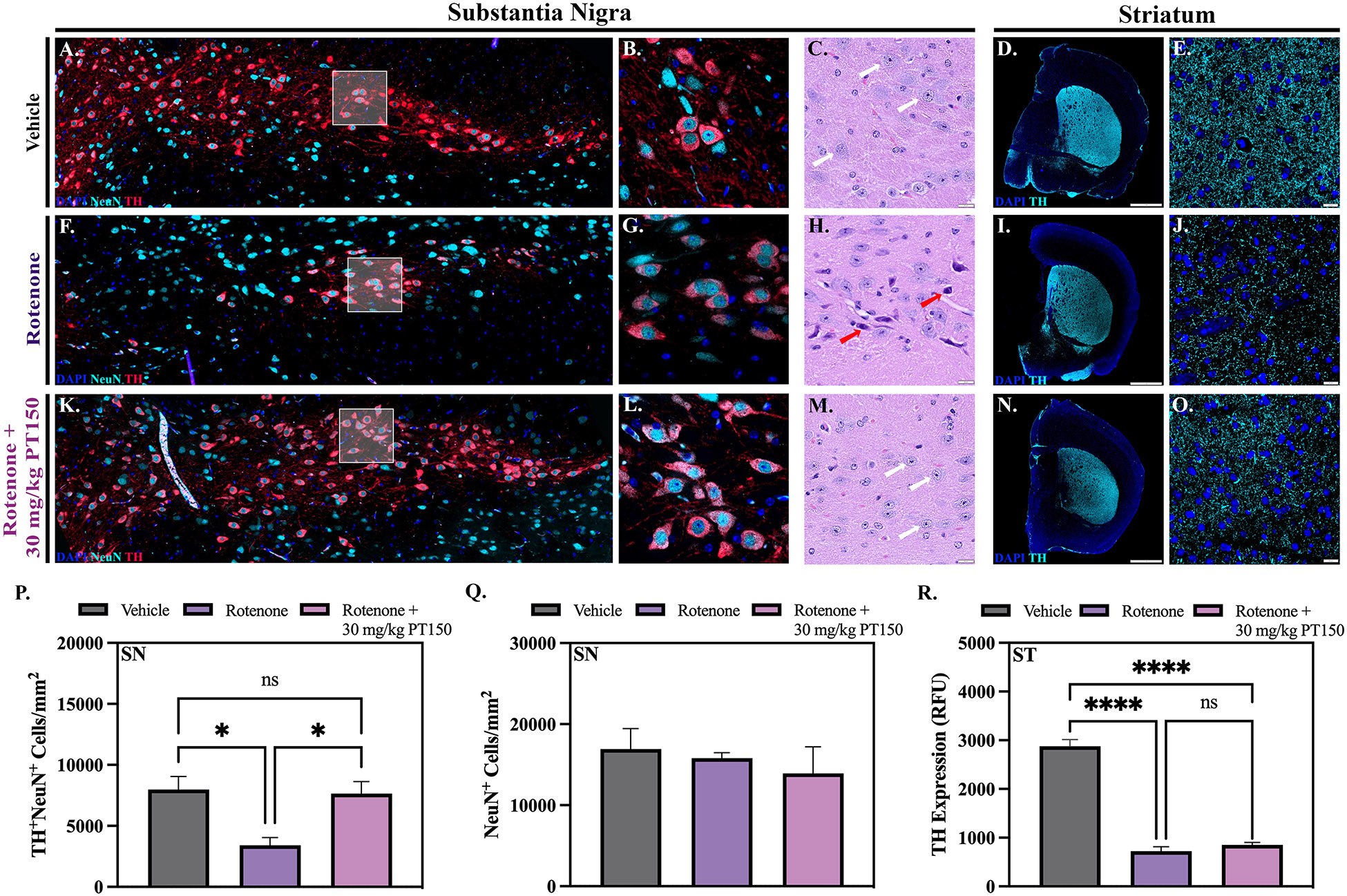
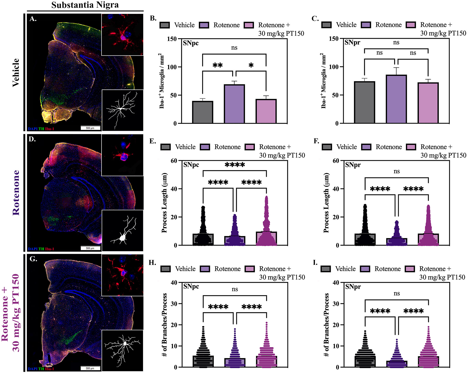
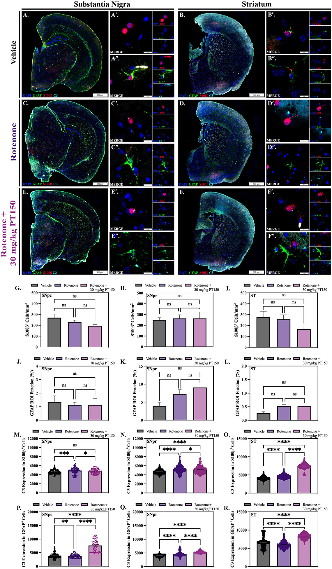
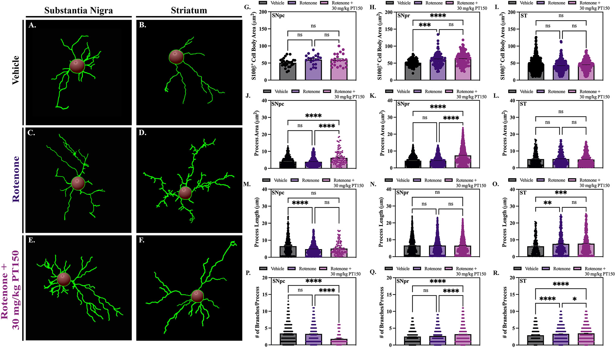
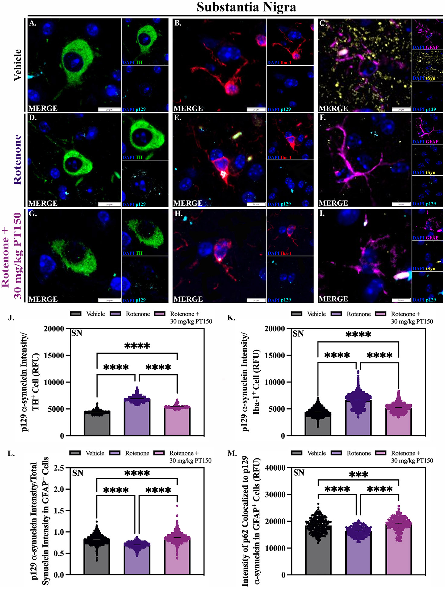
Parkinson's disease (PD) is the most common neurodegenerative movement disorder worldwide. Current treatments for PD largely center around dopamine replacement therapies and fail to prevent the progression of pathology, underscoring the need for neuroprotective interventions. Approaches that target neuroinflammation, which occurs prior to dopaminergic neuron (DAn) loss in the substantia nigra (SN), represent a promising therapeutic strategy. The glucocorticoid receptor (GR) has been implicated in the neuropathology of PD and modulates numerous neuroinflammatory signaling pathways in the brain. Therefore, we investigated the neuroprotective effects of the novel GR modulator, PT150, in the rotenone mouse model of PD, postulating that inhibition of glial inflammation would protect DAn and reduce accumulation of neurotoxic misfolded ⍺-synuclein protein. C57Bl/6 mice were exposed to 2.5 mg/kg/day rotenone by intraperitoneal injection for 14 days. Upon completion of rotenone dosing, mice were orally treated at day 15 with 30 mg/kg/day or 100 mg/kg/day PT150 in the 14-day post-lesioning incubation period, during which the majority of DAn loss and α-synuclein (α-syn) accumulation occurs. Our results indicate that treatment with PT150 reduced both loss of DAn and microgliosis in the nigrostriatal pathway. Although morphologic features of astrogliosis were not attenuated, PT150 treatment promoted potentially neuroprotective activity in these cells, including increased phagocytosis of hyperphosphorylated α-syn. Ultimately, PT150 treatment reduced the loss of DAn cell bodies in the SN, but not the striatum, and prohibited intra-neuronal accumulation of α-syn. Together, these data indicate that PT150 effectively reduced SN pathology in the rotenone mouse model of PD.

Keywords: Astrocyte; Microglia; Molecular modeling; Neurodegeneration; Neuroinflammation; Neuroprotection; Parkinson's disease.


Conflict of interest statement

Declaration of Competing Interest The authors declare the following financial interests/personal relationships which may be considered as potential competing interests: Ronald Tjalkens reports financial support was provided by Colorado State University. If there are other authors, they declare that they have no known competing financial interests or personal relationships that could have appeared to influence the work reported in this paper.

Figures

Figure 1.
Figure 1.. Computational-based molecular docking of PT150 interactions with the ligand binding domain of the human glucocorticoid receptor.
PT150 was docked into the co-activator site and the steroid binding site of the crystal structures of the ligand-binding domain of the glucocorticoid receptor. Restrained minimization of the protein structure was performed and prepared proteins were subjected to SiteMap analysis to identify the available binding sites. The interaction between PT150 and the glucocorticoid receptor demonstrates increased affinity for the co-activating domain (A), with the benzodioxole ring orientated out of the pocket (A’, A”). No anchoring H-bond interactions were observed between PT150 and the co-activator site (B). Assays detecting the fluorescence expression of calcein red-orange and propidium iodide normalized to Hoechst 33342 were used to determine the % viability of cells treated with vehicle (DMSO) or PT150. PT150 treatment at 0.1 μM, 1.0 μM, and 10 μM concentrations increased viability of primary C57Bl/6 mixed glia compared to vehicle (DMSO) controls at 8 hours (C) and 24 hours (D). A luciferase reporter assay was used to evaluate if PT150 reduces NF-κB activation stimulated by TNF (100 pg/mL) in primary NF-κB-GFP/luciferase reporter glia (Created using Biorender) (E). Quantitative graphs demonstrate decreased NF-κB activity via relative luminescence units following 8 hour and 24 hour treatment with 1.0 μM PT150 (F). Parametric one-way ANOVA analysis performed. *p < 0.05; **p < 0.01; ***p < 0.001; ****p < 0.0001.
Figure 2.
Figure 2.. Treatment paradigm and Kaplan-Meier survival curves for each study group.
(A) C57BL/6 mice were treated with 2.5 mg/kg/day rotenone for 14 days, followed by treatment at day 15 with either 30 mg/kg/day or 100 mg/kg/day PT150 by oral gavage for 14 days. Groups: Vehicle control, rotenone, rotenone + 30 mg/kg PT150, rotenone + 100 mg/kg PT150. (B) Survival was reduced in animals treated with 100 mg/kg/day PT150 compared to the other groups. N = 8 mice/group.
Figure 3.
Figure 3.. Stereologic and morphologic characterization of dopaminergic neurons in the nigrostriatal system was determined following exposure to rotenone with and without PT150 treatment.
Representative images are shown for the substantia nigra (A, B, F, G, K, and L) with immunostaining for dopaminergic neurons (TH, red) and total neurons (NeuN, cyan). Stereological analysis was used to determine the quantity of dopaminergic (TH+NeuN+) and total (NeuN+) neuron cell bodies (P and Q). Hematoxylin and eosin staining was utilized to evaluate the presence of intact (white arrows) and pyknotic (red arrows) neuron cell bodies in the substantia nigra (C, H, and M). Intensity measurements for TH+ projecting fibers in the striatum were performed (R); representative images of immunostaining for dopaminergic neurons (TH, cyan) are shown (D, E, I, J, N, and O). Parametric one-way ANOVA analysis performed. N = 3 – 5/group. *p < 0.05; ****p < 0.0001. Scale bars = 1 mm (D, J, and O), 20 μM (C, I, N, E, K, and P).
Figure 4.
Figure 4.. Quantity and morphology of microglia was determined in vehicle, rotenone-exposed, and rotenone + 30 mg/kg PT150-treated animals.
Immunofluorescent labeling of microglia (Iba-1, red) was performed; representative montage and high-magnification images are shown for the SN (A, F, and K) and ST (B, G, and L). Analysis of cell quantity was performed using CellSens (C – E) and Imaris software was used for quantifications of process length (H – J) and the number of process branches (M – O); skeletonized renderings are shown for the SN (A, F, and K) and the ST (B, G, and L). Nonparametric one-way ANOVA with Dunn’s correction analysis performed. N = 3 – 5/group. ns = not significant; *p < .05; **p < 0.01; ***p < 0.001; ****p <0 .0001. Scale bars = 500 μM.
Figure 5.
Figure 5.. Differential effects of rotenone and PT150 on astrogliosis.
The number of astrocytes were quantified in vehicle, rotenone-exposed, and rotenone + 30 mg/kg PT150-treated animals. Immunofluorescent labeling of astrocytes (GFAP, green; S100β, red; C3, cyan) was performed; representative montage and high-magnification images are shown for the SN (A, C, and E) and ST (B, D, and F). The quantity of S100β+ cells is shown for the SNpc (G), SNpr (H), and ST (I). Immunostaining was quantified for the maximum expression of GFAP (% per regional area) in the SNpc (J), SNpr (K), and ST (L). Maximal C3 expression in S100β+ soma and GFAP+ processes were determined within the SNpc (M, P), SNpr (N, Q), and ST (O, R), respectively. Nonparametric one-way ANOVA with Dunn’s correction analysis performed. N = 3 – 5/group. ns = not significant; *p < 0.05; **p <0 .01; ***p < 0.001; ****p <0 .0001. Scale bars = 500 μM (A, C, E, B, D, and F), 20 μM (A’, A”, B’, B”, C’, C”, D’, D”, E’, E”, F’, F”).
Figure 6.
Figure 6.. Astrocyte morphology was evaluated in vehicle, rotenone-exposed, and rotenone + 30 mg/kg PT150-treated animals.
Morphological analysis was performed using CellSens for quantification of cell body area (G – I) and Imaris software for quantifications of process area (J – L), process length (M – O) and the number of process branches (P – R); skeletonized renderings are shown for the SN (A, C, and E) and the ST (B, D, and F). Nonparametric one-way ANOVA with Dunn’s correction analysis performed. N = 3 – 5/group. ns = not significant; **p < 0.05; **p <0 .01; ***p < 0.001; ****p <0 .0001.
Figure 7.
Figure 7.. Accumulation and trafficking of α-synuclein.
The quantity of misfolded α-synuclein was evaluated in dopaminergic neurons, microglia, and astrocytes in vehicle, rotenone-exposed, and rotenone + 30 mg/kg PT150-treated animals. Immunofluorescent labeling of α-synuclein phosphorylated at serine 129 (p129, cyan), dopaminergic neurons (TH, green), microglia (Iba-1, red), and astrocytes (GFAP, purple) was performed; representative high-magnification images are shown for the SN. Maximal p129 expression within dopaminergic neurons (J), microglia (K), and astrocytes (L) was determined. Additional analysis was performed to determine the maximal expression of p62 colocalized to p129 α-synuclein immunolabelling within GFAP+ cells (M). Nonparametric one-way ANOVA with Dunn’s correction analysis performed. N = 3 – 5/group. ****p < 0.0001. Scale bars = 20 μM

Update of

Similar articles

Cited by

References

    1. Global, regional, and national burden of neurological disorders, 1990-2016: a systematic analysis for the Global Burden of Disease Study 2016. Lancet Neurol, 2019. 18(5): p. 459–480. - PMC - PubMed
    1. Poewe W., et al., Parkinson disease. Nature Reviews Disease Primers, 2017. 3(1): p. 17013. - PubMed
    1. Willis AW, et al., Incidence of Parkinson disease in North America. npj Parkinson's Disease, 2022. 8(1): p. 170. - PMC - PubMed
    1. Sveinbjornsdottir S., The clinical symptoms of Parkinson's disease. J Neurochem, 2016. 139 Suppl 1: p. 318–324. - PubMed
    1. Tanner CM, et al., Rotenone, paraquat, and Parkinson's disease. Environ Health Perspect, 2011. 119(6): p. 866–72. - PMC - PubMed

MeSH terms

Substances

LinkOut - more resources