Review
. 2024 Nov 1:258:110061.
doi: 10.1016/j.neuropharm.2024.110061. Epub 2024 Jul 2.

Limitations and potential of κOR biased agonists for pain and itch management

Affiliations
Review

Limitations and potential of κOR biased agonists for pain and itch management

Amal El Daibani et al. Neuropharmacology. .

Abstract

The concept of ligand bias is based on the premise that different agonists can elicit distinct responses by selectively activating the same receptor. These responses often determine whether an agonist has therapeutic or undesirable effects. Therefore, it would be highly advantageous to have agonists that specifically trigger the therapeutic response. The last two decades have seen a growing trend towards the consideration of ligand bias in the development of ligands to target the κ-opioid receptor (κOR). Most of these ligands selectively favor G-protein signaling over β-arrestin signaling to potentially provide effective pain and itch relief without adverse side effects associated with κOR activation. Importantly, the specific role of β-arrestin 2 in mediating κOR agonist-induced side effects remains unknown, and similarly the therapeutic and side-effect profiles of G-protein-biased κOR agonists have not been established. Furthermore, some drugs previously labeled as G-protein-biased may not exhibit true bias but may instead be either low-intrinsic-efficacy or partial agonists. In this review, we discuss the established methods to test ligand bias, their limitations in measuring bias factors for κOR agonists, as well as recommend the consideration of other systematic factors to correlate the degree of bias signaling and pharmacological effects. This article is part of the Special Issue on "Ligand Bias".


Conflict of interest statement

Declaration of competing interest All the authors declare no conflicts of interest.

Figures

Fig. 1.
Fig. 1.
A systematic approach to select the appropriate model to quantify signaling bias for κOR agonists. Adapted from (Kenakin and Christopoulos, 2013). KA is the functional dissociation constant for the agonist; τ is the efficacy of the agonist in the given pathway; pEC50 is the negative logarithm of the EC50; and RA is the relative activity.
Fig. 2.
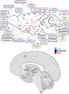
Fig. 2.
Mice κOR expression adapted from Le Merrer et al., 2009) (Le Merrer et al., 2009). ARC arcuate nucleus, hypothalamus, AMY amygdala, BLA basolateral nucleus, amygdala (Knoll et al., 2011), BNST bed nucleus of the stria terminalis (Haun et al., 2020), CeA central nucleus, amygdala (Knoll et al., 2011; Haun et al., 2022; Yakhnitsa et al., 2022; Navratilova et al., 2019), CI claustrum, CL centrolateral thalamus, CM centromedial thalamus, COA cortical nucleus, amygdala, CPU caudate putamen, DMH dorsomedial hypothalamus, DMR dorsal and medial raphe, DR dorsal raphe (Land et al., 2009; Wright et al., 2018), DTN dorsal tegmental nucleus, EN endopiriform cortex, GP globus pallidus, HPC hippocampus (Shirayama et al., 2004), IC inferior colliculus, ICX insular cortex IP interpeduncular nucleus, LC locus coeruleus (Al-Hasani et al., 2013), LH lateral hypothalamus, ME median eminence, MeA median nucleus, amygdala, NAc nucleus accumbens (Al-Hasani et al., 2015; Massaly et al., 2019; Lorente et al., 2024; Coleman et al., 2021; Zhang et al., 2023), NST nucleus tractus solitarius, pituitary, NRGC nucleus reticularis gigantocellularis, OB olfactory bulb, OCX occipital cortex, PAG periaqueductal gray (Tejeda et al., 2013, 2015; Carr et al., 2010), PBN parabrachial nucleus, PCX parietal cortex, PNR pontine reticular, POA preoptic area (Cone et al., 2023), PV paraventricular thalamus, PVN paraventricular hypothalamus, RE reuniens thalamus, RM raphe magnus (Nguyen et al., 2022; Abraham et al., 2018), S septum, SC superior colliculus, SN substantia nigra, STN spinal trigeminal nucleus, TCX temporal cortex, Th thalamus, TU olfactory tubercle, VMH ventromedial hypothalamus, VP ventral pallidum, VR ventral raphe, VTA ventral tegmental area (Robble et al., 2020), Zi zona incerta. Human κOR expression adapted from (Massaly et al., 2019; Liu et al., 2019b; Peng et al., 2012; Hiller and Fan, 1996; Simonin et al., 1995).

References

    1. Abraham AD, et al. , 2018. κ-Opioid receptor activation in dopamine neurons disrupts behavioral inhibition. Neuropsychopharmacology 43, 362–372. - PMC - PubMed
    1. Al-Hasani R, McCall JG, Foshage AM, Bruchas MR, 2013. Locus coeruleus kappa-opioid receptors modulate reinstatement of cocaine place preference through a noradrenergic mechanism. Neuropsychopharmacology 38, 2484–2497. - PMC - PubMed
    1. Al-Hasani R, et al. , 2015. Distinct subpopulations of nucleus accumbens dynorphin neurons drive aversion and reward. Neuron 87, 1063–1077. - PMC - PubMed
    1. Al-Hasani R, et al. , 2017. Circuit dynamics of in vivo dynorphn release in the nucleus accumbens. Alcohol 60, 220.
    1. Appleyard SM, et al. , 1999. Agonist-dependent desensitization of the kappa opioid receptor by G protein receptor kinase and beta-arrestin. J. Biol. Chem. 274, 23802–23807. - PubMed