

Forensic age estimation by MRI of the knee - comparison of two classifications for ossification stages in a German population
- PMID: 38960912
- PMCID: PMC11490462
- DOI: 10.1007/s00414-024-03281-5
Forensic age estimation by MRI of the knee - comparison of two classifications for ossification stages in a German population
Abstract
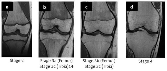
Aim and objectives: In forensic age estimation e.g. for judicial proceedings surpassed age thresholds can be legally relevant. To examine age related differences in skeletal development the recommendations by the Study Group on Forensic Age Diagnostics (AGFAD) are based on ionizing radiation (among others orthopantomograms, plain x-rays of the hand). Vieth et al. and Ottow et al. proposed MRI-classifications for the epiphyseal-diaphyseal fusion of the knee joint to define different age groups in healthy volunteers. The aim of the present study was to directly compare these two classifications in a large German patient population.
Materials and methods: MRI of the knee joint of 900 patients (405 female, 495 male) from 10 to 28 years of age were retrospectively analyzed. Acquired T1-weighted turbo spin-echo sequence (TSE) and T2-weighted sequence with fat suppression by turbo inversion recovery magnitude (TIRM) were analyzed for the two classifications. The different bony fusion stages of the two classifications were determined and the corresponding chronological ages assigned. Differences between the sexes were analyzed. Intra- and inter-observer agreements were determined using Cohen's kappa.
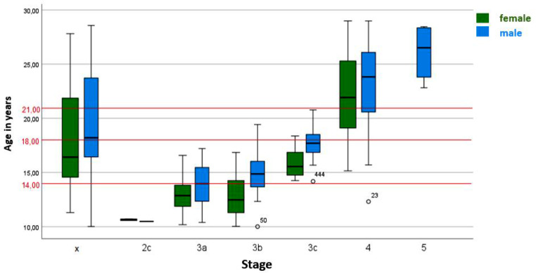
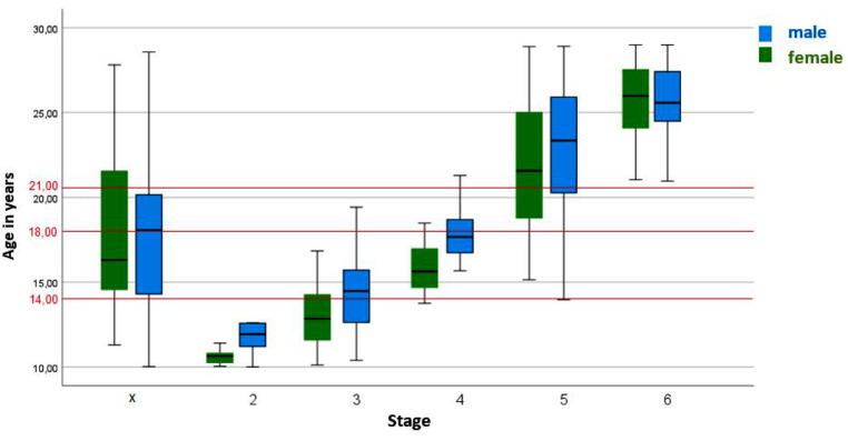
Results: With the classification of Ottow et al. it was possible to determine completion of the 18th and 21st year of life in both sexes. With the classification of Vieth et al. completion of the 18th year of life for female patients and the 14th and 21st year of life in both sexes could be determined. The intra- and inter-observer agreement levels were very good (κ > 0.82).
Conclusion: In the large German patient cohort of this study it was possible to determine the 18th year of life with for both sexes with the classification of Ottow et al. and for female patients with the classification of Vieth et al. It was also possible to determine the 21st year of life for all bones with the classification of Ottow et al. and for the distal femur with the classification of Vieth et al.
Keywords: Bone age; Forensic age estimation; Knee MRI; Ossification stages.
© 2024. The Author(s).
Conflict of interest statement
The authors have no relevant financial or non-financial interests to disclose.
Figures


Similar articles
-
Forensic Age Determination Using MRI Scans of the Ankle: Applying Two Classifications to Assess Ossification.Rofo. 2025 Jul;197(7):791-804. doi: 10.1055/a-2379-8785. Epub 2024 Sep 5. Rofo. 2025. PMID: 39236740 English, German.
-
Forensic age estimation by magnetic resonance imaging of the knee: the definite relevance in bony fusion of the distal femoral- and the proximal tibial epiphyses using closest-to-bone T1 TSE sequence.Eur Radiol. 2017 Dec;27(12):5041-5048. doi: 10.1007/s00330-017-4880-2. Epub 2017 Jul 4. Eur Radiol. 2017. PMID: 28677057
-
Forensic age assessment by 3.0T MRI of the knee: proposal of a new MRI classification of ossification stages.Eur Radiol. 2018 Aug;28(8):3255-3262. doi: 10.1007/s00330-017-5281-2. Epub 2018 Mar 13. Eur Radiol. 2018. PMID: 29536244
-
Forensic age estimation from ossification centres: a comparative investigation of imaging and physical methods.An Acad Bras Cienc. 2024 Oct 7;96(4):e20240181. doi: 10.1590/0001-3765202420240181. eCollection 2024. An Acad Bras Cienc. 2024. PMID: 39383352 Review.
-
Forensic Diagnostics of the Skeletal Age in the Living - Backgrounds and Methodology.Rofo. 2024 Mar;196(3):254-261. doi: 10.1055/a-2130-3162. Epub 2023 Sep 12. Rofo. 2024. PMID: 37699433 Review. English, German.
Cited by
-
Insights into dental age estimation: introducing multiple regression data from a Black South African population on modified gustafson's criteria.Int J Legal Med. 2025 Jan;139(1):143-155. doi: 10.1007/s00414-024-03312-1. Epub 2024 Aug 22. Int J Legal Med. 2025. PMID: 39168896 Free PMC article.
-
Forensic age estimation using Dedouit classification in adolescents of the Southwestern Chinese Han population based on the knee MRI.Int J Legal Med. 2025 Aug 6. doi: 10.1007/s00414-025-03566-3. Online ahead of print. Int J Legal Med. 2025. PMID: 40768048
References
-
- Mauer MAD, Säring D, Stanczus B, Herrmann J, Groth M, Jopp-van Well E (2019) A 2-year follow-up MRI study for the evaluation of an age estimation method based on knee bone development. Int J Legal Med 133:205–215. 10.1007/s00414-018-1826-4 - PubMed
-
- McAuliffe M, Khadria B (2020) 1 report overview: providing perspective on migration and mobility in increasingly uncertain times. World Migration Rep 2020:e00011. 10.1002/wom3.11
-
- Saint-Martin P, Rérolle C, Dedouit F, Bouilleau L, Rousseau H, Rougé D, Telmon N (2013) Age estimation by magnetic resonance imaging of the distal tibial epiphysis and the calcaneum. Int J Legal Med 127:1023–1030. 10.1007/s00414-013-0844-5 - PubMed
-
- Schmeling A, Grundmann C, Fuhrmann A, Kaatsch H-J, Knell B, Ramsthaler F, Reisinger W, Riepert T, Ritz-Timme S, Rösing FW, Rötzscher K, Geserick G (2008) Criteria for age estimation in living individuals. Int J Legal Med 122:457–460. 10.1007/s00414-008-0254-2 - PubMed
-
- Ottow C, Schulz R, Pfeiffer H, Heindel W, Schmeling A, Vieth V (2017) Forensic age estimation by magnetic resonance imaging of the knee: the definite relevance in bony fusion of the distal femoral- and the proximal tibial epiphyses using closest-to-bone T1 TSE sequence. Eur Radiol 27:5041–5048. 10.1007/s00330-017-4880-2 - PubMed
Publication types
MeSH terms
LinkOut - more resources
Full Text Sources
Medical
