. 2024 Nov;52(11):2975-2986.
doi: 10.1007/s10439-024-03560-7. Epub 2024 Jul 3.

OpenHands: An Open-Source Statistical Shape Model of the Finger Bones

Affiliations

OpenHands: An Open-Source Statistical Shape Model of the Finger Bones

T A Munyebvu et al. Ann Biomed Eng. 2024 Nov.

Abstract

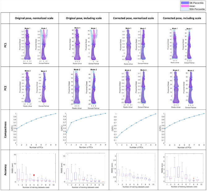
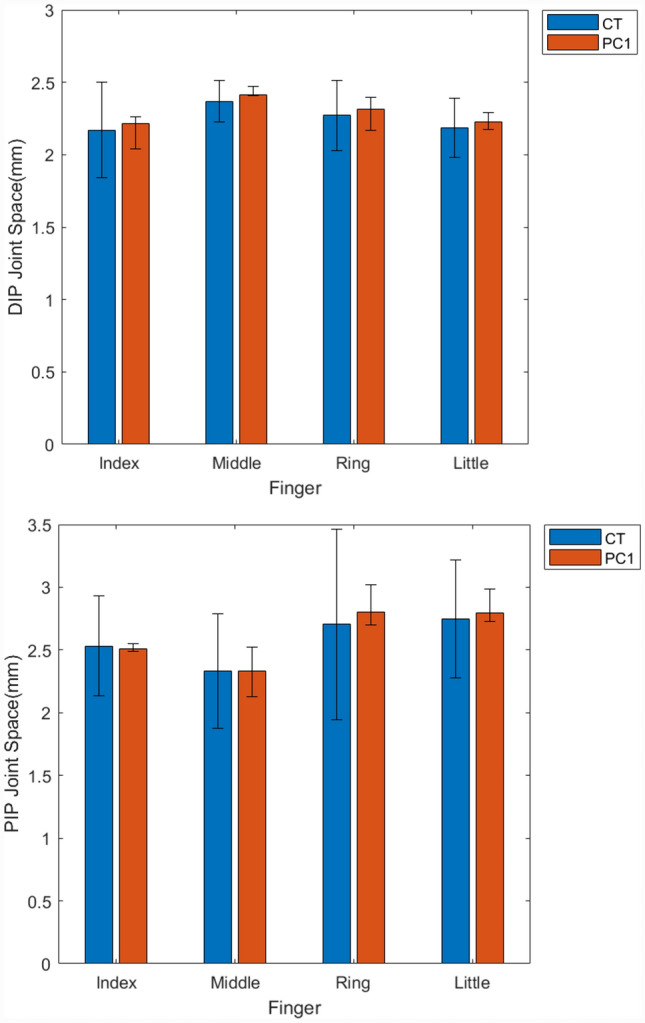
This paper presents statistical shape models of the four fingers of the hand, with an emphasis on anatomic analysis of the proximal and distal interphalangeal joints. A multi-body statistical shape modelling pipeline was implemented on an exemplar training dataset of computed tomography (CT) scans of 10 right hands (5F:5M, 27-37 years, free from disease or injury) imaged at 0.3 mm resolution, segmented, meshed and aligned. Model generated included pose neutralisation to remove joint angle variation during imaging. Repositioning was successful; no joint flexion variation was observed in the resulting model. The first principal component (PC) of morphological variation represented phalanx size in all fingers. Subsequent PCs showed variation in position along the palmar-dorsal axis, and bone breadth: length ratio. Finally, the models were interrogated to provide gross measures of bone lengths and joint spaces. These models have been published for open use to support wider community efforts in hand biomechanical analysis, providing bony anatomy descriptions whilst preserving the security of the underlying imaging data and privacy of the participants. The model describes a small, homogeneous population, and assumptions cannot be made about how it represents individuals outside the training dataset. However, it supplements anthropometric datasets with additional shape information, and may be useful for investigating factors such as joint morphology and design of hand-interfacing devices and products. The model has been shared as an open-source repository ( https://github.com/abel-research/OpenHands ), and we encourage the community to use and contribute to it.

Keywords: Anatomy modelling; Anthropometrics; Distal interphalangeal joint; Ergonomics; Machine Learning; Principal component analysis; Proximal interphalangeal joint.


Conflict of interest statement

All data generated during the study have been made openly available from the University of Southampton repository at 10.5258/SOTON/D2894, on a CC-BY 4.0 licence. Raw datasets analysed during the study under secondary data analysis ethics approval cannot be made publicly available for reasons of individual privacy, and requests to access these datasets should be directed to researchdata@soton.ac.uk. However, the derived OpenHands model has been made openly available at https://github.com/abel-research/openhands, on a CC-BY-SA 4.0 licence. The authors have no conflicts of interest to declare. The funders had no role in the design of the study; in the collection, analyses, or interpretation of data; in the writing of the manuscript, or in the decision to publish the results, and this publication represents the views of the authors only. The funders are not responsible for any use that may be made of the information it contains. Recent work in several fields of science has identified a bias in citation practices such that papers from women and other minority scholars are under cited relative to the number of papers in the field. We recognize this bias and have worked diligently to ensure that we are referencing appropriate papers with fair gender and racial author inclusion.

Figures

Fig. 1
Fig. 1
Flow diagram describing the joint pose neutralisation, to remove bone location variation generated during imaging. Step 1 computes the angles and centroids required for neutralisation. Step 2 involves the pose neutralisation of the PIP and DIP joint in the two reference coordinate systems (CS1 and CS2 respectively)
Fig. 2
Fig. 2
Impact on pose neutralisation and normalising scale on resultant principal components. Note for normalized shapes (column 1 and 3): since the proximal phalanx’s centroid lies at [0 0 0], the mean (against which dimensions are normalized) has a full scale length of one but is represented between approximately − 0.25 and 0.75 along the Proximal-Distal axis.
Fig. 3
Fig. 3
Deviation between the mean and mode extreme shapes generated using n = 10 (Full Shape) and the mean and mode extreme shapes (p represents minimum shape and m represents maximum shape for PC1 – PC9) generated when one dataset is removed (n = 9, LOO SSM Shape)
Fig. 4
Fig. 4
Variance (bar) and cumulative variance (line) captured by all PCs for index (A), middle (B), ring (C) and little (D) finger
Fig. 5
Fig. 5
Index Finger PCs: First principal component showing variation in bone size, the second principal component showing positional variation along the Palmar-Dorsal axis, the third principal component showing orientation variation and the fourth principal component showing variation in bone breadth
Fig. 6
Fig. 6
Average and 5th to 95th percentile range (error bars) finger length in mm across training datasets and PC1 shape instances
Fig. 7
Fig. 7
Average and 5th to 95th percentile range (error bars) for DIP (top) and PIP (bottom) joint space in mm across training datasets and PC1 shape instances

References

    1. Fernandez, J., A. Dickinson, and P. Hunter. Population based approaches to computational musculoskeletal modelling. Biomech. Model. Mechanobiol. 19(4):1165–1168, 2020. 10.1007/s10237-020-01364-x. - PubMed
    1. Steer, J., P. Worsley, M. Browne, and A. Dickinson. Predictive prosthetic socket design: part 1—population-based evaluation of transtibial prosthetic sockets by FEA-driven surrogate modelling. Biomech. Model. Mechanobiol. 19(4):1331–1346, 2020. 10.1007/s10237-019-01195-5. - PMC - PubMed
    1. Saxby, D. J., et al. Machine learning methods to support personalized neuromusculoskeletal modelling. Biomech. Model. Mechanobiol. 19(4):1169–1185, 2020. 10.1007/s10237-020-01367-8. - PubMed
    1. Iyer, K., et al. Statistical shape modeling of multi-organ anatomies with shared boundaries. Front. Bioeng. Biotechnol. 10:1–13, 2023. 10.3389/fbioe.2022.1078800. - PMC - PubMed
    1. Bruse, J. L., et al. A statistical shape modelling framework to extract 3D shape biomarkers from medical imaging data: Assessing arch morphology of repaired coarctation of the aorta. BMC Med. Imaging. 16(1):1–20, 2016. 10.1186/s12880-016-0142-z. - PMC - PubMed