. 2024 Jul 3;10(1):311.
doi: 10.1038/s41420-024-02073-5.

Anti-CCL2 therapy reduces oxygen toxicity to the immature lung

Affiliations

Anti-CCL2 therapy reduces oxygen toxicity to the immature lung

Tayyab Shahzad et al. Cell Death Discov. .

Abstract

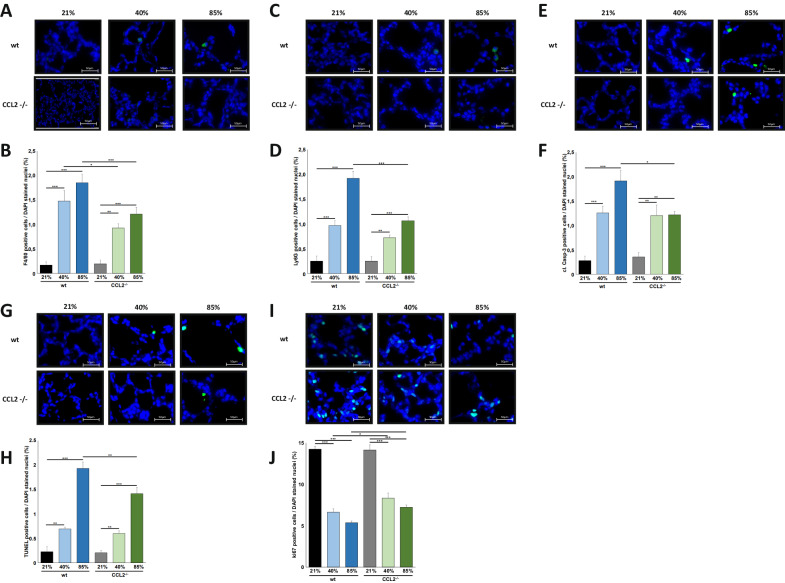
Oxygen toxicity constitutes a key contributor to bronchopulmonary dysplasia (BPD). Critical step in the pathogenesis of BPD is the inflammatory response in the immature lung with the release of pro-inflammatory cytokines and the influx of innate immune cells. Identification of efficient therapies to alleviate the inflammatory response remains an unmet research priority. First, we studied macrophage and neutrophil profiles in tracheal aspirates of n = 103 preterm infants <29 weeks´ gestation requiring mechanical ventilation. While no differences were present at birth, a higher fraction of macrophages, the predominance of the CD14+CD16+ subtype on day 5 of life was associated with moderate/severe BPD. Newborn CCL-2-/- mice insufficient in pulmonary macrophage recruitment had a reduced influx of neutrophils, lower apoptosis induction in the pulmonary tissue and better-preserved lung morphometry with higher counts of type II cells, mesenchymal stem cells and vascular endothelial cells when exposed to hyperoxia for 7 days. To study the benefit of a targeted approach to prevent the pulmonary influx of macrophages, wildtype mice were repeatedly treated with CCL-2 blocking antibodies while exposed to hyperoxia for 7 days. Congruent with the results in CCL-2-/- animals, the therapeutic intervention reduced the pulmonary inflammatory response, attenuated cell death in the lung tissue and better-preserved lung morphometry. Overall, our preclinical and clinical datasets document the predominant role of macrophage recruitment to the pathogenesis of BPD and establish the abrogation of CCL-2 function as novel approach to protect the immature lung from hyperoxic injury.


Conflict of interest statement

The authors declare no competing interests.

Figures

Fig. 1
Fig. 1. Higher fraction of CD14+CD16+ macrophages in tracheal aspirates of preterm infants associated with moderate/severe BPD.
A, B Tracheal aspirates of n = 26 infants with no/mild BPD and n = 42 with moderate/severe BPD requiring prolonged mechanical ventilation until at least day 5 of life were analyzed for the fraction of macrophages (A) and CD14+CD16+ macrophage subtype (B). The fraction of macrophages (A) and the CD14+CD16+ macrophage subtype (B) were significantly higher in infants developing moderate/severe BPD. Data are presented as median, interquartils and range. Statistical analysis was performed using rank sum test. *p < 0.05.
Fig. 2
Fig. 2. Hyperoxia-induced alveolar simplification is reduced in newborn CCL2−/− mice.
A Haematoxylin/eosin staining of representative lung tissue sections from newborn wildtype and CCL2−/− mice exposed to 21%, 40% or 85% of oxygen for 7 days starting after birth. Hyperoxia induced alveolar simplification was visible at 40% and 85% of oxygen. Scale bar: 200 µm. B Enlarged image sections from haematoxylin/eosin stainings as in A. Scale bar: 50 µm. Corresponding lung morphometric analyses for airspace (C), mean linear intercept (D) and septal wall thickness (E) from A. Hyperoxia induced increase in airspace was significantly less pronounced in CCL2−/− compared to wildtype mice for 40% and 85% of oxygen. For mean linear intercept, statistical significance was reached at 85% of oxygen. Data are presented as mean + SEM. Statistical analysis was performed by one way RM ANOVA. *p < 0.05, **p < 0.01, ***p < 0.001, ****p < 0.0001. n ≥ 6 mice/group.
Fig. 3
Fig. 3. Hyperoxia-induced recruitment of innate immune cells and cell death induction are reduced in newborn CCL2−/− mice.
A Immunofluorescence staining for F4/80 positive macrophages in lung tissues from mice from Fig. 2A. Hyperoxia mediated influx of macrophages into the lungs of newborn mice was detectable at 40% and 85% of oxygen but was markedly prohibited in CCL2−/− mice. Scale bar: 50 µm. B Quantification of F4/80 positive macrophages from A. Hyperoxia mediated influx of macrophages was significantly reduced in CCL2−/− compared to wildtype animals at 40% and 85% of oxygen. C Immunofluorescence staining for Ly6G positive neutrophils in lung tissues from mice from Fig. 2A. Hyperoxia mediated influx of neutrophils into the lungs of newborn mice was demonstrated at 40% and 85% of oxygen but was markedly prohibited in CCL2−/− mice at 85% of oxygen. Scale bar: 50 µm. D Quantification of Ly6G positive neutrophils from C. Hyperoxia mediated influx of neutrophils was significantly lower in CCL2−/− compared to wildtype animals at 85% of oxygen. E Immunofluorescence staining for cleaved Caspase-3 (cl-Casp-3) positive cells in lung tissues from mice from Fig. 2A. Cleavage of Caspase-3 was present in the lungs of newborn mice exposed to 40% and 85% of oxygen. Scale bar: 50 µm. F Quantification of cl-Casp-3 positive cells from E. Hyperoxia induced cleavage of Caspase-3 was significantly reduced in CCL2−/− compared to wildtype animals at 85% of oxygen. G Immunofluorescence staining for TUNEL positive cells in lung tissues from mice from Fig. 2A. Hyperoxia induced apoptosis induction in the lungs of newborn mice was apparent at 40% and 85% of oxygen. Scale bar: 50 µm. H Quantification of TUNEL positive cells from G. Hyperoxia mediated apoptosis induction was significantly increased in CCL2−/− compared to wildtype animals at 85% of oxygen. I Immunofluorescence staining for Ki67 positive cells of lung tissues from mice from Fig. 2A. Hyperoxia induced reduction of Ki67 positive cells was present at 40% and 85% of oxygen. Scale bar: 50 µm. J Quantification of Ki67 positive cells from I. Hyperoxia induced reduction of Ki67 positive cells was significantly lower in CCL2−/− compared to wildtype animals at 40% and 85% of oxygen. Data are presented as mean + SEM. Statistical analysis was performed by one way RM ANOVA. *p < 0.05, **p < 0.01, ***p < 0.001, ****p < 0.0001. n ≥ 6 mice/group.
Fig. 4
Fig. 4. Better preserved lung cell populations in newborn CCL2−/− mice exposed to hyperoxia.
A Immunofluorescence staining for surfactant protein C (SPC) positive cells in lung tissues from mice from Fig. 2A. Hyperoxia induced rarefication of SPC cells was detectable at 40% and 85% of oxygen. Scale bar: 50 µm. B Quantification of SPC positive cells from (A). Hyperoxia induced rarefication of SPC positive cells was significantly less pronounced in CCL2−/− compared to wildtype mice for 85% of oxygen. C Immunofluorescence staining for platelet derived growth factor receptor α (PDGFRα) positive cells in lung tissues from mice from Fig. 2A. Hyperoxia induced rarefication of PDGFRα cells was visible at 40% and 85% of oxygen. Scale bar: 50 µm. D Quantification of PDGFRα positive cells from (C). Hyperoxia induced rarefication of PDGFRα positive cells was significantly more pronounced in CCL2−/− compared to wildtype mice for 85% of oxygen. E Immunofluorescence staining for vascular endothelial CD31 positive cells in lung tissues from mice from Fig. 2A. Hyperoxia induced rarefication of CD31 cells was apparent at 40% and 85% of oxygen. Scale bar: 50 µm. F Quantification of CD31 positive cells from (E). Hyperoxia induced rarefication of CD31 positive cells was significantly higher in CCL2−/− compared to wildtype mice for 40% and 85% of oxygen. Data are presented as mean + SEM. Statistical analysis was performed using one way RM ANOVA. *p < 0.05, **p < 0.01, ***p < 0.001, ****p < 0.0001. n ≥ 6 mice/group.
Fig. 5
Fig. 5. Pretreatment with CCL2 blocking antibodies attenuates hyperoxia induced lung injury in newborn wildtype mice.
A Haematoxylin/eosin staining of representative lung tissue sections from newborn wildtype mice pretreated with isotype control or CCL2 blocking antibodies (10 µg/g body weight) within the first 12 h after birth before exposure to 21% or 85% of oxygen for 7 days. Treatment of mice with isotype control or CCL2 blocking antibodies was repeated on P3 and P5 of life. Hyperoxia induced alveolar simplification was visibly reduced in animals treated with CCL2 blocking antibodies. Scale bar: 200 µm. B Enlarged image sections from haematoxylin/eosin stainings as in (A). Scale bar: 50 µm. Corresponding lung morphometric analyses for airspace (C), mean linear intercept (D) and septal wall thickness (E) from (A). Hyperoxia induced increase in airspace and mean linear intercept was reduced in mice treated with CCL2 blocking antibodies. Data are presented as mean + SEM. Statistical analysis was performed using one way RM ANOVA. *p < 0.05, **p < 0.01, ***p < 0.001, ****p < 0.0001. n ≥ 4 mice/group.
Fig. 6
Fig. 6. Reduced innate immune cell recruitment and cell death induction in newborn hyperoxia exposed wildtype mice treated with CCL2 blocking antibodies.
A Immunofluorescence staining for F4/80 positive macrophages in lung tissues from mice from Fig. 5A. Hyperoxia mediated influx of macrophages into the lungs of newborn mice was markedly prohibited in wildtype mice treated with CCL2 blocking antibodies. Scale bar: 50 µm. B Quantification of F4/80 positive macrophages from (A). Hyperoxia mediated influx of macrophages was significantly reduced in wildtype mice treated with CCL2 blocking antibodies compared to isotype control at 85% of oxygen. C Immunofluorescence staining for Ly6G positive neutrophils in lung tissues from mice from Fig. 5A. Hyperoxia mediated influx of neutrophils into the lungs of newborn mice exposed to 85% of oxygen was markedly prohibited in mice treated with CCL2 blocking antibodies. Scale bar: 50 µm. D Quantification of Ly6G positive neutrophils from (C). Hyperoxia mediated influx of neutrophils was significantly lower in wildtype animals treated with CCL2 blocking antibodies and exposure to 85% of oxygen. E Immunofluorescence staining for cleaved Caspase-3 (cl-Casp-3) positive cells in lung tissues from mice from Fig. 5A. Increase in cleavage of Caspase-3 was visibly reduced in the lungs of newborn mice exposed to 85% of oxygen treated with CCL2 blocking antibodies. Scale bar: 50 µm. F Quantification of cl-Casp-3 positive cells from (E). Hyperoxia induced cleavage of Caspase-3 was significantly reduced in wildtype mice treated with CCL2 blocking antibodies compared to isotype control application. G Immunofluorescence staining for Ki67 positive cells in lung tissues from mice from Fig. 5A. Reduction of Ki67 positive cells was present in newborn mice exposed to 85% of oxygen but Ki67 positive cells were visibly better preserved when treated with CCL2 blocking antibodies. Scale bar: 50 µm. H Quantification of Ki67 positive cells from Fig. 5I. Hyperoxia induced reduction of Ki67 positive cells was significantly lower in wildtype animals exposed to 85% of oxygen when treated with CCL2 blocking antibodies. Data are presented as mean + SEM. Statistical analysis was performed by one way RM ANOVA. *p < 0.05, **p < 0.01, ***p < 0.001, ****p < 0.0001. n ≥ 4 mice/group.

Similar articles

Cited by

References

    1. Choi Y, Rekers L, Dong Y, Holzfurtner L, Goetz MJ, Shahzad T, et al. Oxygen toxicity to the immature lung-part I: pathomechanistic understanding and preclinical perspectives. Int J Mol Sci. 2021. 10.3390/ijms222011006. - PMC - PubMed
    1. Behnke J, Dippel CM, Choi Y, Rekers L, Schmidt A, Lauer T, et al. Oxygen toxicity to the immature lung-part II: the unmet clinical need for causal therapy. Int J Mol Sci. 2021. 10.3390/ijms221910694. - PMC - PubMed
    1. Holzfurtner L, Shahzad T, Dong Y, Rekers L, Selting A, Staude B, et al. When inflammation meets lung development-an update on the pathogenesis of bronchopulmonary dysplasia. Mol Cell Pediatr. 2022;9:7. doi: 10.1186/s40348-022-00137-z. - DOI - PMC - PubMed
    1. Albertine KH, Jones GP, Starcher BC, Bohnsack JF, Davis PL, Cho SC, et al. Chronic lung injury in preterm lambs. Disordered respiratory tract development. Am J Respir Crit Care Med. 1999;159:945–58. doi: 10.1164/ajrccm.159.3.9804027. - DOI - PubMed
    1. Bårdsen T, Røksund OD, Eagan TML, Hufthammer KO, Benestad MR, Clemm HSH, et al. Impaired lung function in extremely preterm-born adults in their fourth decade of life. Am J Respir Crit Care Med. 2023;208:493–5. doi: 10.1164/rccm.202303-0448LE. - DOI - PMC - PubMed