Review
. 2024 Jul 3;15(1):5582.
doi: 10.1038/s41467-024-49765-1.

Molecular insights into Sertoli cell function: how do metabolic disorders in childhood and adolescence affect spermatogonial fate?

Affiliations
Review

Molecular insights into Sertoli cell function: how do metabolic disorders in childhood and adolescence affect spermatogonial fate?

Rossella Cannarella et al. Nat Commun. .

Abstract

Male infertility is a major public health concern globally with unknown etiology in approximately half of cases. The decline in total sperm count over the past four decades and the parallel increase in childhood obesity may suggest an association between these two conditions. Here, we review the molecular mechanisms through which obesity during childhood and adolescence may impair future testicular function. Several mechanisms occurring in obesity can interfere with the delicate metabolic processes taking place at the testicular level during childhood and adolescence, providing the molecular substrate to hypothesize a causal relationship between childhood obesity and the risk of low sperm counts in adulthood.


Conflict of interest statement

The authors declare no competing interests.

Figures

Fig. 1
Fig. 1. FSH and IGF1 signaling pathways.
FSH signaling pathways (A). FSH binds FSHR on the cytoplasmic membrane of Sertoli cells. (1) FSHR couples to the Gɑs subunit to activate AC resulting in the activation of the cAMP/PKA pathway. (2) FSHR activates the ERK/MAPK pathway by coupling to the Gɑi and Gɑs subunits. (3) Coupling of FSHR to Gβγ activates the PI3K/AKT/mTOR signaling pathway. Furthermore, (4) the calcium pathway and 5) the PLA2 pathway are activated in a cAMP-dependent manner. IGF1R signaling pathway (B). PP1 is the hub link between the FSHR and IGF1R signaling pathway (C). The binding of FSH to its receptor activates AC through Gɑs. AC converts ATP to cAMP, which in turn activates PKA. PKA phosphorylates MYPT1 that results in an activation of PP1. Subsequently, the latter dephosphorylates inhibitory IRS1 Ser/Thr residues. Finally, IGF1, IGF2, or insulin, interacting with IGF1R, promote the phosphorylation of IRS1 which activates the PI3K/AKT/mTOR signaling pathway. AA arachidonic acid, AC Adenylate cyclase, AKT protein kinase B, ATP adenosine triphosphate, cAMP cyclic adenosine monophosphate, ERK extracellular signal-regulated kinase, FSH follicle-stimulating hormone, FSHR follicle-stimulating hormone receptor, Grb2 growth factor receptor-bound protein 2, IGF1 insulin-like growth factor 1, IGF2 insulin-like growth factor 2, IRS1 insulin receptor substrate 1, MAPK mitogen-activated protein kinase, Mek mitogen-activated protein kinase kinase, mTOR mammalian target of rapamycin, MYPT1 myosin phosphatase target subunit 1, PDK1 3-phosphoinositide-dependent kinase 1, PGE2 Prostaglandin E2, PIP2 phosphatidylinositol 4,5-bisphosphate, PIP3 phosphatidylinositol 3,4,5-trisphosphate, PI3K phosphoinositide 3-kinase, PKA cAMP-dependent protein kinase, PLA2 phospholipase A2, PP1 protein phosphatase 1.
Fig. 2
Fig. 2. Effect of sexual hormones on Sertoli cell differentiation, proliferation, apoptosis.
Androgens directly promote the differentiation of Sertoli cells (A). By binding its receptor in the cytoplasm, A promotes the expression of p21Cip1 and p27Kip gens, which induces cell cycle exit. Furthermore, A can interact with the cell membrane receptor ZIP9, which promotes BTB formation by increasing Claudin-1 and -5 gene expression. Androgens indirectly promote the proliferation of Sertoli cells (B). Androgens bind their receptor, which in turn promote activin A gene expression. Activin A is released in the interstitial space and interacts with ActRII. The latter activates ActRI which phosphorylates SMAD2 and SMAD3. Subsequently, phosphorylated SMAD2-SMAD3 oligomerizes with SMAD4. This oligomer translocates to the nucleus to influence gene transcription. Estrogens promote proliferation and inhibit apoptosis in immature rat Sertoli cells (C). E can influence SC proliferation (pathway indicated by blue arrows) by interacting with mERα, which in turn activates Src. Src phosphorylates EGFR to activate PI3K and ERK1/2 signaling pathways that promote SC proliferation. Furthermore, by binding GPER, E inhibits apoptosis of SCs (pathway indicated by red arrows). GPER activates Src, which stimulates MMP activity. MMP releases HB-EGF from the plasma membrane. HB-EGF interacts with its receptor leading to the activation of the PI3K and ERK1/2 signaling pathways and, subsequently, the expression of genes involved in the inhibition of apoptosis. A androgen, ActRI type I activin A receptor, ActRII type II activin A receptor, AR androgen receptor, ARE androgen response element, BTB blood–testis barrier, cAMP cyclic adenosine monophosphate, CRE cAMP response elements, CREB cAMP-response element binding protein, cyp19a1 cytochrome P450 family 19 subfamily A member 1, E estrogen, EGFR epidermal growth factor receptor, ER endothelial reticulum, ERK1/2 extracellular signal‑regulated protein kinase ½, FSH follicle stimulating hormone, FSHR follicle stimulating hormone receptor, GA Golgi’s apparatus, GPER G protein-coupled estrogen receptor 1, HB-EGF heparin-binding epidermal growth factor-like growth factor, HSP heat shock protein, mERα membrane estrogen receptor α, MMP metalloproteinase, PI3K phosphoinositide 3-kinase, PKA cAMP-dependent protein kinase, PTMC peritubular myoid cells, p21cip1 cyclin-dependent kinase inhibitor 1, p27kip1 cyclin-dependent kinase inhibitor 1B, SC Sertoli cell, ZIP9 zinc transporter SLC39A9.
Fig. 3
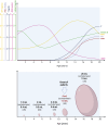
Fig. 3. Testicular growth over time.
The upper panel shows the variation in hormone serum levels from birth to the age of 20. The hormonal peaks that occur during mini-puberty are not represented. The sources used for the reference values are Juul and Skakkebæk for IGF1; Soldin et al. for FSH and LH; Holmes et al. for total testosterone [Edelsztein et al. for AMH; and Kelsey et al. for Inhibin B. The bottom panel shows the increase in testicular volume over time. Values were plotted using the 50th percentile of orchidometric and ultrasound data published by Joustra et al.. AMH anti-Müllerian hormone, FSH follicle-stimulating hormone, IGF1 insulin-like growth factor 1, LH luteinizing hormone, US ultrasonography, T testosterone.
Fig. 4
Fig. 4. Summary of the mechanisms through which obesity can alter Sertoli cell physiology.
(1) Adipokines affect the proliferation of SCs and GCs through the downregulation of FSHR and GDNF in immature and mature SCs, respectively. (2) Inflammatory cytokines (TNF-α and IL-1β) can reduce the T production by LCs. Increased IL-6 induces SC apoptosis via STAT3/FoxO1 signaling pathway. (3) T2DM is associated with increased IL-6 levels, lower T production, and lower VEGF, which in turn can affect testicular microcirculation leading to immature SC apoptosis. (4) Insulin resistance affects proliferation in immature SCs, glucose metabolism, and the production of extrinsic factors such as GDNF by mature SCs. (5) Hyperleptinemia impairs glucose metabolism in SCs and BTB development reducing the expression of the components of tight junction, AR, and other steroidogenic genes. (6) Low T level decreases the expression of Zo-1, claudin, and occluding, which are essential for BTB establishment; reduces the level of GDNF and alters the glucose metabolism in SCs. 7) High E2 negatively affects BTB integrity directly and indirectly, decreases T production, impairs glucose metabolism, increases GC apoptosis increasing the expression of Fas and FasL and reducing c-kit expression, and reduces SOD leading to increased lipid peroxidation and reduced antioxidant defense. (8) Low Ghrelin and GLP-1 are associated with abnormal SC metabolism. (9) Dysbiosis is associated with lower T levels, decreased insulin sensitivity, and disruption of BTB integrity,. More details are given in the text. AR androgen receptor, BTB blood–testis barrier, E2 17-β estradiol, FoxO forkhead box O, Fas Fas cell surface death receptor, FasL Fas ligand, FSHR follicle-stimulating hormone receptor, GC germinal cell, GDNF glial-derived neurotrophic factor, GLP-1 glucagon-like peptide-1, IL interleukin, LC Leydig cell, SC Sertoli cell, SOD superoxide dismutase, T testosterone, TNF-α tumor necrosis factor-α, T2DM type 2 diabetes mellitus, VEGF vascular endothelial growth factor.

Similar articles

Cited by

References

    1. World Health Organization. Obesity and Overweight 2021 (World Health Organization, accessed 16 July 2023); http://www.who.int/mediacentre/factsheets/fs311/en/2021.
    1. Barbagallo F, et al. Molecular mechanisms underlying the relationship between obesity and male infertility. Metabolites. 2021;11:840. doi: 10.3390/metabo11120840. - DOI - PMC - PubMed
    1. Chambers TJ, Richard RA. The impact of obesity on male fertility. Hormones (Athens) 2015;14:563–568. - PubMed
    1. Sermondade N, et al. BMI in relation to sperm count: an updated systematic review and collaborative meta-analysis. Hum. Reprod. Update. 2013;19:221–231. doi: 10.1093/humupd/dms050. - DOI - PMC - PubMed
    1. Guo D, et al. The impact of BMI on sperm parameters and the metabolite changes of seminal plasma concomitantly. Oncotarget. 2017;8:48619–48634. doi: 10.18632/oncotarget.14950. - DOI - PMC - PubMed