. 2024 Aug;43(32):2475-2489.
doi: 10.1038/s41388-024-03094-1. Epub 2024 Jul 3.

Schlafen 11 further sensitizes BRCA-deficient cells to PARP inhibitors through single-strand DNA gap accumulation behind replication forks

Affiliations

Schlafen 11 further sensitizes BRCA-deficient cells to PARP inhibitors through single-strand DNA gap accumulation behind replication forks

Hiroshi Onji et al. Oncogene. 2024 Aug.

Abstract

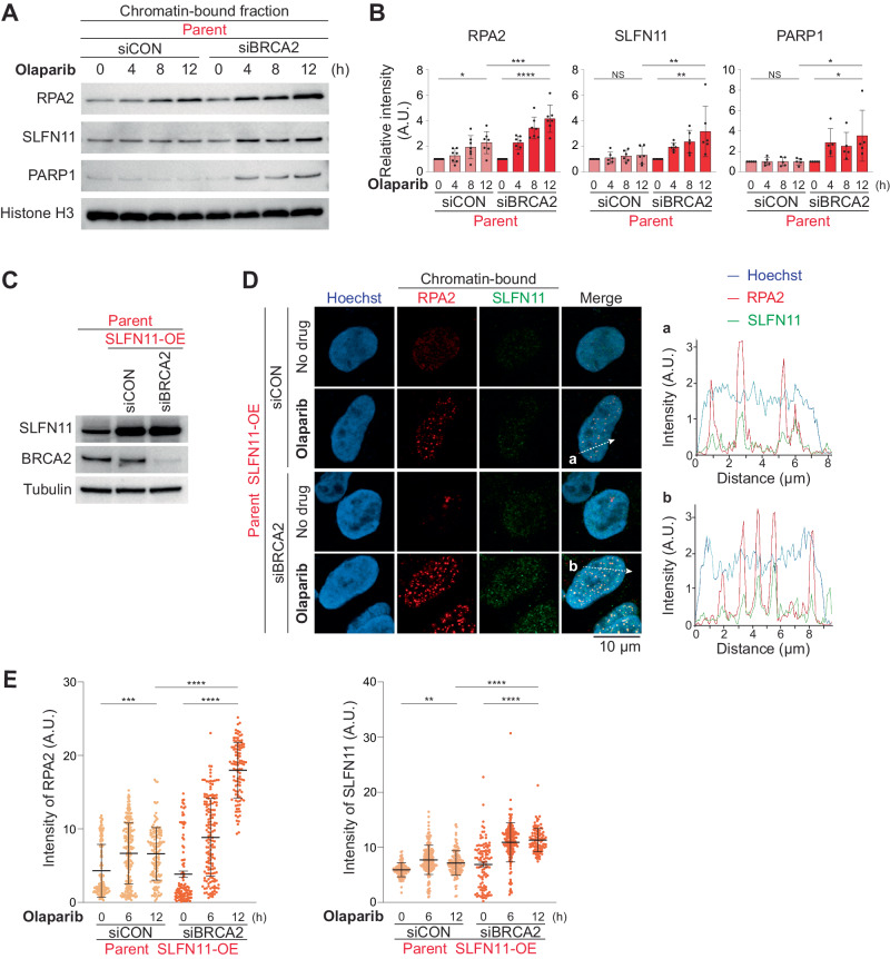
The preferential response to PARP inhibitors (PARPis) in BRCA-deficient and Schlafen 11 (SLFN11)-expressing ovarian cancers has been documented, yet the underlying molecular mechanisms remain unclear. As the accumulation of single-strand DNA (ssDNA) gaps behind replication forks is key for the lethality effect of PARPis, we investigated the combined effects of SLFN11 expression and BRCA deficiency on PARPi sensitivity and ssDNA gap formation in human cancer cells. PARPis increased chromatin-bound RPA2 and ssDNA gaps in SLFN11-expressing cells and even more in cells with BRCA1 or BRCA2 deficiency. SLFN11 was co-localized with chromatin-bound RPA2 under PARPis treatment, with enhanced recruitment in BRCA2-deficient cells. Notably, the chromatin-bound SLFN11 under PARPis did not block replication, contrary to its function under replication stress. SLFN11 recruitment was attenuated by the inactivation of MRE11. Hence, under PARPi treatment, MRE11 expression and BRCA deficiency lead to ssDNA gaps behind replication forks, where SLFN11 binds and increases their accumulation. As ovarian cancer patients who responded (progression-free survival >2 years) to olaparib maintenance therapy had a significantly higher SLFN11-positivity than short-responders (<6 months), our findings provide a mechanistic understanding of the favorable responses to PARPis in SLFN11-expressing and BRCA-deficient tumors. It highlight the clinical implications of SLFN11.


Conflict of interest statement

This work is supported by Ehime University PROS Grant for Intramural Collaboration (2022) (to J.M. and T.S.), and by the Japan Society for the Promotion of Science, Grants-in-Aid for Scientific Research (JP19H03505 and JP23H02768 to J.M.) and by the Japan Science and Technology Agency (JST) grant number JPMJFR2056 (JST FOREST Program to J.M). J.M. receives lecture fees from AstraZeneca plc and Takeda Pharmaceutical Company Limited. Y.K. receives lecture fees from AstraZeneca plc, Takeda Pharmaceutical Company Limited and ACTmed Company Limited. Other authors declare no potential competing interests.

Figures

Fig. 1
Fig. 1. SLFN11 enhances cellular sensitivity to PARP inhibitors in BRCA1/2-deficient cells.
A Immunoblots of whole cell lysates from genetically modified TOV-112D cells: SLFN11-proficient (parent), SLFN11-KO, control siRNA (siCON) or BRCA2 siRNA (siBRCA2). Blots were probed with the indicated antibodies. B, C Viability of TOV-112D cells under each condition after 48 h of continuous drug treatment. Cellular ATP activity was used to measure cell viability. The viability of untreated cells was set as 100%. Data are means ± standard deviations (n = 3, technical replicates) and represent one of two independent experiments. D Apoptosis analysis of TOV-112D cells by flow cytometry. The indicated cell lines were treated continuously with 2.5 μM olaparib for 0, 24, or 48 h. Data represents one of two independent experiments. E Immunoblots showing BRCA1 knockdown by siRNA for BRCA1. Data are shown as in A. F Cell proliferation curves of the indicated TOV-112D cells. G Viability of TOV-112D cells under the indicated conditions after 48 and 72 h of continuous olaparib treatment. NS not significant, *P < 0.05, **P < 0.01, ***P < 0.001, ****P < 0.0001 (one-way analysis of variance with Tukey’s post-hoc multiple comparisons test).
Fig. 2
Fig. 2. SLFN11 expression and BRCA2 deficiency increase chromatin-bound RPA2 under PARPi treatments.
A, C Representative immunoblots of chromatin-bound fractions prepared from the indicated TOV-112D cells treated with 10 μM olaparib or 100 nM CPT for 0, 6, or 12 h. Blots were probed with the indicated antibodies. B Quantification of data from A. Data were normalized to the untreated cells (0 h). Data are means ± standard deviations (n = 3, biological replicates). D Representative confocal microscopy images; chromatin-bound RPA2 (red) and Hoechst (blue) in TOV-112D cells. Cells were treated with or without 10 μM olaparib for 12 h. E Quantification of data from D. Scatter plots show the mean signal intensity of RPA2. Data are means ± standard deviations (n = 116–199, one-time experiment). Data represents one of two independent experiments. NS not significant, *P < 0.05, **P < 0.01, ***P < 0.001, ****P < 0.0001 (one-way analysis of variance with Tukey’s post-hoc multiple comparisons test).
Fig. 3
Fig. 3. SLFN11 expression and BRCA1/2 deficiency increase ssDNA gaps induced by PARPis.
A Scheme of the alkaline BrdU comet assay. B Representative alkaline BrdU comet assay images of TOV-112D cells treated with or without 10 μM olaparib. Scatter plots showing BrdU tail moments in TOV-112D cells under each drug treatment (C: 10 μM olaparib, D: 10 μM olaparib, F: 100 nM olaparib, G: 100 nM CPT). Data are means ± standard deviations (C: n = 54–59, D: n = 78–112, F: n = 54–77, G: n = 91–139, one-time experiment). E Immunoblots of PAR levels in whole cell lysates from parent TOV-112D cells treated as indicated for 30 min. Blots were probed with the indicated antibodies. The asterisk indicates a nonspecific band. NS not significant, *P < 0.05, **P < 0.01, ***P < 0.001, ****P < 0.0001 (one-way analysis of variance with Tukey’s post-hoc multiple comparisons test).
Fig. 4
Fig. 4. BRCA2 deficiency enhances the recruitment of SLFN11 on chromatin in PARPi-treated cells.
A Representative immunoblots of chromatin-bound fractions prepared from TOV-112D cells treated with 10 μM olaparib for 0, 4, 8, or 12 h. Blots were probed with the indicated antibodies. B Quantification of data from A. Data were normalized to untreated cells (0 h). Data are means ± standard deviations (n = 5–7, biological replicates). C Immunoblots of whole cell lysates prepared from the indicated TOV-112D cells. Blots were probed with the indicated antibodies. D Representative confocal microscopy images; Hoechst (blue), chromatin-bound RPA2 (red), and SLFN11 (green) in TOV-112D cells. Cells were treated with or without 10 μM olaparib for 12 h. Representative tracings of the distribution of signals along the white dashed arrow (a and b) are shown in the merged panel. E Quantification of data from D. Scatter plots show mean signal intensities of RPA2 and SLFN11. Data are means ± standard deviations (n = 104–195, one-time experiment). NS not significant, *P < 0.05, **P < 0.01, ***P < 0.001, ****P < 0.0001 (one-way analysis of variance with Tukey’s post-hoc multiple comparisons test).
Fig. 5
Fig. 5. SLFN11 recruitment behind replication forks does not block replication.
Representative flow cytometry cell cycle data in TOV-112D cells treated with 100 nM CPT (A) or 10 μM olaparib (D) for 6, 12, or 24 h. The percentage of highly replicating cells is annotated. Data represents one of two independent experiments. EdU 5-ethynil-2′-deoxyuridine, PI propidium iodide. B, E Representative confocal microscopy images; EdU (purple), Hoechst (blue), chromatin-bound RPA2 (red), and SLFN11 (green) in TOV-112D cells. Cells were treated with or without 100 nM CPT (B) and 10 μM olaparib (E) for 12 h. C Quantification of data from B, E. Scatter plots show the mean signal intensity of EdU. Data are means ± standard deviations (CPT: n = 58–102, olaparib: n = 126–164, one-time experiment). Data represents one of two independent experiments. NS not significant, ****P < 0.0001 (one-way analysis of variance with Tukey’s post-hoc multiple comparisons test).
Fig. 6
Fig. 6. Resection by MRE11 is required for SLFN11 to be recruited on chromatin.
A Immunoblots of chromatin-bound fractions prepared from TOV-112D (SLFN11-KO) cells treated with 10 μM olaparib and with or without 50 μM mirin for 0, 6, or 12 h. Blots were probed with the indicated antibodies. B Quantification of data from A. Data were normalized to untreated cells (0 h). Data are means ± standard deviations (n = 3–4, biological replicates). C Representative immunoblots of chromatin-bound fractions prepared from TOV-112D (parent) treated with 10 μM olaparib with or without 50 μM mirin for 0, 6, or 12 h. Blots were probed with the indicated antibodies. D Quantification of data from C. Data were normalized to untreated cells (0 h). Data are means ± standard deviations (n = 3–4, biological replicates). E Immunoblots of whole cell lysates prepared from the indicated TOV-112D cells: siCON or MRE11 siRNA (siMRE11). Blots were probed with the indicated antibodies. F Representative immunoblots of chromatin-bound fractions prepared from TOV-112D (parent) treated with 10 μM olaparib for 0, 6, or 12 h. Blots were probed with the indicated antibodies. G Quantification of data from F. Data were normalized to untreated cells (0 h). Data are means ± standard deviations (n = 7, biological replicates). H Representative immunoblots of whole cell lysates from parent TOV-112D cells treated with 10 μM etoposide or 10 μM olaparib for 0, 3, or 6 h. Blots were probed with the indicated antibodies. I Immunoblots of whole cell lysates prepared from the indicated TOV-112D cells: siCON or CtIP siRNA (siCtIP). Blots were probed with the indicated antibodies. J Representative immunoblots of chromatin-bound fractions prepared from TOV-112D (parent) treated with 10 μM olaparib for 0, 6, or 12 h. Blots were probed with the indicated antibodies. K Quantification of data from J. Data were normalized to untreated cells (0 h). Data are means ± standard deviations (n = 5, biological replicates). NS not significant, *P < 0.05, **P < 0.01, ***P < 0.001, ****P < 0.0001 (one-way analysis of variance with Tukey’s post-hoc multiple comparisons test).
Fig. 7
Fig. 7. SLFN11-positive ovarian cancers exhibit better responses to olaparib.
A Participant flow. B Representative immunohistochemical images of SLFN11-positive and SLFN11-negative ovarian cancer samples (magnification ×400). C The chart depicts SLFN11 expression level in two groups: super-responders and short-responders (N = 11 each). P < 0.005 (chi-square test). Information on BRCA status is provided in parentheses. D Proposed model describing the involvement of SLFN11 and BRCA for CPT (as a typical DNA damaging agent) vs. PARPi treatment. Please see the Discussion section for details.

Similar articles

Cited by

References

    1. Lord CJ, Ashworth A. PARP inhibitors: Synthetic lethality in the clinic. Science. 2017;355:1152–8. 10.1126/science.aam7344 - DOI - PMC - PubMed
    1. Bryant HE, Schultz N, Thomas HD, Parker KM, Flower D, Lopez E, et al. Specific killing of BRCA2-deficient tumours with inhibitors of poly(ADP-ribose) polymerase. Nature. 2005;434:913–7. 10.1038/nature03443 - DOI - PubMed
    1. Farmer H, McCabe N, Lord CJ, Tutt AN, Johnson DA, Richardson TB, et al. Targeting the DNA repair defect in BRCA mutant cells as a therapeutic strategy. Nature. 2005;434:917–21. 10.1038/nature03445 - DOI - PubMed
    1. Sullivan-Reed K, Bolton-Gillespie E, Dasgupta Y, Langer S, Siciliano M, Nieborowska-Skorska M, et al. Simultaneous Targeting of PARP1 and RAD52 Triggers Dual Synthetic Lethality in BRCA-Deficient Tumor Cells. Cell Rep. 2018;23:3127–36. 10.1016/j.celrep.2018.05.034 - DOI - PMC - PubMed
    1. Belan O, Sebald M, Adamowicz M, Anand R, Vancevska A, Neves J, et al. POLQ seals post-replicative ssDNA gaps to maintain genome stability in BRCA-deficient cancer cells. Mol Cell. 2022;82:4664–80.e4669. 10.1016/j.molcel.2022.11.008 - DOI - PubMed

MeSH terms