. 2025 Jan;30(1):13-28.
doi: 10.1038/s41380-024-02648-9. Epub 2024 Jul 3.

Influenza A virus during pregnancy disrupts maternal intestinal immunity and fetal cortical development in a dose- and time-dependent manner

Affiliations

Influenza A virus during pregnancy disrupts maternal intestinal immunity and fetal cortical development in a dose- and time-dependent manner

Ashley M Otero et al. Mol Psychiatry. 2025 Jan.

Abstract

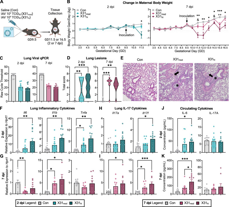
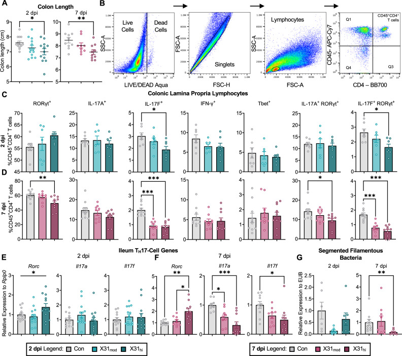
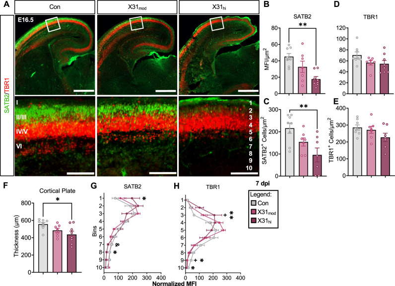
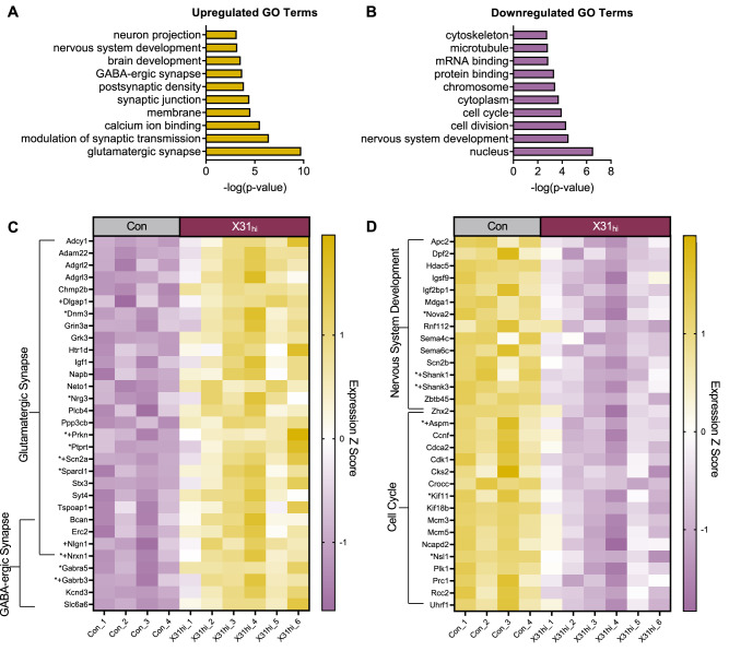
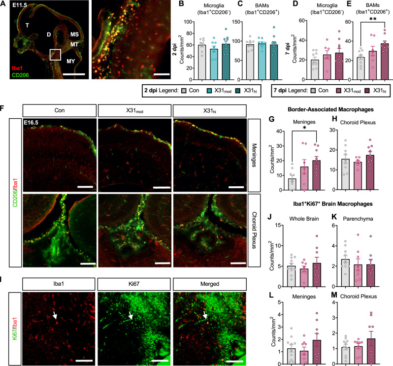
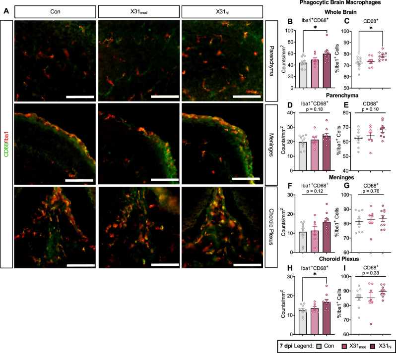
Epidemiological studies link exposure to viral infection during pregnancy, including influenza A virus (IAV) infection, with increased incidence of neurodevelopmental disorders (NDDs) in offspring. Models of maternal immune activation (MIA) using viral mimetics demonstrate that activation of maternal intestinal T helper 17 (TH17) cells, which produce effector cytokine interleukin (IL)-17, leads to aberrant fetal brain development, such as neocortical malformations. Fetal microglia and border-associated macrophages (BAMs) also serve as potential cellular mediators of MIA-induced cortical abnormalities. However, neither the inflammation-induced TH17 cell pathway nor fetal brain-resident macrophages have been thoroughly examined in models of live viral infection during pregnancy. Here, we inoculated pregnant mice with two infectious doses of IAV and evaluated peak innate and adaptive immune responses in the dam and fetus. While respiratory IAV infection led to dose-dependent maternal colonic shortening and microbial dysregulation, there was no elevation in intestinal TH17 cells nor IL-17. Systemically, IAV resulted in consistent dose- and time-dependent increases in IL-6 and IFN-γ. Fetal cortical abnormalities and global changes in fetal brain transcripts were observable in the high-but not the moderate-dose IAV group. Profiling of fetal microglia and BAMs revealed dose- and time-dependent differences in the numbers of meningeal but not choroid plexus BAMs, while microglial numbers and proliferative capacity of Iba1+ cells remained constant. Fetal brain-resident macrophages increased phagocytic CD68 expression, also in a dose- and time-dependent fashion. Taken together, our findings indicate that certain features of MIA are conserved between mimetic and live virus models, while others are not. Overall, we provide consistent evidence of an infection severity threshold for downstream maternal inflammation and fetal cortical abnormalities, which recapitulates a key feature of the epidemiological data and further underscores the importance of using live pathogens in NDD modeling to better evaluate the complete immune response and to improve translation to the clinic.


Conflict of interest statement

Competing interests: The authors declare no competing interests.

Figures

Fig. 1
Fig. 1. Respiratory IAV infection alters maternal lung inflammation and circulating cytokines in a dose- and time-dependent manner.
A Experimental schematic. B IAV inoculation at GD9.5 suppressed body weight gain per day from 2-to-7 dpi in X31hi dams only (repeated measures 2-way ANOVA, the main effect of time = p < 0.001; * = Con vs X31hi, & = X31mod vs X31hi, + = Con vs X31mod). C The presence of IAV-X31 in lungs at 2 and 7 dpi was evaluated using qPCR with a cycle threshold of ≤ 30 cycles as confirmed infection (dotted line). D Quantification of H&E pathological scoring showed elevated lung lesion scores in infected dams. The scoring criteria are listed in the methods with additional scoring values in Supplementary Table S3. E Representative photomicrographs of H&E-stained lung sections. Asterisks (*) indicate bronchi filled with clusters of neutrophils with cellular debris, and arrows (→) indicate arterial and venous endothelia with rolling neutrophils. Genes encoding for classic pro-inflammatory cytokines IL-6, IL-1β, and TNF-α in maternal lungs were F upregulated in a dose-dependent manner at 2 dpi whereas G only Il1b was upregulated at 7 dpi. IL-17 genes were upregulated in the maternal lung at H 2 and I 7 dpi in a dose-dependent manner. Maternal cytokines in circulation at J 2 and K 7 dpi. Pro-inflammatory cytokine IL-6 was upregulated in moderate- and high-dose dams proportional to dosage at both time points. IL-17A was not upregulated in circulation at either endpoint. IAV = influenza A virus, GD = gestational day, dpi = days post-inoculation, Con = saline control, X31mod = IAV-X31 103 TCID50, X31hi = IAV-X31 104 TCID50. Groups were compared using one-way ANOVA with Tukey post hoc for multiple comparisons. For data containing residuals with unequal variance, Brown-Forsythe and Welch’s ANOVA with Dunnett T3 post hoc multiple comparisons was used. For non-parametric data, Kruskal–Wallis ANOVA with Dunn’s correction for multiple comparisons was used. Data are means ± SEM; one symbol = p < 0.05, two symbols = p < 0.01, three symbols = p < 0.001; dots represent individual dams; n = 9–14 per treatment group. See Supplementary Tables S3–5 for complete statistical analysis of all data collected for this figure (individual mean ± SEM per group, p-values, hypothesis test used, and test statistic).
Fig. 2
Fig. 2. Respiratory IAV infection dysregulates maternal intestinal immunity in a dose- and time-dependent manner.
A IAV reduced colon length in high-dose dams as early as 2 dpi, and this persisted at 7 dpi. B Example flow cytometry gating for colonic LPLs. Gating on CD45+CD4+ T cells in the colon revealed C downregulation of IL-17F+ and IL-17F+RORγt+ T cells at 2 dpi which persisted into D 7 dpi in addition to downregulation of RORγt+ and IL-17A+RORγt+ T cells. qPCR of the ileum, the terminal end of the small intestine, confirms findings in the colon where E little changes were observed at 2 dpi and F downregulation in Il17a and Il17f was seen at 7 dpi. Notably, Rorc transcription did not coincide with decreased RORγt protein expression. G Relative gene expression via qPCR showed a decrease in SFB, a bacterial regulator of TH17 cells, in the colon contents of high-dose dams at 7 dpi. IAV = influenza A virus, dpi = days post-inoculation, LPL = lamina propria lymphocytes, Con = saline control, X31mod = IAV-X31 103 TCID50, X31hi = IAV-X31 104 TCID50, SSC-A = side scatter-area, FSC-A = forward scatter-area. Groups were compared with one-way ANOVA with Tukey post hoc for multiple comparisons. For data containing residuals with unequal variance, Brown-Forsythe and Welch’s ANOVA with Dunnett T3 post hoc multiple comparisons was used. For non-parametric data, Kruskal-Wallis ANOVA with Dunn’s correction for multiple comparisons was used. Data are means ± SEM; *p < 0.05, **p < 0.01, ***p < 0.001; dots represent individual dams; n = 7–14 per treatment group. See Supplementary Tables S6–7 for complete statistical analysis of all data collected for this figure (individual mean ± SEM per group, p-values, hypothesis test used, and test statistic).
Fig. 3
Fig. 3. Respiratory IAV infection during pregnancy impacts cortical development in fetal brains from high- but not moderate-dose dams at E16.5, 7 dpi.
A Representative images of E16.5, 7 dpi fetal brains stained for SATB2 (green) and TBR1 (red). Close-up images were taken in the right-hemisphere somatosensory cortex in a 300 × 300 µm2 ROI. Top scale bars = 500 µm; bottom scale bars = 100 µm. I–VI in the bottom left image represents cortical layers 1–6, and 1–10 in the bottom right image represents the bins. B, C MFI and cell count per µm2 of SATB2, an upper excitatory neuronal marker, is decreased in the fetal brains of high-dose mothers. D, E MFI and cell counts of TBR1, a deep excitatory neuronal marker, were not statistically different between groups (p-value = 0.10). F Prenatal exposure to IAV-X31 reduced cortical thickness in fetal brains from X31hi dams. Dividing the ROI into 10 equal cortical laminar bins showed altered cortical lamination in G SATB2 bins 1, 7, and 8 and H TBR1 bins 3, 9, and 10 (* = Con vs X31hi, & = X31mod vs X31hi,+= Con vs X31mod). IAV = influenza A virus, dpi = days post-inoculation, E = embryonic day, ROI = region of interest, MFI = mean fluorescence intensity, Con = saline control, X31mod = IAV-X31 103 TCID50, X31hi = IAV-X31 104 TCID50. Groups were compared with one-way ANOVA with Tukey post hoc for multiple comparisons. For data containing residuals with unequal variance, Brown-Forsythe and Welch’s ANOVA with Dunnett T3 post hoc multiple comparisons was used. For non-parametric data, Kruskal–Wallis ANOVA with Dunn’s correction for multiple comparisons was used. Data are means ± SEM; one symbol = p < 0.05, two symbols = p < 0.01; dots represent one representative fetus per litter; n = 9–10 per treatment group. See Supplementary Table S8 for complete statistical analysis of all data collected for this figure (individual mean ± SEM per group, p-values, hypothesis test used, and test statistic).
Fig. 4
Fig. 4. Bulk RNA-seq reveals genes enriched in neuronal development and synaptic signaling in fetal brains exposed to a high dose of prenatal IAV infection at E16.5, 7 dpi.
Top ten significantly enriched A upregulated and B downregulated GO pathways in fetal brains prenatally exposed to a high dose of IAV, quantified by –log (p-value). Pathways were generated from the Database for Annotation, Visualization, and Integrated Discovery terms (DAVID). C Heatmap of genes differentially upregulated in the glutamatergic and GABA-ergic synapse pathways. D Heatmap of genes differentially downregulated in the nervous system development and cell cycle pathways. Genes with * represent candidate genes for neurodevelopmental disorders based on the Developmental Brain Disorder Gene Database (DBD). Genes with + represent candidate genes for Autism Spectrum Disorder based on the Simons Foundation Autism Research Initiative (SFARI) genes database. IAV = influenza A virus, dpi = days post-inoculation, Con = saline control, X31hi = IAV-X31 104 TCID50, GO = gene ontology. Benjamini–Hochberg’s correction for false discovery (p < 0.1) was used to identify differentially expressed genes. Data are standardized logCPM values (Z score); dots represent one representative fetus per litter; n = 4–6 per treatment group. See Supplementary Tables S9–10 for additional statistical analysis of data collected for this figure.
Fig. 5
Fig. 5. Border-associated macrophages but not microglia are upregulated in fetal brains from high-dose IAV dams.
A Representative sagittal sections of the E11.5 fetal brain stained with Iba1 (red) and CD206 (green). Microglia are Iba1+CD206- and BAMs are Iba1+CD206+. Left scale bar = 1000 µm; right scale bar = 100 µm. There were no changes in B microglia or C BAM count per mm2 at E11.5, 2 dpi. While there were no changes in D microglia count, E BAM count was upregulated in E16.5, 7 dpi fetal brains exposed to a high dose of IAV. F Representative coronal sections of the meninges and choroid plexus from each treatment group of Iba1 and CD206 co-stained E16.5 fetal brains. Scale bars = 100 µm. G, H Separation of choroid plexus and meningeal BAMs showed that only G meningeal BAMs were increased at 7 dpi. I Evaluation of E16.5 brain macrophage proliferation via Ki67 and Iba1 co-staining revealed no changes in double-positive cells in the J whole brain, K parenchyma, L meninges, or M choroid plexus. Arrows = representative co-staining; scale bars = 100 µm. IAV = influenza A virus, dpi = days post-inoculation, E = embryonic day, MFI = mean fluorescence intensity, T = telencephalon, D = diencephalon, MS = mesencephalon, MT = metencephalon MY = myencephalon, Con = saline control, X31mod = IAV-X31 103 TCID50, X31hi = IAV-X31 104 TCID50. Groups were compared using one-way ANOVA with Tukey post hoc for multiple comparisons. For data containing residuals with unequal variance, Brown-Forsythe and Welch’s ANOVA with Dunnett T3 post hoc multiple comparisons was used. Data are means ± SEM; *p < 0.05, **p < 0.01; dots represent one representative fetus per litter; n = 9–14 per treatment group. See Supplementary Table S11 for complete statistical analysis of all data collected for this figure (individual mean ± SEM per group, p-values, hypothesis test used, and test statistic).
Fig. 6
Fig. 6. Brain-resident macrophages exhibit elevated levels of phagocytic markers in fetal brains from high-dose IAV dams.
A Representative coronal sections of E16.5 fetal brains stained with Iba1 (red) and CD68 (green). Scale bars = 100 µm. Brain-resident macrophages (Iba1+ cells) showed increased phagocytic capacity in X31hi fetal brains when evaluating B total counts of Iba1+CD68+ cells per mm2 and C percentage of Iba1+ cells that were also CD68+. DG Parenchymal Iba1+ cells, which approximate microglia, and meningeal Iba1+ cells (meningeal BAMs) did not demonstrate a dose-dependent increase in phagocytic capacity. H Elevated counts of Iba1+CD68+ choroid plexus BAMs did not persist when evaluated as a I percentage of Iba1+ cells that were also CD68+. IAV = influenza A virus, dpi = days post-inoculation, E = embryonic day, BAM = border-associated macrophage, Con = saline control, X31mod = IAV-X31 103 TCID50, X31hi = IAV-X31 104 TCID50, Groups were compared using one-way ANOVA with Tukey post hoc for multiple comparisons. Data are means ± SEM; * = p < 0.05; dots represent one representative fetus per litter; n = 9–10 per treatment group. See Supplementary Table S11 for complete statistical analysis of all data collected for this figure (individual mean ± SEM per group, p-values, hypothesis test used, and test statistic).

Similar articles

Cited by

References

    1. Oseghale O, Vlahos R, O’Leary JJ, Brooks RD, Brooks DA, Liong S, et al. Influenza virus infection during pregnancy as a trigger of acute and chronic complications. Viruses. 2022;14:2729. - PMC - PubMed
    1. Raj RS, Bonney EA, Phillippe M. Influenza, immune system, and pregnancy. Reprod Sci. 2014;21:1434–51. - PMC - PubMed
    1. Mertz D, Geraci J, Winkup J, Gessner BD, Ortiz JR, Loeb M. Pregnancy as a risk factor for severe outcomes from influenza virus infection: a systematic review and meta-analysis of observational studies. Vaccine. 2017;35:521–8. - PMC - PubMed
    1. Dawood FS, Kittikraisak W, Patel A, Rentz Hunt D, Suntarattiwong P, Wesley MG, et al. Incidence of influenza during pregnancy and association with pregnancy and perinatal outcomes in three middle-income countries: a multisite prospective longitudinal cohort study. Lancet Infect Dis. 2021;21:97–106. - PMC - PubMed
    1. Abu-Raya B, Michalski C, Sadarangani M, Lavoie PM. Maternal immunological adaptation during normal pregnancy. Front Immunol. 2020;11:575197. - PMC - PubMed

MeSH terms