

Image features and clinical analysis of retroperitoneal pelvic schwannoma: a case report
- PMID: 38961371
- PMCID: PMC11221090
- DOI: 10.1186/s12883-024-03715-y
Image features and clinical analysis of retroperitoneal pelvic schwannoma: a case report
Abstract
Background: Schwannomas are benign usually encapsulated nerve sheath tumors derived from the Schwann cells, and affecting single or multiple nerves. The tumors commonly arise from the cranial nerves as acoustic neurinomas but they are extremely rare in the pelvis and the retroperitoneal area. Retroperitoneal pelvic schwannomas often present with non-specific symptoms leading to misdiagnosis and prolonged morbidity.
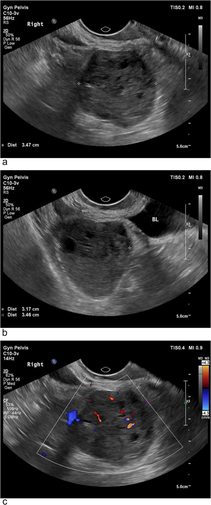
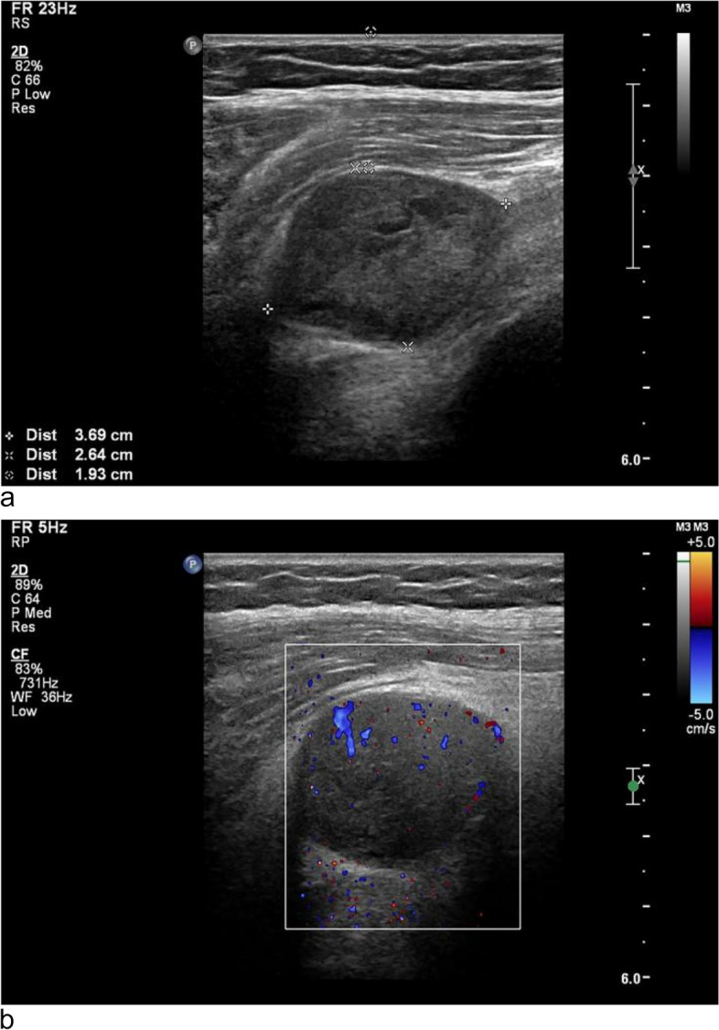
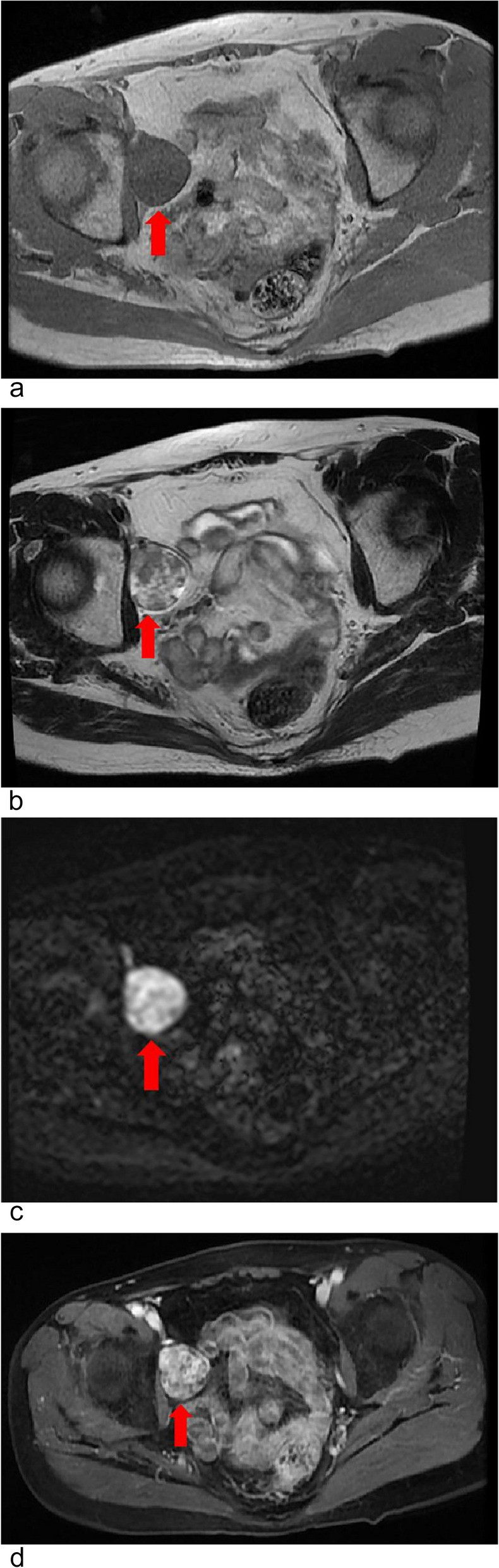
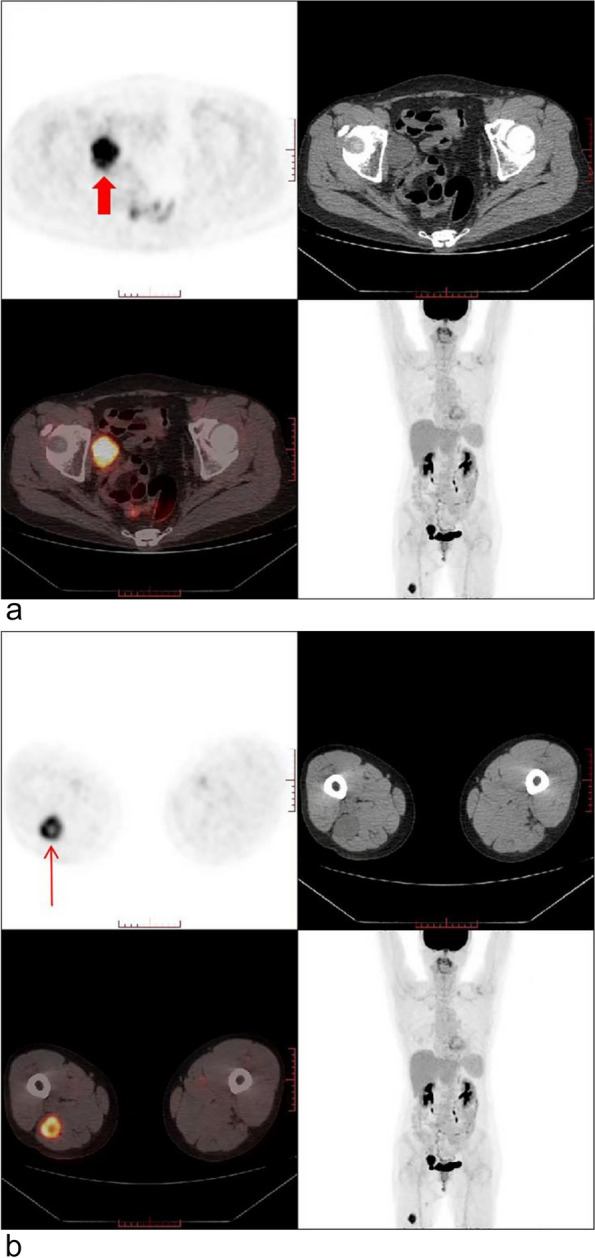
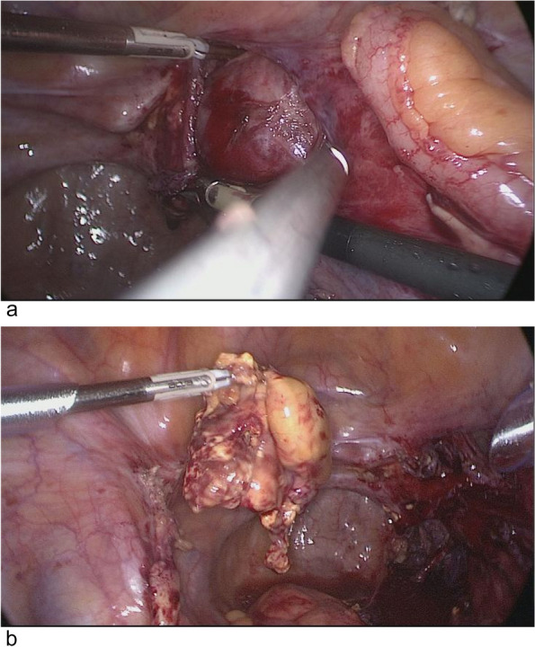
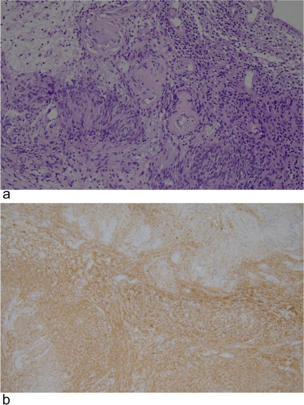
Case presentation: We report the case of a 59-year-old woman presenting with a feeling of heaviness in the lower abdomen who was found to have a retroperitoneal pelvic schwannoma originating from the right femoral nerve. She had a history of two resections of peripheral schwannomas at four different sites of limbs. After conducting magnetic resonance imaging, this pelvic schwannoma was misdiagnosed as a gynecological malignancy. The tumor was successfully removed by laparoscopic surgery. Pathological analysis of the mass revealed a benign schwannoma of the femoral nerve sheath with demonstrating strong, diffuse positivity for S-100 protein.
Conclusions: Although retroperitoneal pelvic schwannoma is rare, it should be considered in the differential diagnosis of pelvic masses, especially in patients with a history of neurogenic mass or the presence of neurogenic mass elsewhere.
Keywords: Femoral; Pelvic; Retroperitoneal; Schwannoma.
© 2024. The Author(s).
Conflict of interest statement
The authors declare that they have no conflicts of interest and nothing to disclose.
Figures
Similar articles
-
Pelvic retroperitoneal schwannoma presenting as a gynecologic mass: case report.Eur J Gynaecol Oncol. 2005;26(1):117-9. Eur J Gynaecol Oncol. 2005. PMID: 15755018
-
Laparoscopic resection of a retroperitoneal ancient schwannoma: a case report and review of the literature.Anticancer Res. 2008 Sep-Oct;28(5B):2889-91. Anticancer Res. 2008. PMID: 19031930
-
A 68-Year-Old Woman Presenting with Recurrent Abdominal Pain and a Diagnosis of a Presacral Retroperitoneal Benign Schwannoma that Mimicked an Ovarian Tumor on Pelvic Magnetic Resonance Imagining.Am J Case Rep. 2022 Jul 20;23:e935985. doi: 10.12659/AJCR.935985. Am J Case Rep. 2022. PMID: 35854634 Free PMC article.
-
Imaging in gynecological disease (26): clinical and ultrasound characteristics of benign retroperitoneal pelvic peripheral-nerve-sheath tumors.Ultrasound Obstet Gynecol. 2023 Nov;62(5):727-738. doi: 10.1002/uog.26223. Ultrasound Obstet Gynecol. 2023. PMID: 37058402 Review.
-
Pelvic schwannoma in pregnancy. A case report.J Reprod Med. 1993 Oct;38(10):826-8. J Reprod Med. 1993. PMID: 8263877 Review.
Cited by
-
Traumatic retroperitoneal neuroma at the site of prior radical nephrectomy: A case report.Urol Case Rep. 2025 Apr 21;61:103048. doi: 10.1016/j.eucr.2025.103048. eCollection 2025 Jul. Urol Case Rep. 2025. PMID: 40342688 Free PMC article.
References
-
- Erdoğan F, Say F, Barış YS. Schwannomatosis of the sciatic nerve: a case report. Br J Neurosurg. 2021;18:1–4. - PubMed
Publication types
MeSH terms
LinkOut - more resources
Full Text Sources
