. 2024 Jul 3;22(1):621.
doi: 10.1186/s12967-024-05428-9.

Accumulation of liposomes in metastatic tumor sites is not necessary for anti-cancer drug efficacy

Affiliations

Accumulation of liposomes in metastatic tumor sites is not necessary for anti-cancer drug efficacy

Jessica Kalra et al. J Transl Med. .

Abstract

Background: The tumor microenvironment is profoundly heterogeneous particularly when comparing sites of metastases. Establishing the extent of this heterogeneity may provide guidance on how best to design lipid-based drug delivery systems to treat metastatic disease. Building on our previous research, the current study employs a murine model of metastatic cancer to explore the distribution of ~ 100 nm liposomes.

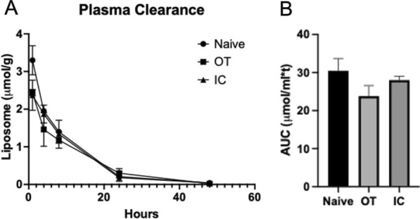
Methods: Female NCr nude mice were inoculated with a fluorescently labeled, Her2/neu-positive, trastuzumab-resistant breast cancer cell line, JIMT-1mkate, either in the mammary fat pad to create an orthotopic tumor (OT), or via intracardiac injection (IC) to establish tumors throughout the body. Animals were dosed with fluorescent and radio-labeled liposomes. In vivo and ex vivo fluorescent imaging was used to track liposome distribution over a period of 48 h. Liposome distribution in orthotopic tumors was compared to sites of tumor growth that arose following IC injection.

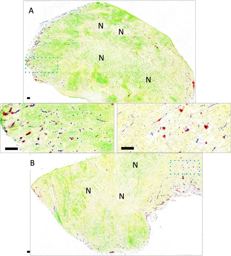
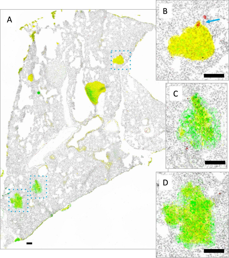
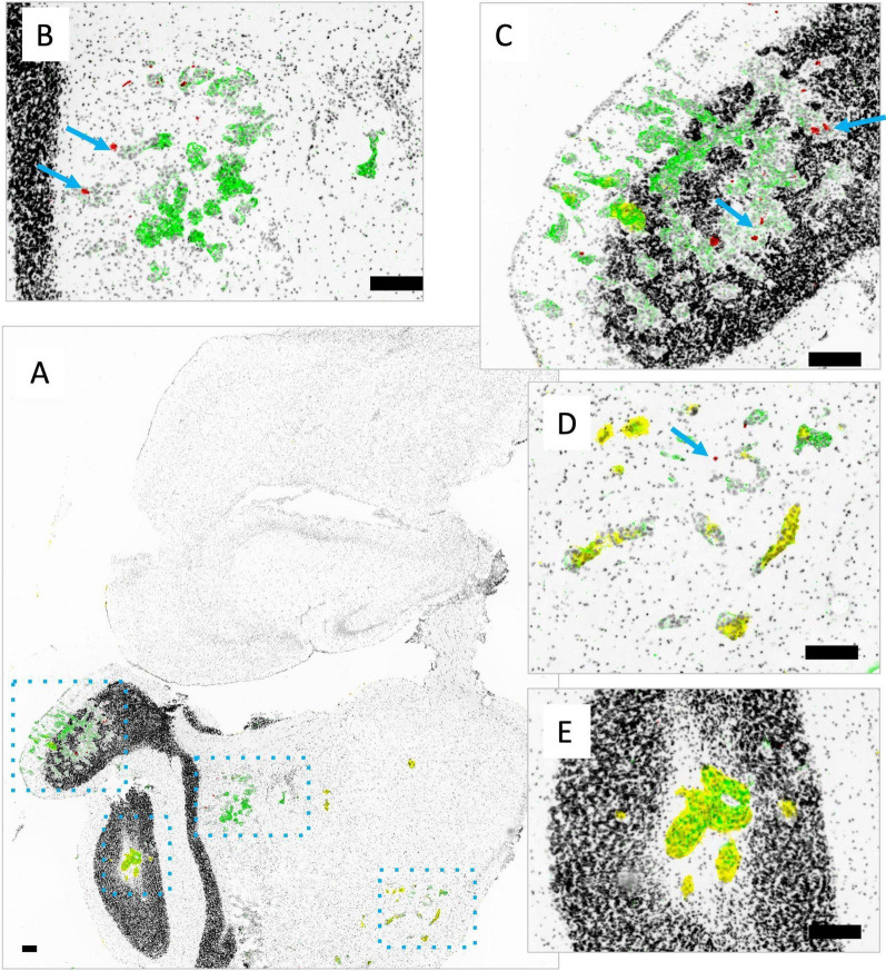
Results: A significant amount of inter-vessel heterogeneity for DiR distribution was observed, with most tumor blood vessels showing little to no presence of the DiR-labelled liposomes. Further, there was limited extravascular distribution of DiR liposomes in the perivascular regions around DiR-positive vessels. While all OT tumors contained at least some DiR-positive vessels, many metastases had very little or none. Despite the apparent limited distribution of liposomes within metastases, two liposomal drug formulations, Irinophore C and Doxil, showed similar efficacy for both the OT and IC JIMT-1mkate models.

Conclusion: These findings suggest that liposomal formulations achieve therapeutic benefits through mechanisms that extend beyond the enhanced permeability and retention effect.

Keywords: Enhanced permeability and retention effect; Inter- and intra-tumor heterogeneity; Liposomes; Metastases/metastasis; Pre-clinical models; Translational research; Tumor microenvironment.


Conflict of interest statement

The authors declare no competing interests.

Figures

Fig. 1
Fig. 1
Tumor naïve mice were treated with a single i.v. injection of DiR labelled liposomes at a lipid dose of 100 mg/kg. In vivo fluorescence imaging of the distribution of DiR over 48 h is shown (A). Fluorescence signal intensity in the whole animal was quantified (B) and demonstrated to have decreased by 58%, 48 h post injection. DiR labelled liposome levels following injection was measured using liquid scintillation counting ([3H]-CHE) and the plasma elimination (C) and AUC0-48Hr for each organ was calculated (D). Ex vivo imaging of tumor naïve organs and related fluorescence signal intensity over 48 h is shown (E)
Fig. 2
Fig. 2
Mice bearing orthotopic (OT) JIMT-1mKate tumors were given a single i.v. injection of DiR labelled liposomes at a liposomal lipid dose of 100 mg/kg. In vivo fluorescence imaging of DiR was performed at 1, 4, 8, 24 and 48 h. Representative images are shown (A). Ex vivo imaging of DiR in the OT JIMT-1 tumors at 1, 4, 8, 24 and 48 h is shown (B). Plasma elimination (C) and OT tumor accumulation (D) of injected DiR labelled liposomes in mice with established JIMT-1 OT tumor bearing animals was measured using liquid scintillation counting
Fig. 3
Fig. 3
Mice bearing tumors arising following IC injection of JIMT-1mKate cells were given a single i.v. injection of DiR labelled liposomes at a liposomal lipid dose of 100 mg/kg. In vivo fluorescence imaging of DiR was performed at 1, 4, 8, 24 and 48 h. Representative images are shown (A). Ex vivo imaging of DiR (green) in JIMT-1mKate tumor positive (red) lung, liver, kidney, adrenal gland, ovary and brain, 24 h post injection (bottom panel) are compared to tumor naive organs (top panel) (B). Co-localization (yellow) of DiR and mKate was observed
Fig. 4
Fig. 4
Mice with mKate positive tumors following IC injection JIMT-1mKate cells were given a single i.v. injection of DiR labelled liposomes at a liposomal lipid dose of 100 mg/kg. Plasma elimination (A) and the estimated AUC0-48Hr (B) of the liposomes from control (non-tumor bearing) mice, mice with OT JIMT-1mKate tumors and mice with IC JIMT-1mKate tumors are compared
Fig. 5
Fig. 5
Tumors grown orthotopically in the mammary fat pad are shown with mKate (yellow) and Her2/neu expression (green) overlayed on Hoechst nuclear dye (grey), with CD31 immunostaining (blue) and DiR fluorescence (red; overlapped with CD31 in black). Tumor A (top) was excised 8 h following DiR administration, and tumor B (bottom) at 24 h. Both tumors show heterogeneous Her2/neu and mkate with some necrotic areas (marked with N). Insets illustrate the high degree of inter-vessel heterogeneity, where DiR colocalizes with only a select proportion of CD31 positive blood vessels, often with neighbouring vessels showing alternate patterns. No significant increase in extravascular distribution of DiR is noticeable at the longer 24 h time-point (B) relative to 8 h (A). Scale bars = 150 µm
Fig. 6
Fig. 6
Representative whole adrenal gland (left), ovary (middle) and kidney (right) from non-tumor bearing animals (top) compared with organs bearing metastatic lesions (bottom; insets with enlarged views) are shown. Tumor tissue is identified via mKate fluorescent signal (yellow) and immunostaining of Her2/neu (green); either mKate or Her2/neu positivity suggests tumor growth. Both Her2/neu and mKate expression show heterogeneity, requiring visualization of both markers in addition to pathological review of the tissue in order to identify tumor sites. DiR (red) and nuclear dye Hoechst 33342 (grey) are also included in the image overlays. Organs such as the adrenal gland and ovary are largely overtaken with tumor growth, whereas the kidneys were more likely to have discrete lesions as shown. Liposomal DiR is seen in a few isolated spots of adrenal glands and ovaries, without additional uptake in the tumor-bearing sites within the organs. As expected, the glomeruli of the kidney show greatest DiR signal distributed throughout the organ, with little to no uptake observed within the Her2/neu and mKate positive tumor region. Scale bars = 150 µm
Fig. 7
Fig. 7
Metastatic tumor nodules in the liver grow as discrete nodules as shown in a representative image of one liver section (A). The metastatic lesions are heterogeneous in their degree of Her2/neu (green) and mKate (yellow) expression. Shown in B is one nodule with little to no Her2/neu or mKate and in C another nodule with both markers. DiR fluorescence (red) is evenly distributed throughout the liver, while tumor lesions are largely zones of exclusion for the liposome. One nodule has some central DiR positivity (B, blue arrow) while the other has possible DiR positivity at the margins of normal tissue (C). Scale bars = 150 µm
Fig. 8
Fig. 8
Metastatic tumor nodules in the lung grow as discrete nodules within the organ as shown in a representative image of one lung section (A). The metastatic lesions all show mKate (yellow; high for all 3 nodules shown) but are heterogeneous in their degree of Her2/neu expression (green; low for B; high for C, D). DiR fluorescence (red) is generally very low in the lung. Except for two locations adjacent to a single nodule (B; blue arrow), no other nodules show DiR positivity. Scale bars = 150 µm
Fig. 9
Fig. 9
Metastatic tumor nodules in the brain grow as small clusters of cells throughout a whole brain as seen in the representative image from one brain section (A). Lesions are identified as dense structures of nuclei stained with Hoechst 33342 (grey) that may be Her2/neu positive (green; parts of B), mKate positive (yellow; parts of D) or sometimes both (overlap green and yellow; E and parts of C). Liposomal DiR fluorescence (red) is also shown, with very limited positivity, highlighted using blue arrows (B, C, D). Scale bars = 150 µm
Fig. 10
Fig. 10
Mice bearing OT and IC JIMT-1mKate tumors were treated with liposomal doxorubicin (Doxil®) (1 mg/kg; Q14Dx2), or Irinotecan (Irinophore C) (20 mg/kg; Q4Dx3) such that the final liposomal lipid concentration was 100 mg/kg). Kaplan–Meier curves are shown where mice were euthanized due to tumor progression or poor health status

Similar articles

References

    1. Kalra J, Baker J, Song J, Kyle A, Minchinton A, Bally M. Inter-metastatic heterogeneity of tumor marker expression and microenvironment architecture in a preclinical cancer model. Int J Mol Sci. 2021 doi: 10.3390/ijms22126336. - DOI - PMC - PubMed
    1. Allen TM, Cullis PR. Drug delivery systems: entering the mainstream. Science. 2004;303:1818–1822. doi: 10.1126/science.1095833. - DOI - PubMed
    1. Leonard RCF, Williams S, Tulpule A, Levine AM, Oliveros S. Improving the therapeutic index of anthracycline chemotherapy: focus on liposomal doxorubicin (Myocet) Breast. 2009;18:218–224. doi: 10.1016/j.breast.2009.05.004. - DOI - PubMed
    1. Gordon AN, Tonda M, Sun S, Rackoff W, Doxil Study 30-49 Investigators Long-term survival advantage for women treated with pegylated liposomal doxorubicin compared with topotecan in a phase 3 randomized study of recurrent and refractory epithelial ovarian cancer. Gynecol Oncol. 2004;95:1–8. doi: 10.1016/j.ygyno.2004.07.011. - DOI - PubMed
    1. O’Brien MER, Wigler N, Inbar M, Rosso R, Grischke E, Santoro A, et al. Reduced cardiotoxicity and comparable efficacy in a phase III trial of pegylated liposomal doxorubicin HCl (CAELYX/Doxil) versus conventional doxorubicin for first-line treatment of metastatic breast cancer. Ann Oncol. 2004;15:440–449. doi: 10.1093/annonc/mdh097. - DOI - PubMed