

Primary lung chordoma: a case report
- PMID: 38961474
- PMCID: PMC11223418
- DOI: 10.1186/s13000-024-01522-0
Primary lung chordoma: a case report
Abstract
Background: Chordoma, a rare malignant tumor arising from notochordal tissue, usually occurs along the spinal axis. Only a few published reports of primary lung chordomas exist. Herein, we present a case of primary lung chordoma and discuss important considerations for diagnosing rare chordomas.
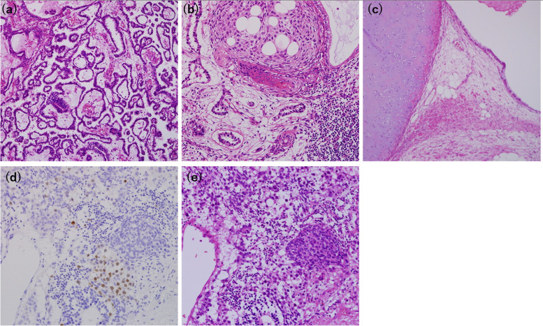
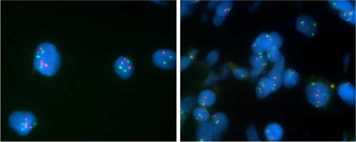
Case presentation: We report a case of primary lung chordoma in a 39-year-old male with a history of testicular mixed germ-cell tumor of yolk sac and teratoma. Computed tomography revealed slow-growing solid lesions in the left lower lobe. We performed wedge resection for suspected germ-cell tumor lung metastasis. Histologically, large round or oval cells with eosinophilic cytoplasm were surrounded by large cells with granular, lightly eosinophilic cytoplasm. Tumor cells were physaliphorous. Immunohistochemistry was positive for brachyury, S-100 protein, epithelial membrane antigen, vimentin, and cytokeratin AE1/AE3, suggesting pulmonary chordoma. Re-examination of the testicular mixed germ-cell tumor revealed no notochordal elements. Although some areas were positive for brachyury staining, hematoxylin and eosin (HE) staining did not show morphological features typical of chordoma. Complementary fluorescence in situ hybridization (FISH) of the lung tumor confirmed the absence of isochromosome 12p and 12p amplification. Thus, a final diagnosis of primary lung chordoma was established.
Conclusions: In patients with a history of testicular mixed germ cell tumors, comparison of histomorphology using HE and Brachyury staining of lung and testicular tumors, and analyzing isochromosome 12p and 12p amplification in lung tumors using FISH is pivotal for the diagnosis of rare lung chordomas.
Keywords: Brachyury; FISH; Isochromosome 12p; Lung chordoma; Testicular mixed germ-cell tumor.
© 2024. The Author(s).
Conflict of interest statement
The authors declare no competing interests.
Figures


Similar articles
-
[Diagnostic value of immunohistochemistry and FISH for chromosome 12p in type Ⅱ testicular germ cell tumors].Zhonghua Nan Ke Xue. 2016 Aug;22(8):692-697. Zhonghua Nan Ke Xue. 2016. PMID: 29019224 Chinese.
-
Retroperitoneal Sarcomatoid Yolk Sac Tumor in a Chemotherapy-Naive Patient With Testicular Postpubertal Type Teratoma: A Rare Case Report With Emphasis on Molecular Features.Int J Surg Pathol. 2024 Dec;32(8):1537-1543. doi: 10.1177/10668969241231973. Epub 2024 Feb 20. Int J Surg Pathol. 2024. PMID: 38377960
-
Clinicopathologic features of four rare types of chordomas, confirmed by brachyury immunostaining.Indian J Pathol Microbiol. 2017 Jul-Sep;60(3):350-354. doi: 10.4103/IJPM.IJPM_409_16. Indian J Pathol Microbiol. 2017. PMID: 28937370
-
Germ cell tumors of the gonads: a selective review emphasizing problems in differential diagnosis, newly appreciated, and controversial issues.Mod Pathol. 2005 Feb;18 Suppl 2:S61-79. doi: 10.1038/modpathol.3800310. Mod Pathol. 2005. PMID: 15761467 Review.
-
Perspectives on testicular germ cell neoplasms.Hum Pathol. 2017 Jan;59:10-25. doi: 10.1016/j.humpath.2016.08.002. Epub 2016 Aug 26. Hum Pathol. 2017. PMID: 27569298 Review.
References
-
- Mirra JM, Nelson SD, Della Rocca C, Mertens F. Chordoma. In: Fletcher CDM, Unni KK, Mertens F, editors. Pathology and genetics of tumors of soft tissue and bone World Health Organization classification of tumors. Lyon: IARC Press; 2002. pp. 316–7.
Publication types
MeSH terms
Substances
Supplementary concepts
LinkOut - more resources
Full Text Sources
Medical
Miscellaneous
