. 2024 Jul 3;21(1):168.
doi: 10.1186/s12974-024-03110-x.

Redefining the ontogeny of hyalocytes as yolk sac-derived tissue-resident macrophages of the vitreous body

Affiliations

Redefining the ontogeny of hyalocytes as yolk sac-derived tissue-resident macrophages of the vitreous body

Dennis-Dominik Rosmus et al. J Neuroinflammation. .

Abstract

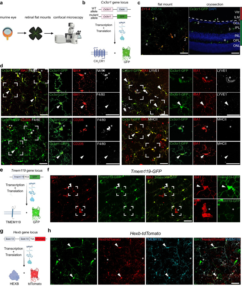
Background: The eye is a highly specialized sensory organ which encompasses the retina as a part of the central nervous system, but also non-neural compartments such as the transparent vitreous body ensuring stability of the eye globe and a clear optical axis. Hyalocytes are the tissue-resident macrophages of the vitreous body and are considered to play pivotal roles in health and diseases of the vitreoretinal interface, such as proliferative vitreoretinopathy or diabetic retinopathy. However, in contrast to other ocular macrophages, their embryonic origin as well as the extent to which these myeloid cells might be replenished by circulating monocytes remains elusive.

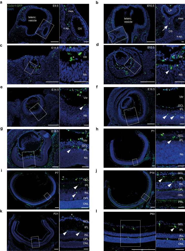
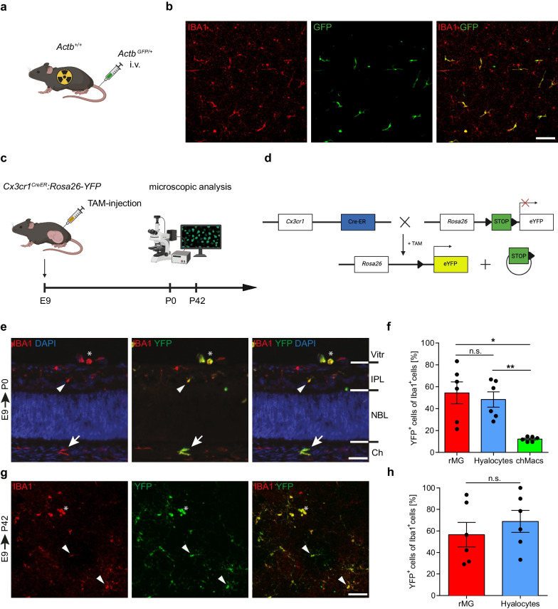
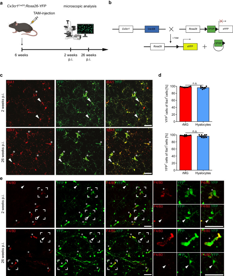
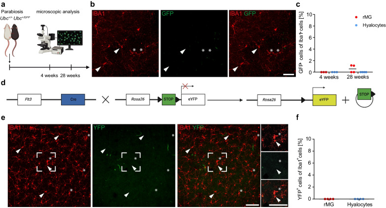
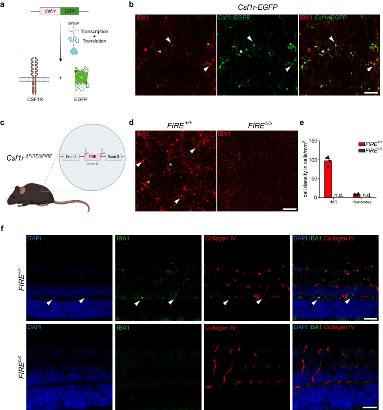
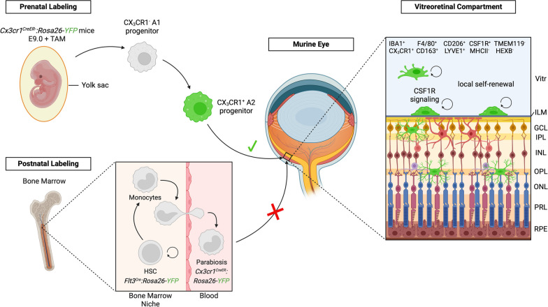
Results: In this study, we combine transgenic reporter mice, embryonic and adult fate mapping approaches as well as parabiosis experiments with multicolor immunofluorescence labeling and confocal laser-scanning microscopy to comprehensively characterize the murine hyalocyte population throughout development and in adulthood. We found that murine hyalocytes express numerous well-known myeloid cell markers, but concomitantly display a distinct immunophenotype that sets them apart from retinal microglia. Embryonic pulse labeling revealed a yolk sac-derived origin of murine hyalocytes, whose precursors seed the developing eye prenatally. Finally, postnatal labeling and parabiosis established the longevity of hyalocytes which rely on Colony Stimulating Factor 1 Receptor (CSF1R) signaling for their maintenance, independent of blood-derived monocytes.

Conclusion: Our study identifies hyalocytes as long-living progeny of the yolk sac hematopoiesis and highlights their role as integral members of the innate immune system of the eye. As a consequence of their longevity, immunosenescence processes may culminate in hyalocyte dysfunction, thereby contributing to the development of vitreoretinal diseases. Therefore, myeloid cell-targeted therapies that convey their effects through the modification of hyalocyte properties may represent an interesting approach to alleviate the burden imposed by diseases of the vitreoretinal interface.

Keywords: CSF1R; Cx3cr1; Development; Fate mapping; Hyalocytes; Macrophages; Turnover; Vitreous body.


Conflict of interest statement

The authors declare that they have no competing interests.

Figures

Fig. 1
Fig. 1
Immunophenotyping of murine hyalocytes underlines their myeloid cell Identity. a Graphical illustration of the experimental setup. Retinal flat mounts were prepared of eyes of several transgenic reporter mouse models and analyzed using immunofluorescence labeling and confocal laser-scanning microscopy. b Graphical scheme of gene targeting in Cx3cr1-GFP mice where one allele of the Cx3cr1 locus is replaced by green fluorescent protein (GFP). c Representative images from retinal flat mount (left) and cryosection (right) from Cx3cr1-GFP mice depicting the anatomical localization of hyalocytes and retinal microglia (rMG). Planes 1-4 (red) include hyalocytes (1) residing above the inner limiting membrane (ILM, dashed line). Planes 7-14 (green) comprise rMG positioned in the inner plexiform layer (IPL) (2, 3) and outer plexifom layer (OPL) (4). The z- step size is 1,5 µm. Vitr: vitreous, GCL: ganglion cell layer, INL: inner nuclear layer, ONL: outer nuclear layer. d Representative images of retinal flat mounts from Cx3cr1-GFP mice. In contrast to rMG (arrowheads), murine hyalocytes (asterisks) express F4/80, CD206 and LYVE1. Both cell types did not show MHCII-immunoreactivity. Pictures are representative for three (CD206, LYVE1, MHCII) or five (F4/80) mice, respectively. e Graphical scheme depicting gene targeting in Tmem119-GFP mice. GFP was introduced in the stop codon of the Tmem119 locus separated by a P2A-site enabling functional expression of both proteins. f Representative images of retinal flat mounts from Tmem119-GFP mice. Hyalocytes were GFP (asterisks) while rMG (arrowheads) are consistently GFP+. Images are representative for three mice. g Graphical illustration of gene targeting in Hexb-tdTomato reporter mice. A tdTomato cassette was introduced downstream of Exon 14 of the Hexb locus separated through a T2A-site enabling the expression of functional HEXB protein and tdTomato. h Representative images of retinal flat mounts from Hexb-tdTomato mice. Hyalocytes were identified as tdTomatoTMEM119 cells (asterisks) compared to tdTomato+TMEM119+ rMG (arrowheads). Images are representative for two mice. All scale bars equal 50 µm
Fig. 2
Fig. 2
Histological analysis of the spatiotemporal development of myeloid cell populations in the murine eye. ag Cryosections from Cx3cr1-GFP mice at different time points during prenatal development. GFP+ myeloid cells can be found in the periocular mesenchyme as early as embryonic day (E) E9.5 and enter the optic cup through the optic stalk at E10.5 (arrow), whereas hyalocytes (asterisks) can be distinguished from microglia (arrowheads) for the first time at E11.5 by localization. Scale bar = 200 µm (overview) or 50 µm (magnification). h–l Representative images from cryosections of Cx3cr1-GFP mice during early postnatal development. Hyalocytes can be found in the vitreous (asterisk), retinal microglia (arrowheads) are evenly distributed across the emerging plexiform layers. Scale bar = 200 µm (overview) or 50 µm (magnification). telenc. vesicle telencephalic vesicle, mes—mesenchyme, n.ep.—neuroepithelium, OV—optic vesicle, OS—optic stalk, LP—lens placode, L—lens, Nb—neuroblast layer, Vitr—vitreous, GCL—ganglion cell layer, IPL—inner plexiform layer, INL—inner nuclear layer, OPL—outer plexiform layer, ONL—outer nuclear layer, PRL—photoreceptor layer. Images are representative for n = 2 mice per time point
Fig. 3
Fig. 3
Embryonic pulse labeling reveals a prenatal origin of murine hyalocytes. a Schematic of bone marrow chimera creation. Wildtype mice (Actb+/+) were whole-body irradiated and reconstituted intravenously (i.v.). with bone marrow of ActbGFP/+ mice. b Images of retinal flat mounts of ActbGFP/+:Actb+/+ bone marrow chimeras. GFP+ donor-derived hyalocytes are present in the vitreous. Images are representative for three mice. Scale bar = 100 µm. c Schematic of embryonic pulse labeling experiment. Cx3cr1CreER:Rosa26-YFP mice were injected with tamoxifen (TAM) at embryonic day (E) 9 and analyzed at postnatal day (P) 0 or P42. d Illustration of embryonic pulse labeling approach. TAM administration activates inducible Cre-recombinase which irreversibly removes a LoxP-site-flanked STOP-sequence in Cx3cr1-expressing cells, causing consistent YFP-expression and labeling CX3CR1+ cells and their progeny. e Images of E9.0-labeled hyalocytes (asterisk), rMG (arrowhead) and choroidal macrophages (chMacs) (arrow) in a cryosection from Cx3cr1CreER:Rosa26-YFP mice at P0. Images are representative for six mice from three independent experiments. Scale bar = 50 µm. Vitr—vitreous, IPL—inner plexiform layer, NBL—neuroblast layer, Ch—choroid. f Quantification of YFP+ cells among IBA1+ hyalocytes, rMG and chMacs in E9.0-labeled Cx3cr1CreER:Rosa26-YFP mice at P0. Graphs depict mean ± S.E.M for six mice from three independent experiments. Statistics: one-way repeated measure ANOVA with post-hoc Tukey’s multiple-comparison test (* p < 0.05, ** p < 0.01). g Images of retinal flat mounts depicting E9.0-labeled hyalocytes (asterisk) and rMG (arrowheads) from Cx3cr1CreER:Rosa26-YFP mice at P42. Images are representative for six mice. Scale bar = 50 µm. h Percentage of YFP+ cells among IBA1+ hyalocytes and rMG in E9.0-labeled Cx3cr1CreER:Rosa26-YFP mice at P42. Graphs depict mean ± S.E.M for six mice. Statistics: paired t-test (n.s., p > 0.05)
Fig. 4
Fig. 4
Hyalocytes represent a long-living tissue-resident macrophage population. a Graphical scheme of the experimental setup. Six-week-old Cx3cr1CreER:Rosa26-YFP mice were injected with tamoxifen (TAM) and retinal whole mounts subsequently analyzed by fluorescence microscopy at 2 and 26 weeks post-injection (p.i.). b Graphical scheme illustrating the rationale of the adult turnover approach. TAM administration leads to nuclear translocation of cytosolic Cre-ER fusion protein and subsequent Cre-mediated irreversible excision of a LoxP-site-flanked STOP-cassette in Cx3cr1-expressing cells. This causes a consistent level of YFP-expression under the control of the constitutively active Rosa26 promoter and labeling of CX3CR1-positive cells and their progeny. c Confocal images of YFP+ and IBA1+ hyalocytes (asterisks) and microglia (arrowheads) in Cx3cr1CreER:Rosa26-YFP at 2 weeks and 26 weeks after injection of TAM. Images are representative for eight animals (2 weeks) from three independent experiments and ten animals (26 weeks) from two independent experiments, respectively. Scale bar = 50 µm. d Percentages of YFP+ cells among IBA1+ hyalocytes and rMG in Cx3cr1CreER:Rosa26-YFP mice 2 weeks (upper plot, N = 8, n.s., p > 0.05, paired t-test) and 26 weeks (lower plot, N = 10, n.s., p > 0.05, Wilcoxon signed-rank test) post-injection. Recombination efficiency, as determined by the percentage of YFP+ brain microglia using flow cytometry, was 90.38 ± 2.98% (2 weeks) and 97.17 ± 1.49% (26 weeks) among viable doublet-excluded CD45loCD11b+ cells. Data are presented as mean ± S.E.M. e Representative confocal images of F4/80+YFP+ hyalocytes (asterisks) and F4/80YFP+ rMG in retinal whole mounts from Cx3cr1CreER:Rosa26-YFP at 2 weeks and 26 weeks after injection of TAM. Images are representative for four mice per timepoint. Scale bar = 50 µm
Fig. 5
Fig. 5
Circulating monocytes from adult hematopoiesis do not contribute to the resident hyalocyte pool under homeostasis. a Graphical scheme of parabiosis experiments. UbcGFP/+ donor mice were surgically connected to Ubc+/+ wildtype mice for parabiosis and retinal whole mounts subsequently analyzed by fluorescence microscopy after 4 and 28 weeks, respectively. b Confocal images of retinal flat mounts from Ubc+/+ acceptor parabionts. Hyalocytes (asterisks) and rMG (arrowheads) in the inner plexiform layer can be identified. Images are representative for three mice (4 weeks) and four mice (28 weeks), respectively. Scale bar = 50 µm. c Quantification of GFP+ cells among IBA1+ hyalocytes and rMG in Ubc+/+ parabiotic mice 4 (N = 3) and 28 (N = 4) weeks after surgery. Each symbol represents one animal. Blood chimerism of CD11b+ myeloid blood cells in Ubc+/+ recipient parabionts, as assessed by flow cytometry, was 46.93 ± 5.6% for 4 weeks and 52.98 ± 6.9% for 28 weeks post surgery. d Graphical illustration describing the experimental setup. In Flt3Cre:Rosa26-YFP mice, constitutive activity of Cre-recombinase leads to YFP expression, under the control of the Rosa26-promoter, in all FLT3+ hematopoietic cells during fetal and postnatal hematopoiesis and their progeny. e Fluorescent microscopic visualization of IBA1 (red) and YFP (green) in hyalocytes (asterisks) and rMG (arrowheads) in Flt3Cre:Rosa26-YFP mice. Images are representative for four mice. Scale bar = 100 µm. f Quantification of YFP+ cells among IBA1+ hyalocytes and rMG in Flt3Cre:Rosa26-YFP mice (N = 4). Each symbol represents one animal from one litter
Fig. 6
Fig. 6
CSF1R-dependence of murine hyalocytes and retinal microglia. a Graphical scheme depicting the experimental setup. In Csf1r-EGFP mice, enhanced green fluorescent protein (EGFP) is expressed under the control of the transgenic Csf1r promoter. Subsequent protein biosynthesis leads to the simultaneous expression of CSF1R and EGFP in these mice. b Confocal images of IBA1 and anti-GFP immunofluorescence co-staining on retinal flat mounts from Csf1r-EGFP mice. EGFP+ hyalocytes (asterisks) and rMG (arrowheads) can be regularly identified. Images are representative for three mice. Scale bar = 50 µm. c Graphical illustration depicting the gene targeting approach in Csf1r∆FIRE/∆FIRE mice. CRISPR/Cas9-based gene editing was applied to delete the fms-intronic regulatory element (FIRE) in the second intron of the Csf1r gene locus. d Confocal images of IBA1 immunofluorescence labeling on Csf1r∆FIRE/∆FIRE mice and wildtype controls. Hyalocytes (asterisks) and rMG (arrowheads) can be found in wildtype mice, whereas IBA1+ myeloid cells are completely absent in Csf1r∆FIRE/∆FIRE mice. Images are representative for four mice per group and two independent experiments. Scale bar = 50 µm. e Quantification of microglia and hyalocyte density in Csf1r∆FIRE/∆FIRE (N = 4) and wildtype controls (N = 4). Data are presented as mean ± S.E.M. f Images from Collagen IV and IBA1 immunofluorescence co-staining on cryo-sections of eyes from wildtype controls (upper panel) and Csf1r∆FIRE/∆FIRE mice (lower panel). Images are representative for four mice per group. Scale bar = 50 µm
Fig. 7
Fig. 7
Origin, turnover and phenotype of murine hyalocytes. Graphical summary of the findings in this study. Prenatally, the local hyalocyte and microglial pool is recruited from yolk sac-derived CX3CR1+ A2 progenitors that are labeled with YFP (green) in Cx3cr1CreER:Rosa26-YFP mice after tamoxifen (TAM) injection at E9.0 and enter the vitreous cavity through the blood stream. Postnatally, fate mapping in adult Cx3cr1CreER:Rosa26-YFP mice has shown that hyalocytes, which exhibit a unique immunophenotype and rely on CSF1R-signaling for their maintenance, are long-living cells independent of replenishment from circulating peripheral myeloid cells from the definitive hematopoiesis. Hyalocytes are largely maintained by local self-renewal and reside above the inner limiting membrane while retinal microglia are located below in the neuroretina. The inner limiting membrane is constituted of both vitreal and retinal laminae. The vitreal side has a dense collagen fibril meshwork connected via extracellular matrices to the retinal glia limitans, built by the endfeet of the astrocytes (purple) residing in the nerve fiber and ganglion cell layer, separating hyalocytes and retinal microglia in two distinct compartments of the eye. Vitr—vitreous, ILM—inner limiting membrane, GCL—ganglion cell layer, IPL—inner plexiform layer, INL—inner nuclear layer, OPL—outer plexiform layer, ONL—outer nuclear layer, PRL—photoreceptor layer, RPE—retinal pigment epithelium

Similar articles

Cited by

References

    1. van Furth R, Cohn ZA, Hirsch JG, Humphrey JH, Spector WG, Langevoort HL. The mononuclear phagocyte system: a new classification of macrophages, monocytes, and their precursor cells. Bull World Health Organ. 1972;46(6):845–852. - PMC - PubMed
    1. Ginhoux F, Guilliams M. Tissue-resident macrophage ontogeny and homeostasis. Immunity. 2016;44(3):439–449. doi: 10.1016/j.immuni.2016.02.024. - DOI - PubMed
    1. Mass E, Nimmerjahn F, Kierdorf K, Schlitzer A. Tissue-specific macrophages: how they develop and choreograph tissue biology. Nat Rev Immunol. 2023;15:1–17. - PMC - PubMed
    1. Prinz M, Jung S, Priller J. Microglia biology: one century of evolving concepts. Cell. 2019;179(2):292–311. doi: 10.1016/j.cell.2019.08.053. - DOI - PubMed
    1. Prinz M, Masuda T, Wheeler MA, Quintana FJ. Microglia and central nervous system-associated macrophages-from origin to disease modulation. Annu Rev Immunol. 2021;26(39):251–277. doi: 10.1146/annurev-immunol-093019-110159. - DOI - PMC - PubMed

MeSH terms

Substances