

Differences in the pupillary responses to evening light between children and adolescents
- PMID: 38961509
- PMCID: PMC11221120
- DOI: 10.1186/s40101-024-00363-6
Differences in the pupillary responses to evening light between children and adolescents
Abstract
Background: In the mammalian retina, intrinsically-photosensitive retinal ganglion cells (ipRGC) detect light and integrate signals from rods and cones to drive multiple non-visual functions including circadian entrainment and the pupillary light response (PLR). Non-visual photoreception and consequently non-visual sensitivity to light may change across child development. The PLR represents a quick and reliable method for examining non-visual responses to light in children. The purpose of this study was to assess differences in the PLRs to blue and red stimuli, measured one hour prior to bedtime, between children and adolescents.
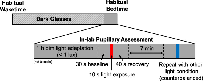
Methods: Forty healthy participants (8-9 years, n = 21; 15-16 years, n = 19) completed a PLR assessment 1 h before their habitual bedtime. After a 1 h dim-light adaptation period (< 1 lx), baseline pupil diameter was measured in darkness for 30 s, followed by a 10 s exposure to 3.0 × 10¹³ photons/cm2/s of either red (627 nm) or blue (459 nm) light, and a 40 s recovery in darkness to assess pupillary re-dilation. Subsequently, participants underwent 7 min of dim-light re-adaptation followed by an exposure to the other light condition. Lights were counterbalanced across participants.
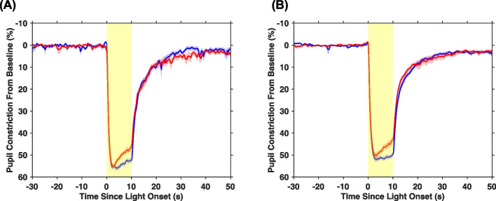
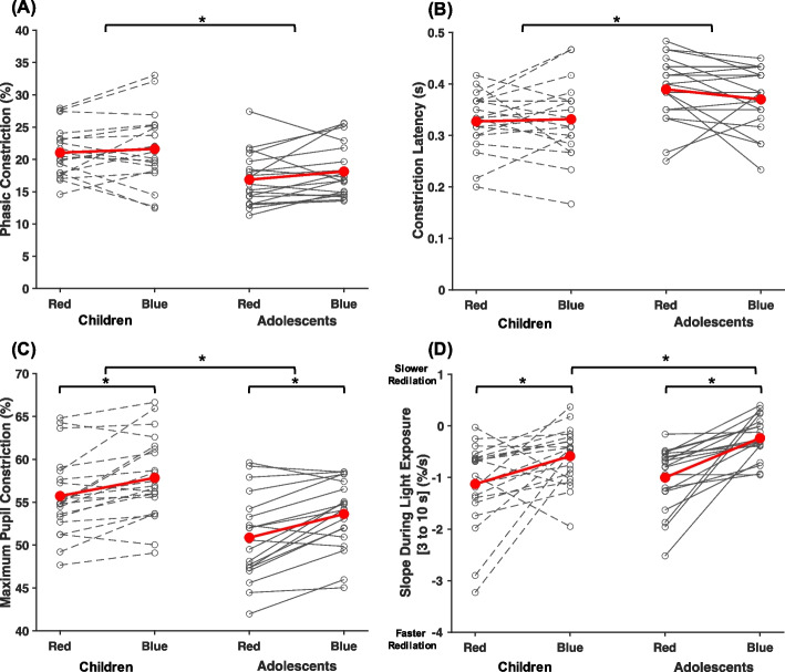
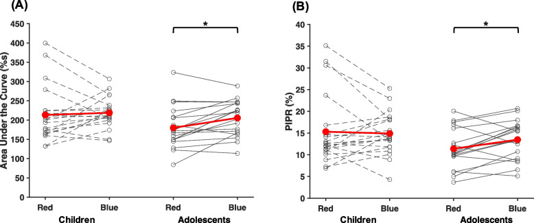
Results: Across both age groups, maximum pupil constriction was significantly greater (p < 0.001, ηp2 = 0.48) and more sustained (p < 0.001, ηp2 = 0.41) during exposure to blue compared to red light. For adolescents, the post-illumination pupillary response (PIPR), a hallmark of melanopsin function, was larger after blue compared with red light (p = 0.02, d = 0.60). This difference was not observed in children. Across light exposures, children had larger phasic (p < 0.01, ηp2 = 0.20) and maximal (p < 0.01, ηp2 = 0.22) pupil constrictions compared to adolescents.
Conclusions: Blue light elicited a greater and more sustained pupillary response than red light in children and adolescents. However, the overall amplitude of the rod/cone-driven phasic response was greater in children than in adolescents. Our findings using the PLR highlight a higher sensitivity to evening light in children compared to adolescents, and continued maturation of the human non-visual photoreception/system throughout development.
Keywords: Adolescents; Children; Intrinsically photosensitive retinal ganglion cells; Light exposure; Pupillary light reflex.
© 2024. The Author(s).
Conflict of interest statement
LEH and MTD have no financial or personal conflicts to declare. MKL reports receiving travel funds from the Australian Research Council and research support from the National Institutes of Health, beyond the submitted work. RPN has a patent application for a handheld pupillometer (PCT/SG2018/050204): Handheld ophthalmic and neurological screening device. The device was not used in this study.
Figures
Update of
-
Differences in the Pupillary Responses to Evening Light between Children and Adolescents.bioRxiv [Preprint]. 2023 Aug 14:2023.08.09.552691. doi: 10.1101/2023.08.09.552691. bioRxiv. 2023. Update in: J Physiol Anthropol. 2024 Jul 3;43(1):16. doi: 10.1186/s40101-024-00363-6. PMID: 37645820 Free PMC article. Updated. Preprint.
Similar articles
-
Differences in the Pupillary Responses to Evening Light between Children and Adolescents.bioRxiv [Preprint]. 2023 Aug 14:2023.08.09.552691. doi: 10.1101/2023.08.09.552691. bioRxiv. 2023. Update in: J Physiol Anthropol. 2024 Jul 3;43(1):16. doi: 10.1186/s40101-024-00363-6. PMID: 37645820 Free PMC article. Updated. Preprint.
-
Binocular Summation in Postillumination Pupil Response Driven by Melanopsin-Containing Retinal Ganglion Cells.Invest Ophthalmol Vis Sci. 2018 Oct 1;59(12):4968-4977. doi: 10.1167/iovs.18-24639. Invest Ophthalmol Vis Sci. 2018. PMID: 30326065
-
Temporal characteristics of melanopsin inputs to the human pupil light reflex.Vision Res. 2015 Feb;107:58-66. doi: 10.1016/j.visres.2014.12.001. Epub 2014 Dec 10. Vision Res. 2015. PMID: 25497360 Free PMC article.
-
[Pupil and melanopsin photoreception].Nippon Ganka Gakkai Zasshi. 2013 Mar;117(3):246-68; discussion 269. Nippon Ganka Gakkai Zasshi. 2013. PMID: 23631256 Review. Japanese.
-
Chromatic Pupillometry Methods for Assessing Photoreceptor Health in Retinal and Optic Nerve Diseases.Front Neurol. 2019 Feb 12;10:76. doi: 10.3389/fneur.2019.00076. eCollection 2019. Front Neurol. 2019. PMID: 30809186 Free PMC article. Review.
Cited by
-
Light at night and circadian rhythms: from the perspective of physiological anthropology research.J Physiol Anthropol. 2024 Dec 26;43(1):32. doi: 10.1186/s40101-024-00380-5. J Physiol Anthropol. 2024. PMID: 39726017 Free PMC article. No abstract available.
-
The Circadian Response to Evening Light Spectra in Early Childhood: Preliminary Insights.J Biol Rhythms. 2025 Apr;40(2):181-193. doi: 10.1177/07487304241311652. Epub 2025 Jan 8. J Biol Rhythms. 2025. PMID: 39773135 Free PMC article.
References
MeSH terms
Grants and funding
- T32 HL149646/HL/NHLBI NIH HHS/United States
- R01 HD087707/HD/NICHD NIH HHS/United States
- F32-HD103390/Eunice Kennedy Shriver National Institute of Child Health and Human Development
- R01-HD087707/Eunice Kennedy Shriver National Institute of Child Health and Human Development
- T32-HL149646/HL/NHLBI NIH HHS/United States
LinkOut - more resources
Full Text Sources
