Review
. 2024 Aug;36(33):e2401750.
doi: 10.1002/adma.202401750. Epub 2024 Jul 3.

Interface-Mediated Neurogenic Signaling: The Impact of Surface Geometry and Chemistry on Neural Cell Behavior for Regenerative and Brain-Machine Interfacing Applications

Affiliations
Review

Interface-Mediated Neurogenic Signaling: The Impact of Surface Geometry and Chemistry on Neural Cell Behavior for Regenerative and Brain-Machine Interfacing Applications

Ian Sands et al. Adv Mater. 2024 Aug.

Abstract

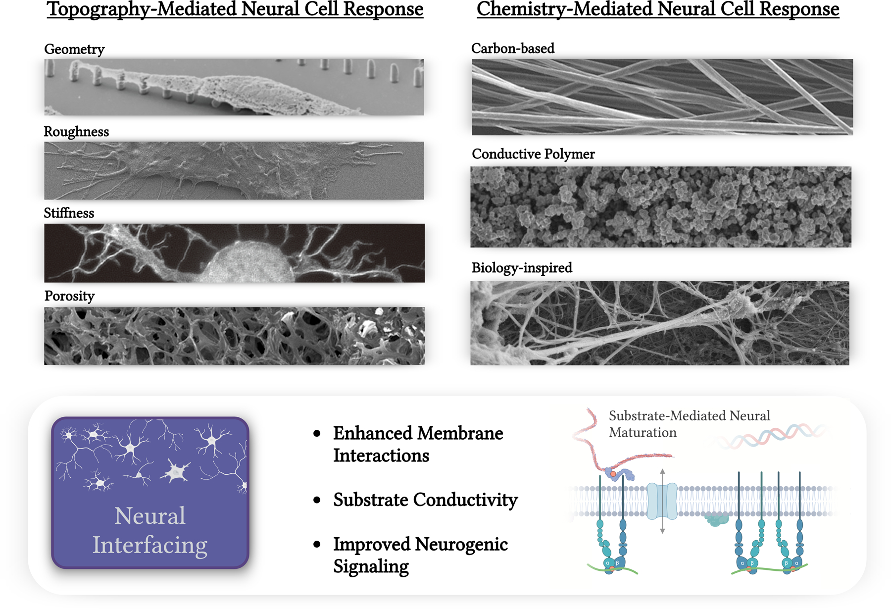
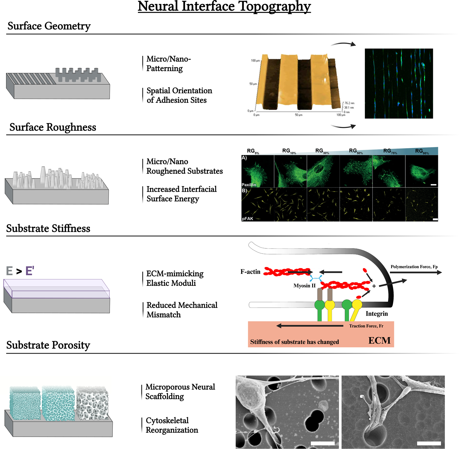
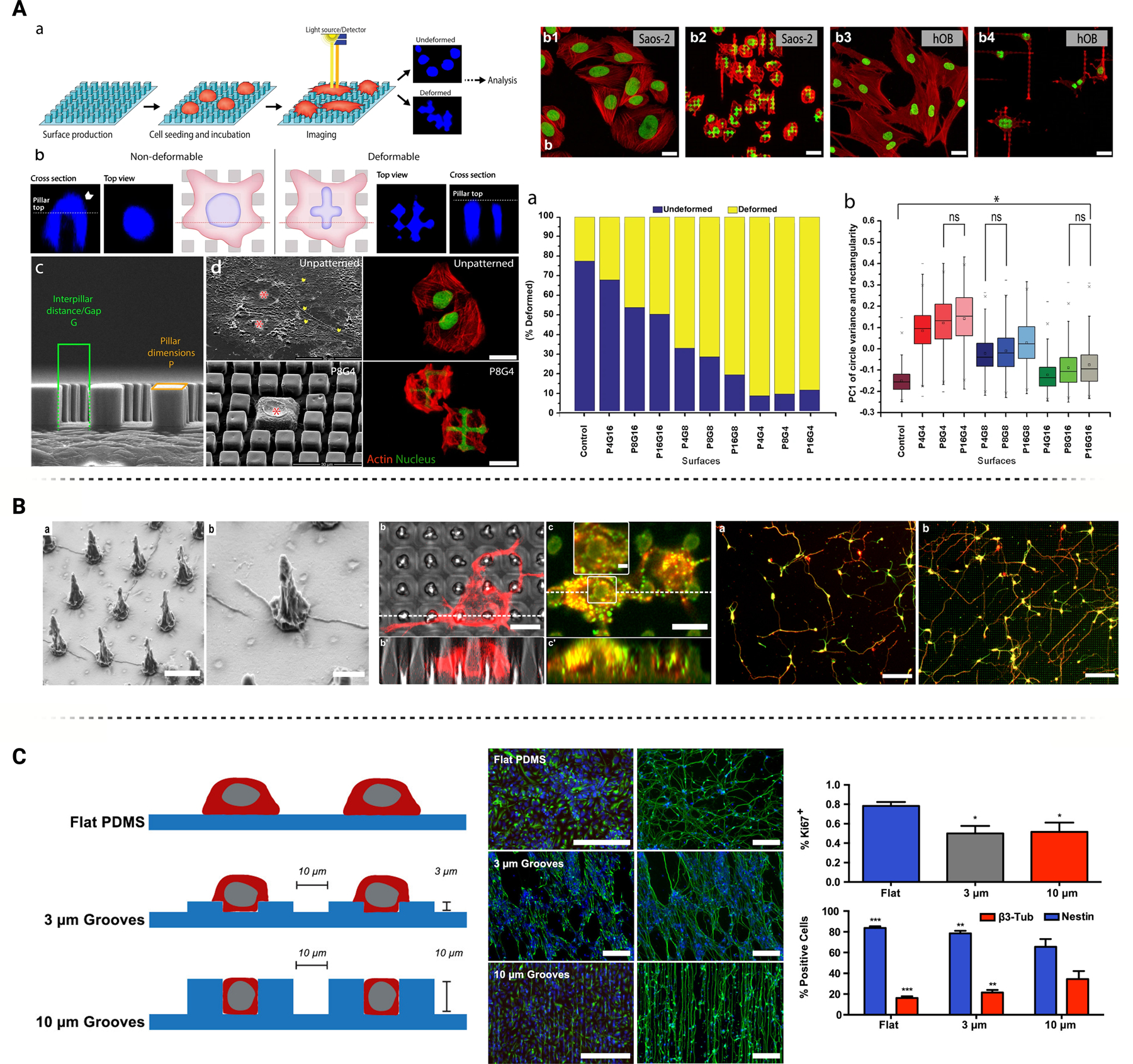
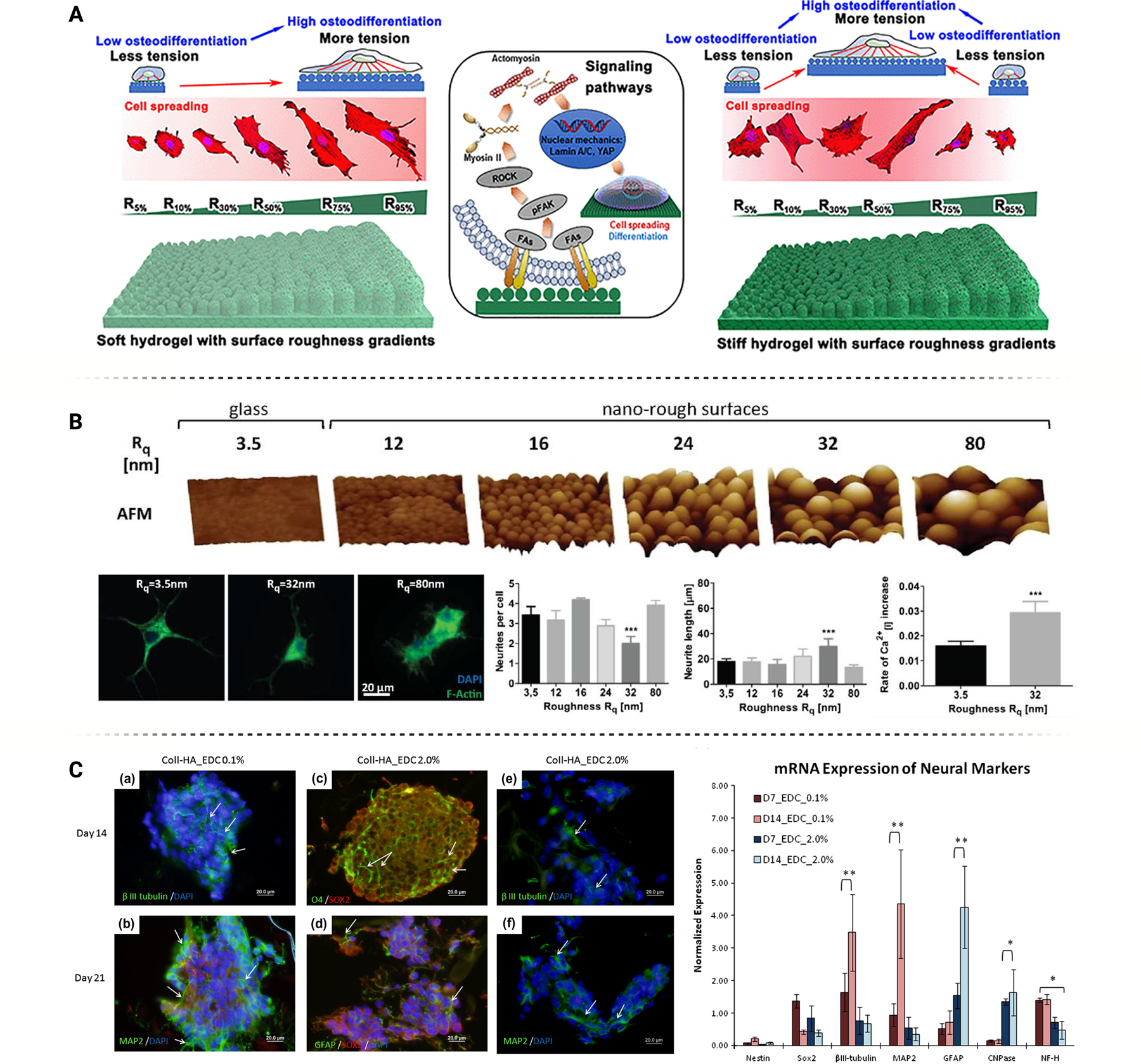
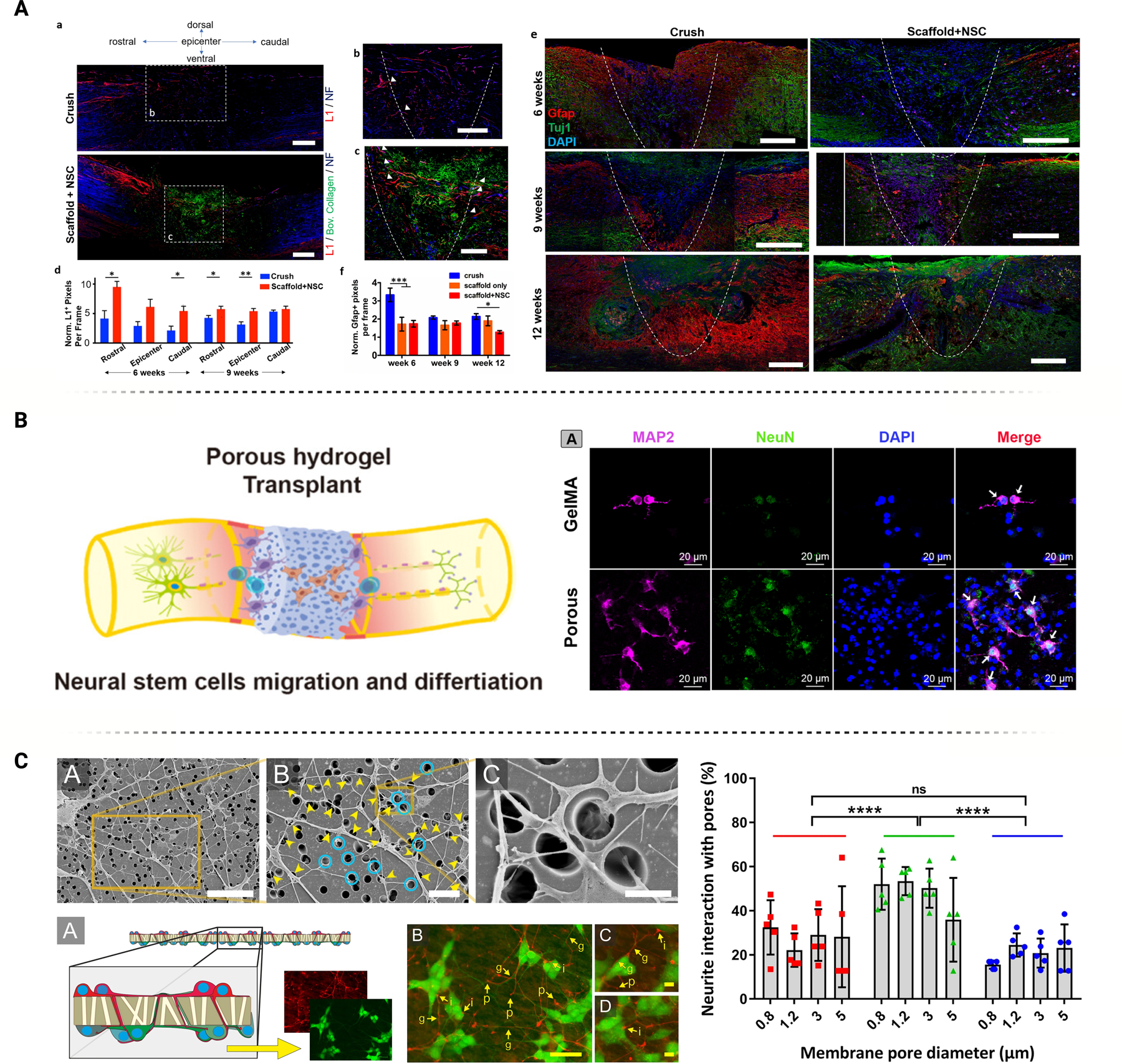
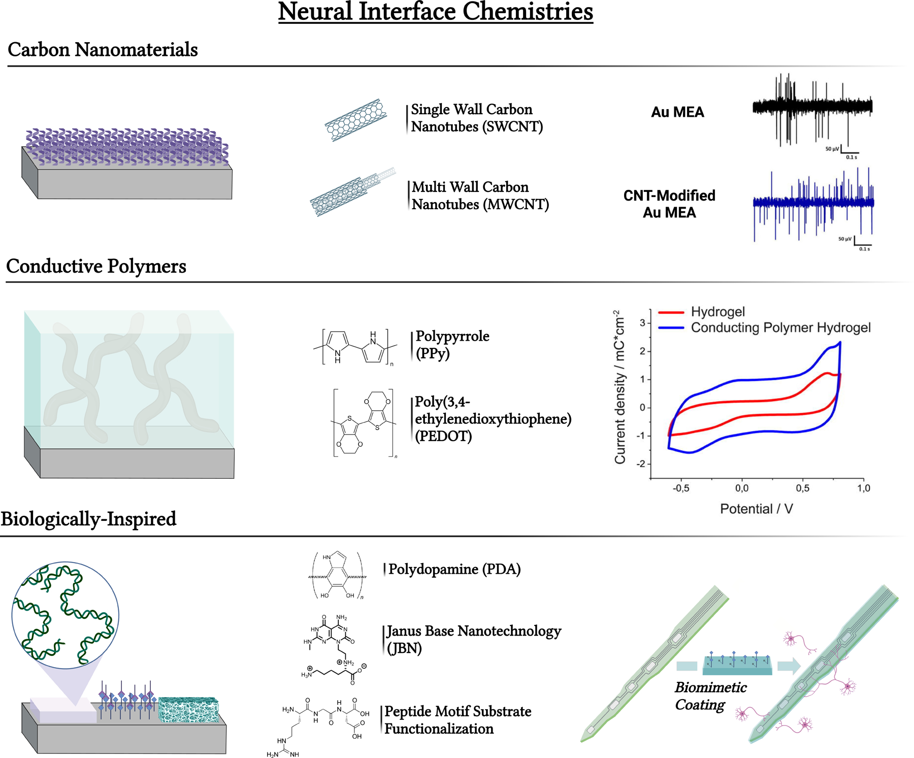
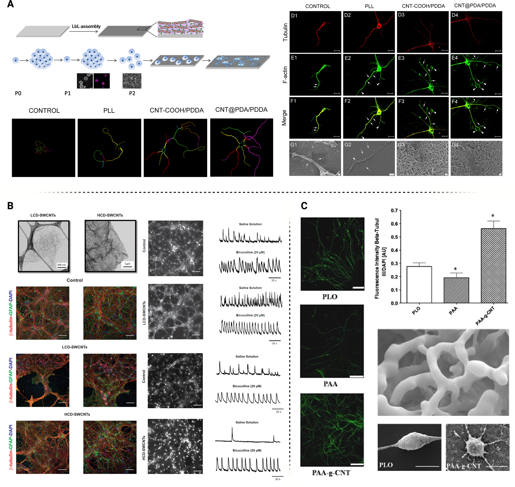
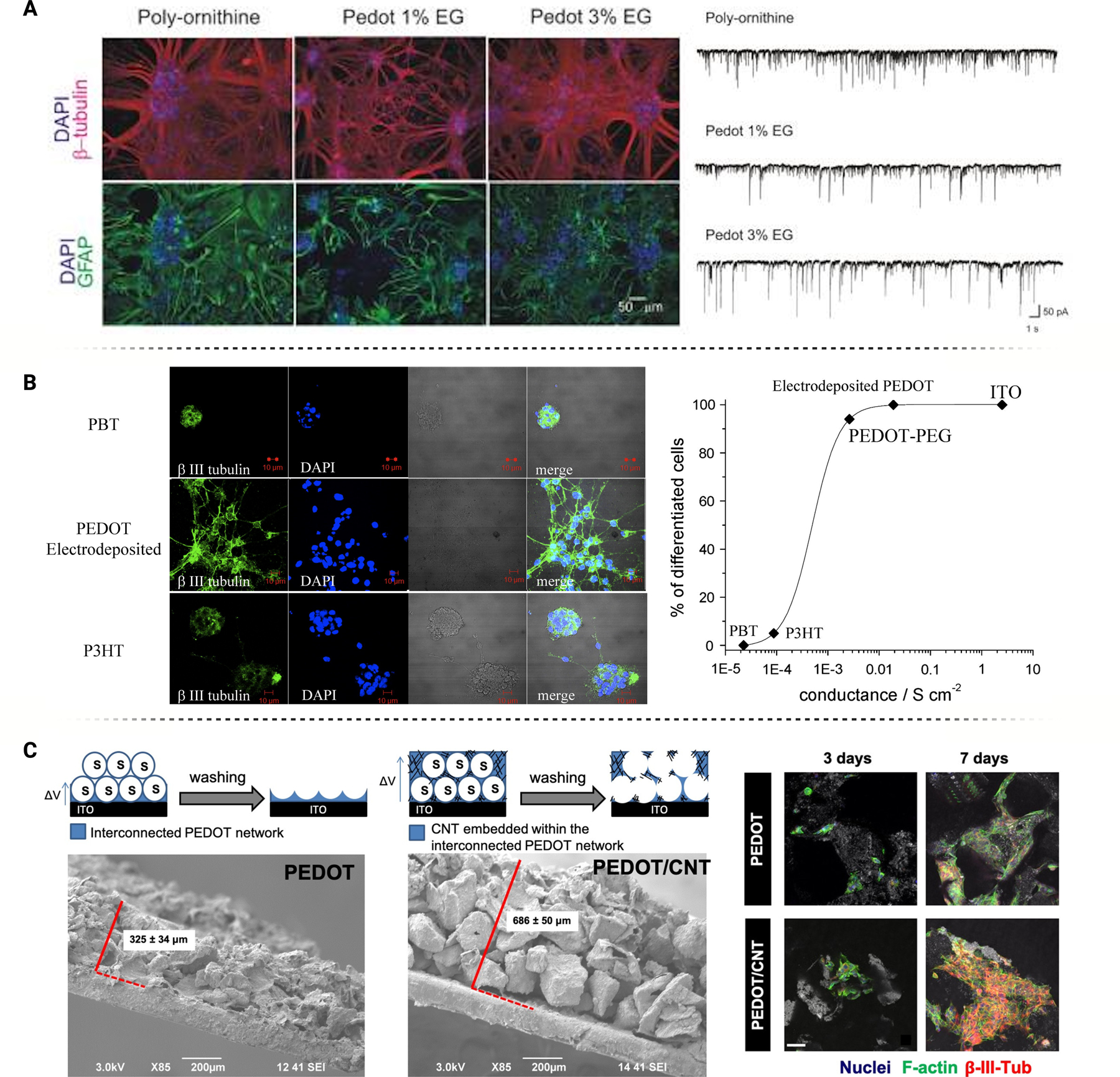
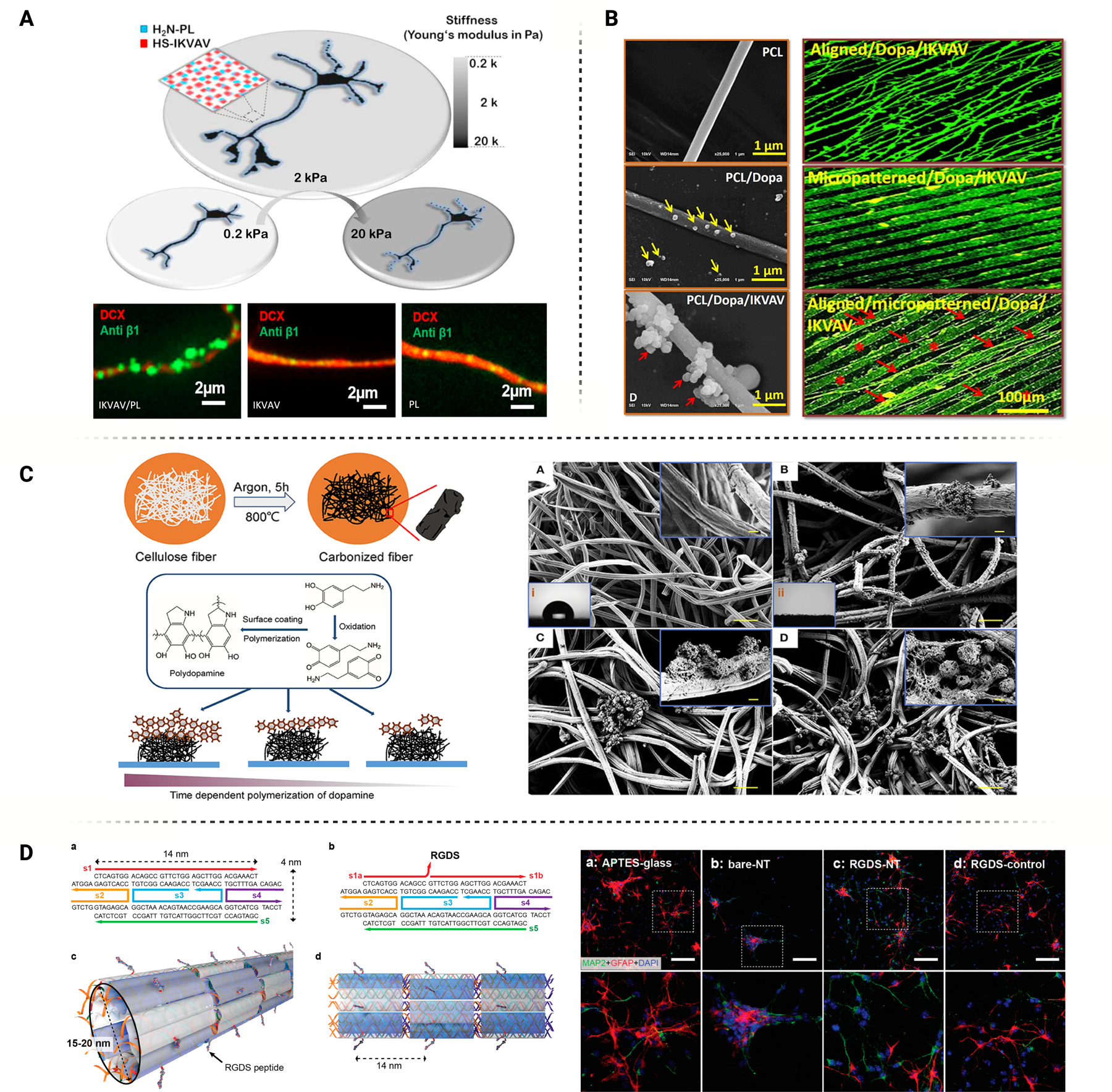
Nanomaterial advancements have driven progress in central and peripheral nervous system applications such as tissue regeneration and brain-machine interfacing. Ideally, neural interfaces with native tissue shall seamlessly integrate, a process that is often mediated by the interfacial material properties. Surface topography and material chemistry are significant extracellular stimuli that can influence neural cell behavior to facilitate tissue integration and augment therapeutic outcomes. This review characterizes topographical modifications, including micropillars, microchannels, surface roughness, and porosity, implemented on regenerative scaffolding and brain-machine interfaces. Their impact on neural cell response is summarized through neurogenic outcome and mechanistic analysis. The effects of surface chemistry on neural cell signaling with common interfacing compounds like carbon-based nanomaterials, conductive polymers, and biologically inspired matrices are also reviewed. Finally, the impact of these extracellular mediated neural cues on intracellular signaling cascades is discussed to provide perspective on the manipulation of neuron and neuroglia cell microenvironments to drive therapeutic outcomes.

Keywords: cell signaling; neural Interfaces; neurogenesis; substrate chemistry; surface topographies.


Figures

Figure 1
Figure 1
Summary of physical modifications to neural interfaces including porosity [12], geometry [13], stiffness [14], and roughness. All cited categories are from works adapted to this figure under the Creative Commons license (CC BY 3.0 or 4.0). Additional chemical substrate modifications are categorized into carbon-based [15], conductive polymer [16], or biologically-inspired chemistries. All cited categories are from works adapted to this figure under the Creative Commons license (CC BY 3.0 or 4.0). Neural interface applications discussed within include brain machine interfacing and CNS/PNS regenerative scaffolding.
Figure 2
Figure 2
Neural interface modifications including the alteration of substrate geometry using micro/nano-patterning techniques. These can result in microgrooves, microchannels, micropillars, or other modifications that impact the spatial orientation of adhesion sites. Figure adapted with permission from Pardo-Figuerez et al. [29] under the Creative Commons license (CC BY 3.0 or 4.0). Alteration of interfacial energy by altering surface roughness also impacts neural cell response. Figure adapted from [30] under the Creative Commons license (CC BY 3.0 or 4.0). Neural cell interactions may also be impacted by altering surface porosity for network growth. Figure adapted from [31] under the Creative Commons license (CC BY 3.0 or 4.0). Substrate stiffness similar to native tissue ECM can facilitate a superior device-tissue integration. Figure adapted from [32] under the Creative Commons license (CC BY 3.0 or 4.0).
Figure 3
Figure 3
Modifications to neural interface surface geometries including micropillars and etched microgroove/microchannels demonstrate key influence on neural cell behavior. (A) Micropillar induced nuclear deformations over patterned surfaces demonstrate heightened nuclear elasticity and actin activity over Saos-2 cells compared to unpatterned topographies. Figure adopted with permission from Ermis et al. [37] under the Creative Commons license (CC BY 3.0 or 4.0). (B) PH3T micropillar geometries combined with light excitation demonstrate critical upregulation of key neuronal markers MAP2 and TUJ1. Figure adopted with permission from Milos et al. [40] under the Creative Commons license (CC BY 3.0 or 4.0). (C) PDMS microchannels modulate neuronal-linked epigenetic factors at different key depths and channel widths, as well as significant impact on NOTCH pathway capabilities. Surface topographies promote neural lineage and maturation amongst neural stem cell cultures. Figures adopted with permission from Milos et.al. [47]
Figure 4
Figure 4
(A) Soft and stiff hydrogels, each with the same surface roughness gradients, differentially impact cellular behavior. Figure adapted from Hou et al. [55] with permission. (B) Surfaces with an optimal stochastic nanoroughness, Rq = ~ 23 nm, induce increased neuronal differentiation and longer neurite outgrowth, as compared to smooth surfaces, Rq = ~ 3.5 nm. Figure adapted from Blumenthal et al. [65] under Creative Commons License (CC BY 3.0 or 4.0). (C) Hydrogel stiffness, controlled through concentration of crosslinking agent (EDC), influences stem cell differentiation towards different neural lineages. Figure adapted from Her et al. [83] with permission.
Figure 5
Figure 5
Porosity, including individual pore size and overall material porosity, can impact neural behavior in a variety of different ways. (A) Porous collagen scaffolds deliver neural stem cells to lesion sites of spinal cord injuries, improving axonal elongation and reducing astrogliosis. Figure adapted from Kourgiantaki et al. [121] under Creative Commons License (CC BY 3.0 or 4.0). (B) GelMA hydrogel with inner connective pores enables cell infiltration, in turn promoting NSC differentiation. Figure adapted from Shi et al. [58] with permission. (C) Neurons can interact with the pores in many ways, including entering the pore itself, crossing over it, or skirting around the edge. Pore diameter can impact these interactions. Figure adapted from George et al. [21] under Creative Commons License (CC BY 3.0 or 4.0).
Figure 6
Figure 6
Common neural interface chemistries can impact therapeutic outcomes depending on conductivity, bioactivity, toxicity, etc. Chemical composition alone can influence cell behavior which can further impact the therapeutic success of the interface. Common interface chemistries include carbon nanomaterials, conductive polymers, and biologically inspired materials. Figures adapted with permission from Vafaiee et al. and Kleber et al., respectively [132, 155].
Figure 7
Figure 7
A) Neural stem cell behavior may be actuated through interfacing with CNT composite scaffolds, improving adhesion, viability, neurite outgrowth, and differentiation. Figure adapted with permission from Shao et al. [158] B) Standalone CNT films can impact the development of neural networks and augment signal transduction. The degree of crosslinking between CNTs also impact the strength of the neural circuitry. Figure adapted from Barrejòn et al. with permission [164] C) Poly(acrylic acid) modified with CNTs demonstrate similar improvements in neural viability and neurite outgrowth. Figure adapted with permission from Chao et al. [165]
Figure 8
Figure 8
A) Poly-ornithine substrates modified with PEDOT demonstrated reduced neuroglial binding indicated by a decrease in GFAP expression in neural cultures. Single cell electrophysiology confirmed robust signal transfer on PEDOT-modified films in vitro. Figure adapted with permission from Cellot et al. [198] B) A decrease in substrate conductivity led to a decrease in neural cell differentiation. Similar to RA-induced differentiation, proliferative pathways were inhibited on PEDOT films leading to enhanced neurite outgrowth. Figure adapted with permission from Ostrakhovitch et al. [199] C) PEDOT vs. PEDOT/CNT composite interfaces demonstrate an enhanced neural response on PEDOT/CNT substrates, further linking substrate conductivity and differentiation potential. Figure adapted with permission from Dominguez-Alfaro [200].
Figure 9
Figure 9
A) Standalone benefits of IKVAV and PL were insignificant, however synergistic benefits of IKVAV and PL functional groups for neural binding were identified through integrin IHC. Figure adapted with permission from Farrukh et al. [211] B) PCL electrospun fibers functionalized with DOPA and IKVAV provided extremely well controlled directional growth and myelination of Schwann cells. Figure adapted with permission from Li et al. [212] C) Polydopamine-modified carbonized microfibers increase neural stem cell adhesion, organization, and intracellular coupling. Figure adapted with permission from Yang et al. [219] D) DNA nanotubes covalently functionalized with RGDS peptides for cellular recognition and binding. DNA nanotube substrates preferentially differentiated neural stem cells into neurons rather than astrocytes. Figure adapted with permission from Stephanopoulos et al. [222]
Figure 10
Figure 10
The potential applications for neural interface engineering will improve chronic stability, balance bioactivity and conductivity, and provide easier-to-implement practices in biomedical research. These improvements will help to accelerate the development and success of therapeutic modalities that rely on complex and intimate interfacing with nervous tissue.

Similar articles

Cited by

References

    1. Strumwasser F, “Long-term recording from single neurons in brain of unrestrained mammals,” Science, vol. 127, no. 3296, pp. 469–470, 1958. - PubMed
    1. Dow B, Vautin R, and Bauer R, “The mapping of visual space onto foveal striate cortex in the macaque monkey,” Journal of Neuroscience, vol. 5, no. 4, pp. 890–902, 1985. - PMC - PubMed
    1. Dario P et al. , “Neural interfaces for regenerated nerve stimulation and recording,” IEEE Transactions on Rehabilitation Engineering, vol. 6, no. 4, pp. 353–363, 1998, doi: 10.1109/86.736149. - DOI - PubMed
    1. Kipke DR et al. , “Advanced neurotechnologies for chronic neural interfaces: new horizons and clinical opportunities,” Journal of Neuroscience, vol. 28, no. 46, pp. 11830–11838, 2008. - PMC - PubMed
    1. Kozai TDY, “The History and Horizons of Microscale Neural Interfaces,” (in eng), Micromachines (Basel), vol. 9, no. 9, Sep 6 2018, doi: 10.3390/mi9090445. - DOI - PMC - PubMed

Substances

LinkOut - more resources