. 2024 Aug 12;25(8):5121-5137.
doi: 10.1021/acs.biomac.4c00508. Epub 2024 Jul 3.

A Rheological Study on the Effect of Tethering Pro- and Anti-Inflammatory Cytokines into Hydrogels on Human Mesenchymal Stem Cell Migration, Degradation, and Morphology

Affiliations

A Rheological Study on the Effect of Tethering Pro- and Anti-Inflammatory Cytokines into Hydrogels on Human Mesenchymal Stem Cell Migration, Degradation, and Morphology

Thomas C O'Shea et al. Biomacromolecules. .

Abstract

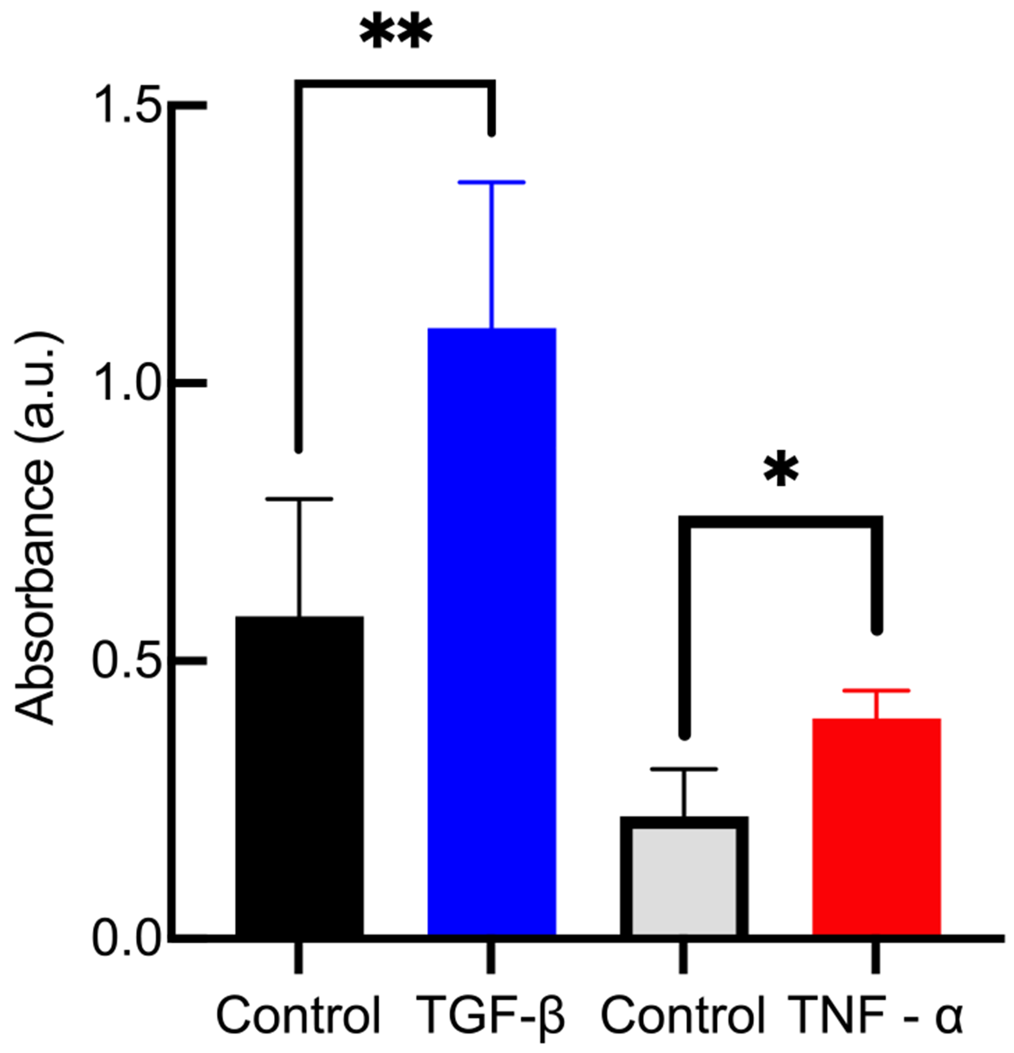
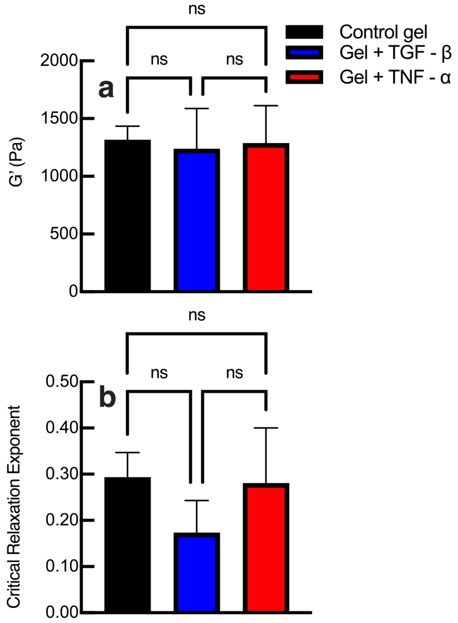
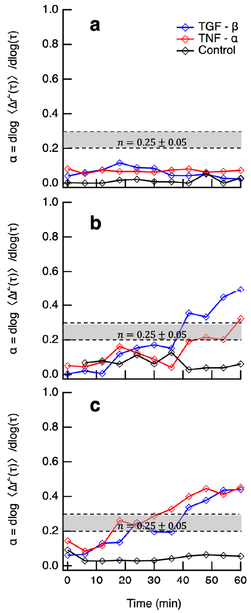
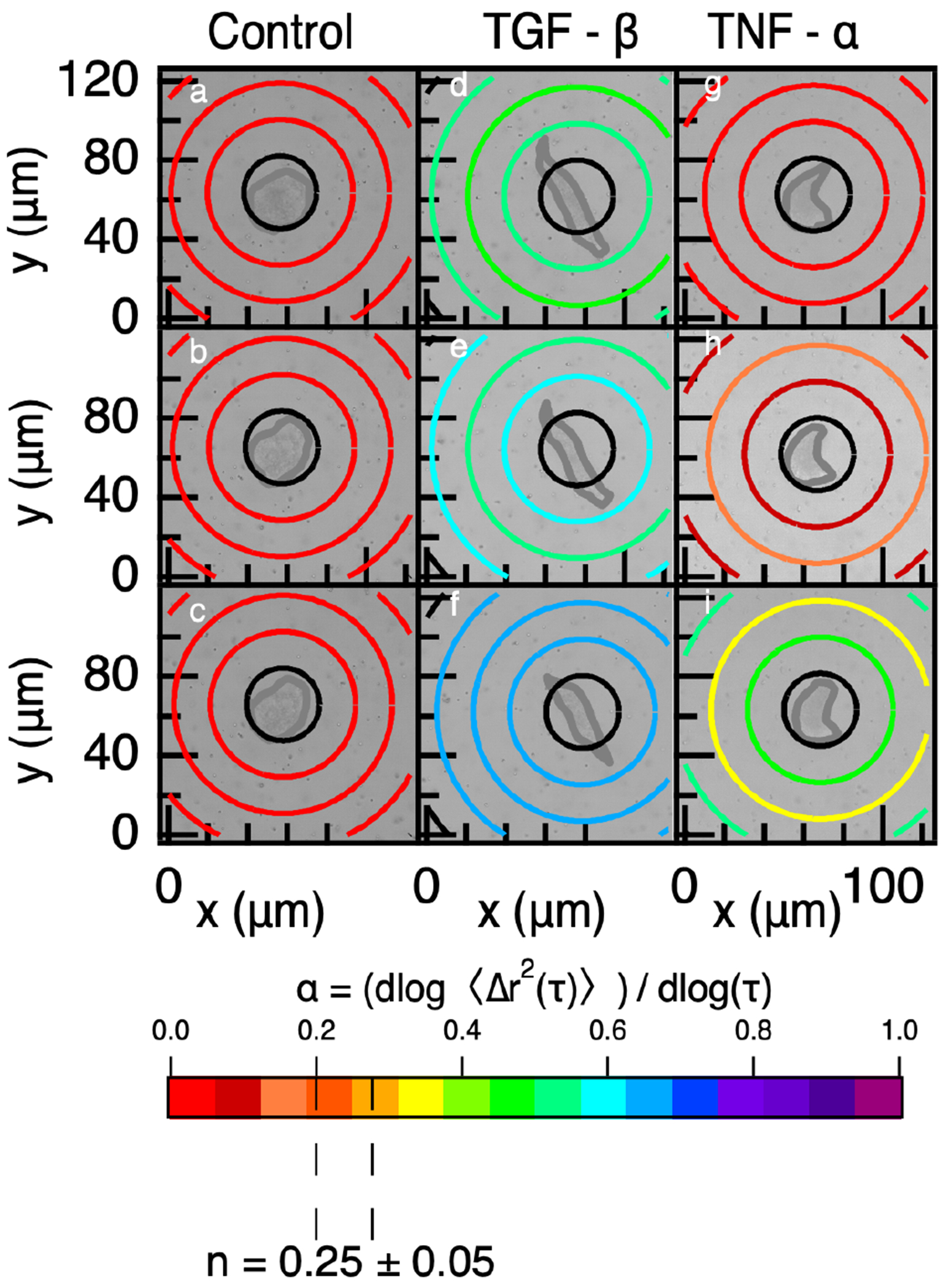
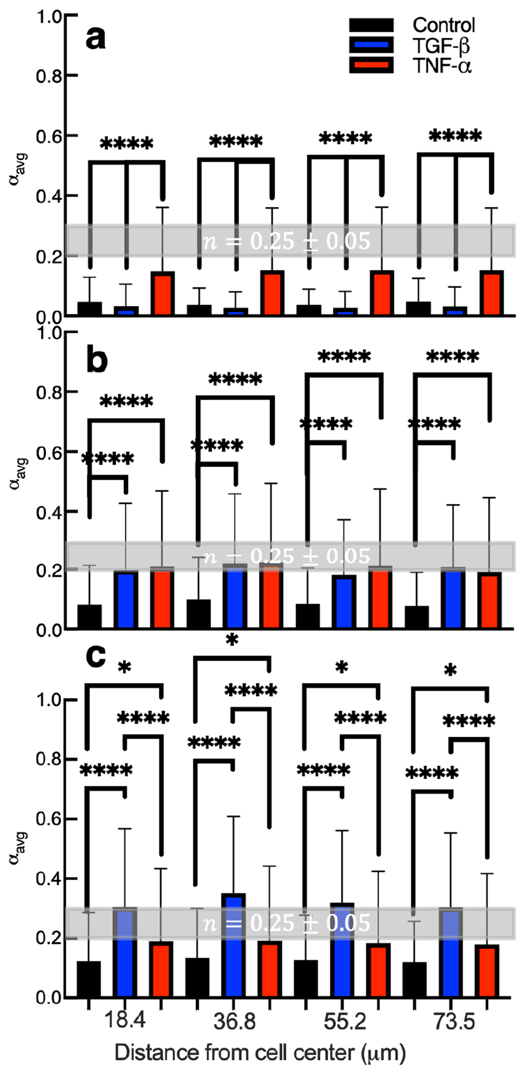
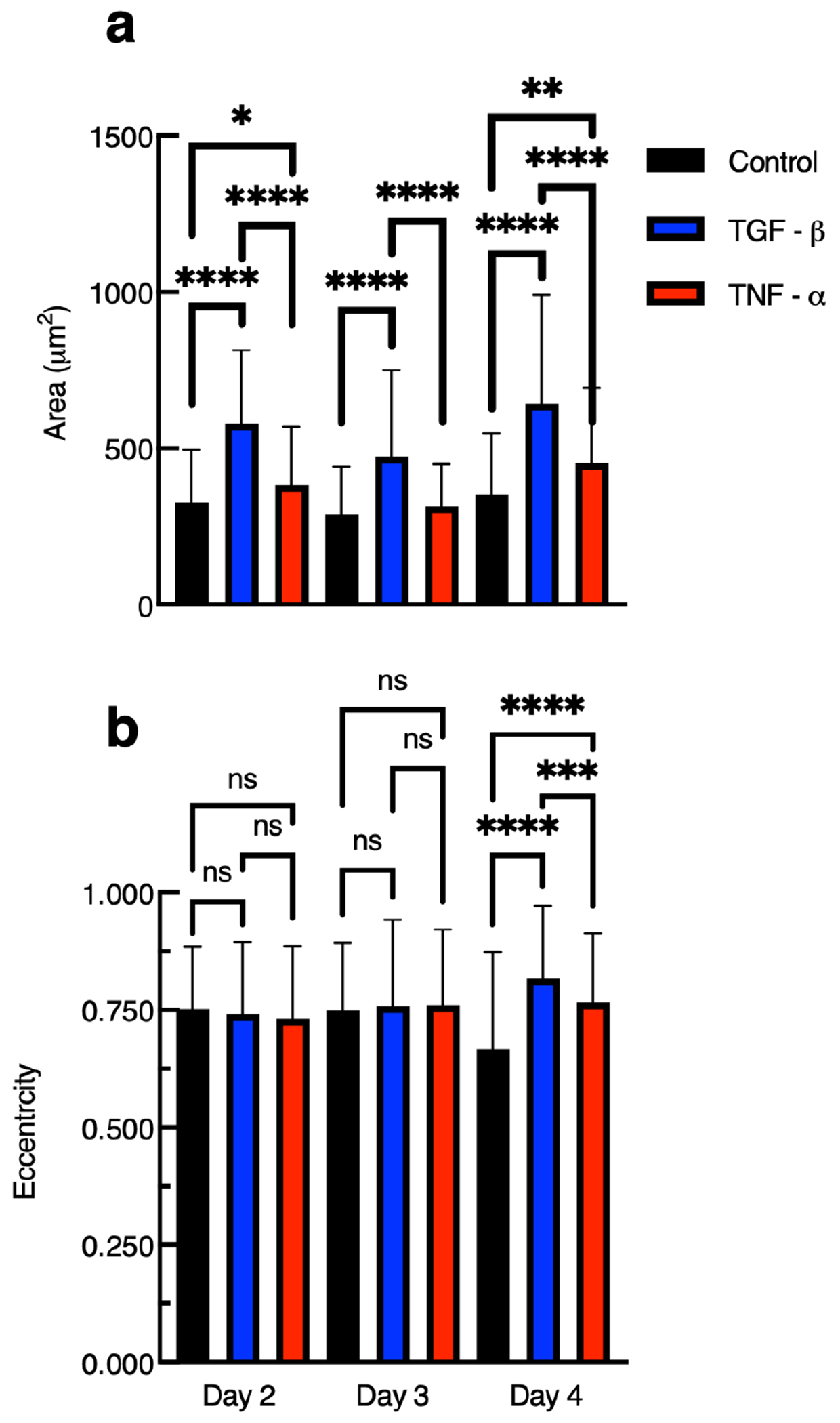
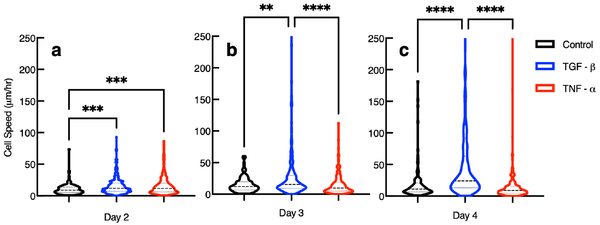
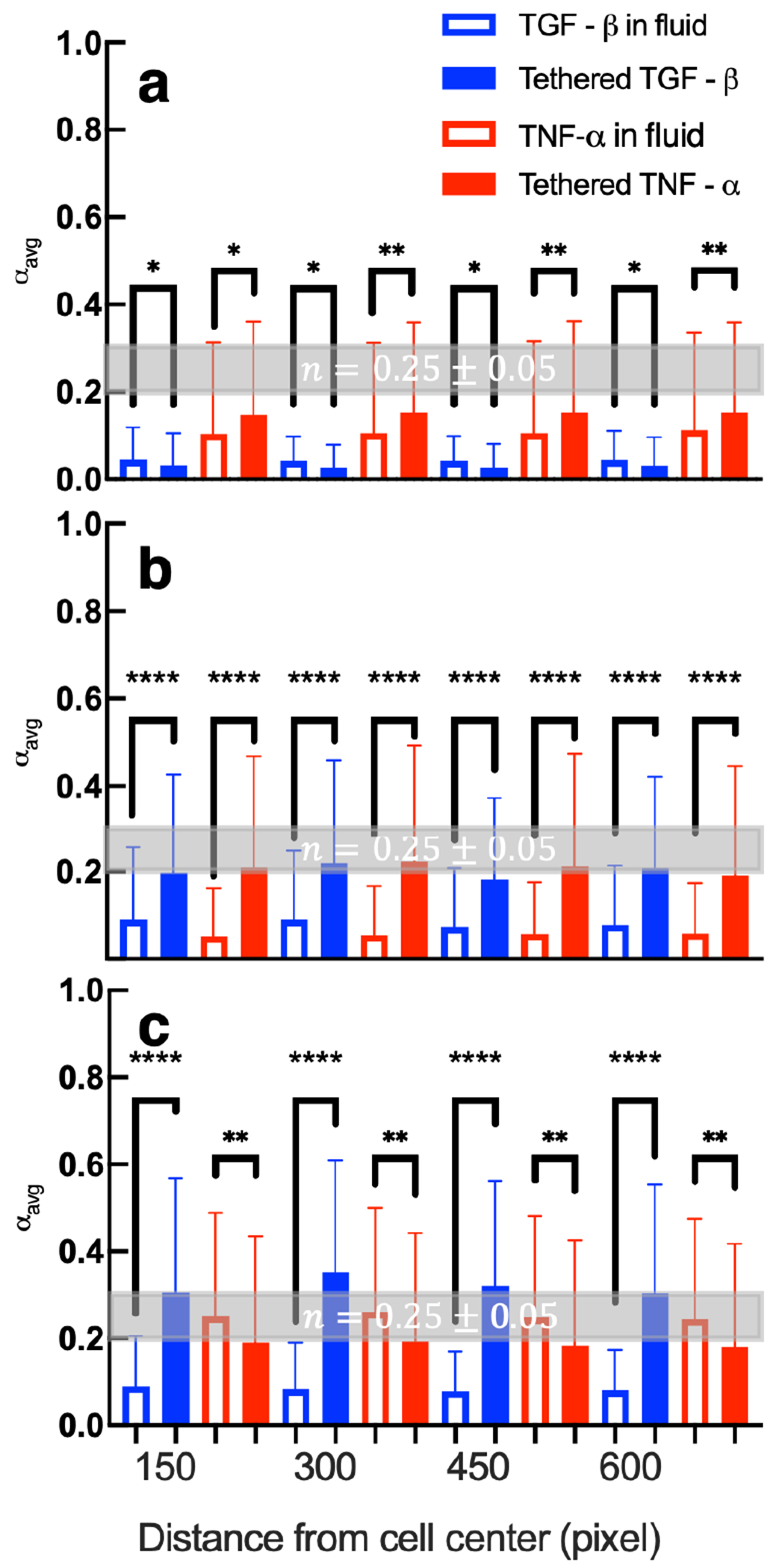
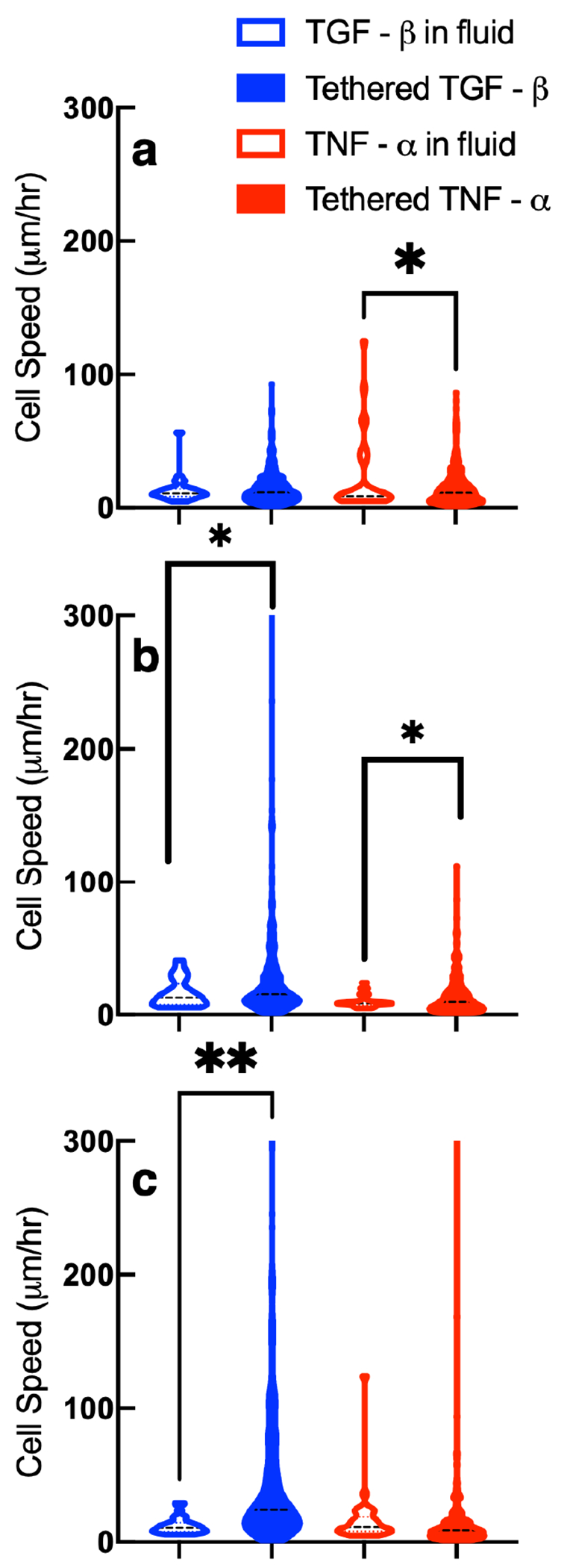
Polymer-peptide hydrogels are being designed as implantable materials that deliver human mesenchymal stem cells (hMSCs) to treat wounds. Most wounds can progress through the healing process without intervention. During the normal healing process, cytokines are released from the wound to create a concentration gradient, which causes directed cell migration from the native niche to the wound site. Our work takes inspiration from this process and uniformly tethers cytokines into the scaffold to measure changes in cell-mediated degradation and motility. This is the first step in designing cytokine concentration gradients into the material to direct cell migration. We measure changes in rheological properties, encapsulated cell-mediated pericellular degradation and migration in a hydrogel scaffold with covalently tethered cytokines, either tumor necrosis factor-α (TNF-α) or transforming growth factor-β (TGF-β). TNF-α is expressed in early stages of wound healing causing an inflammatory response. TGF-β is released in later stages of wound healing causing an anti-inflammatory response in the surrounding tissue. Both cytokines cause directed cell migration. We measure no statistically significant difference in modulus or the critical relaxation exponent when tethering either cytokine in the polymeric network without encapsulated hMSCs. This indicates that the scaffold structure and rheology is unchanged by the addition of tethered cytokines. Increases in hMSC motility, morphology and cell-mediated degradation are measured using a combination of multiple particle tracking microrheology (MPT) and live-cell imaging in hydrogels with tethered cytokines. We measure that tethering TNF-α into the hydrogel increases cellular remodeling on earlier days postencapsulation and tethering TGF-β into the scaffold increases cellular remodeling on later days. We measure tethering either TGF-β or TNF-α enhances cell stretching and, subsequently, migration. This work provides rheological characterization that can be used to design new materials that present chemical cues in the pericellular region to direct cell migration.


Conflict of interest statement

The authors declare no competing financial interest.

Figures

Figure 1.
Figure 1.
Absorbance measurements after modified indirect ELISA for hydrogels with either tethered TGF-β or tethered TNF-α. Comparing gels with cytokines with control gels that contain no cytokines, there is a statistically significant increase in absorbance, which measures the presence of the cytokines in the hydrogels. Data are presented as the mean ± the standard deviation (number of replicates i = 3). Significance is denoted by adjusted p-values (*p < 0.0032 and **p < 0.0021).
Figure 2.
Figure 2.
(a) Bulk rheological and (b) microrheological characterization of hydrogels without cytokines (control) and with tethered TGF-β or tethered TNF-α. Bulk properties and the scaffold microstructure at the phase transition, characterized by the critical relaxation exponent, do not change when cytokines are tethered into the network. Data are presented as the mean ± the standard deviation (number of replicates i = 3 for each condition). Significance is denoted by adjusted p-values (*p < 0.0032, **p < 0.0021, and p > 0.0032 is not significant and denoted as ns).
Figure 3.
Figure 3.
Logarithmic slope of the MSD, α=dlogΔr2(τ)dlogτ, is measured for pericellular regions every 6 min around hMSCs 3D encapsulated in hydrogels with no cytokine (control), tethered TGF-β and tethered TNF-α. The critical relaxation exponent, n, where the gel–sol phase transition occurs is shaded in gray at n=0.25±0.05. Representative data are provided of pericellular rheology days (a) 2, (b) 3, and (c) 4 postencapsulation. Each line is a temporal MPT measurement of a single pericellular region (shown in these graphs is number of cells i=1 per condition per day).
Figure 4.
Figure 4.
Spatiotemporal MPT measurements in (a–c) a control gel, (d–f) gel with tethered TGF-β, and (g–i) gel with tethered TNF-α 4 days postencapsulation. The rheology of the material is shown as colored rings with the color representing the value of the logarithmic slope of the MSD, α=dlogΔr2(τ)dlogτ, for particles in that ring. Warm colors are low values of α indicating little to no particle motion, and cool colors are high values of β indicating restricted to freely diffusive particle motion. The critical relaxation exponent, n, where the gel–sol phase transition occurs is highlighted at n = 0.25 ± 0.05. Data are collected at (a,d,g) 0, (b,e,h) 30, and (c,f,i) 60 minutes (shown in these graphs is number of cells i = 1 per condition).
Figure 5.
Figure 5.
Calculated values of αavg for all measurements in hydrogel pericellular regions (a) 2, (b) 3, and (c) 4 days postencapsulation. The critical relaxation exponent, n, where the gel–sol phase transition occurs is shaded in gray at n = 0.25 ± 0.05. Data are presented as the mean ± the standard deviation as a function of increasing distance from the cell center, and i is the number of data points for each condition. For hydrogels with no cytokines, (a) i = 176, (b) i = 168, and (c) i = 139. For hydrogels with tethered TGF-β, (a) i = 322, (b) i = 333, and (c) i = 315. For hydrogels with tethered TNF-α, (a) i = 330, (b) i = 322, and (c) i = 345. Significance is denoted by adjusted p-values where *p < 0.0032, **p < 0.0021, ***p < 0.0002, and ****p < 0.0001.
Figure 6.
Figure 6.
Measurements of 3D encapsulated hMSC (a) area and (b) eccentricity in gels with and without tethered cytokines 2, 3, and 4 days postencapsulation. Data are presented as the mean ± the standard deviation, and i is the number of data points. For measurements of cells in hydrogels with tethered TGF-β, on day 2 postencapsulation the number of points are i = 351, day 3 postencapsulation are i = 303, and day 4 postencapsulation are i = 283. For measurements of cells in hydrogels with tethered TNF-α, on day 2 postencapsulation are i = 301, day 3 postencapsulation are i = 308, and day postencapsulation are 4 postencapsulation are i = 407. For measurements of cells in control gels, on day 2 postencapsulation i = 122, day 3 postencapsulation are i = 118, and day 4 postencapsulation i = 119. Significance is denoted by adjusted p-values (*p < 0.0032, **p < 0.0021, ***p < 0.0002, ****p < 0.0001, and p > 0.0032 is denoted as ns or not significant).
Figure 7.
Figure 7.
Speed of hMSCs 3D encapsulated in hydrogels without cytokines with tethered TGF-β or with tethered TNF-α days 2, 3, and 4 postencapsulation. Data are presented as the mean ± the standard deviation, and i is the number of data points for each condition. For measurements of cells in hydrogels with tethered TGF-β, (a) i = 351, (b) i = 303, and (c) i = 283. For measurements of cells in hydrogels with tethered TNF-α, (a) i = 301, (b) i = 308, and (c) i = 407. For measurements of cells in control hydrogels, (a) i = 122, (b) i = 118, and (c) i = 119. Significance is denoted by adjusted p-values (*p < 0.0032, **p < 0.0021, ***p < 0.0002, and ****p < 0.0001).
Figure 8.
Figure 8.
A comparison of calculated values of αavg in hydrogels with pro- or anti-inflammatory cytokines tethered into the network to hydrogels with pro- or anti-inflammatory in the surrounding fluid (a) 2, (b) 3, and (c) 4 days postencapsulation. The critical relaxation exponent, n, where the gel–sol phase transition occurs is shaded in gray at n = 0.25 ± 0.05. Data are presented as the mean ± the standard deviation and the number of data points i ≥ 214 for each condition on each day at each distance. Significance is denoted by p-values (*p < 0.0032, **p < 0.0021, ***p < 0.0002, and ****p < 0.0001). Data collected with cytokines in the fluid environment are reprinted with permission from ref . Copyright 2021 American Chemical Society.
Figure 9.
Figure 9.
A comparison of calculated values of cell speed in hydrogels with pro- or anti-inflammatory cytokines tethered in the network or with pro- or anti-inflammatory cytokines in the surrounding fluid (a) 2, (b) 3, and (c) 4 days postencapsulation. Significance is denoted by p-values (*p < 0.0032, **p < 0.0021, ***p < 0.0002, and ****p < 0.0001). i is the number of measurements with i ≥ 29 for each condition on each day. Data collected with cytokines in the fluid environment are reprinted with permission from ref . Copyright 2021 American Chemical Society.

Similar articles

References

    1. Dash BC; Xu Z; Lin L; Koo A; Ndon S; Berthiaume F; Dardik A; Hsia H Stem cells and engineered scaffolds for regenerative wound healing. Bioengineering 2018, 5, 23. - PMC - PubMed
    1. Lee PY; Cobain E; Huard J; Huang L Thermosensitive hydrogel PEG–PLGA–PEG enhances engraftment of muscle-derived stem cells and promotes healing in diabetic wound. Mol. Ther 2007, 15, 1189–1194. - PubMed
    1. Lutolf MP; Lauer-Fields JL; Schmoekel HG; Metters AT; Weber FE; Fields GB; Hubbell JA Synthetic matrix metalloproteinase-sensitive hydrogels for the conduction of tissue regeneration: engineering cell-invasion characteristics. Proc. Natl. Acad. Sci. U.S.A 2003, 100, 5413–5418. - PMC - PubMed
    1. Ennis WJ; Sui A; Bartholomew A Stem cells and healing: impact on inflammation. Adv. Wound Care 2013, 2, 369–378. - PMC - PubMed
    1. Maxson S; Lopez EA; Yoo D; Danilkovitch-Miagkova A; LeRoux MA Concise review: role of mesenchymal stem cells in wound repair. Stem Cells Transl. Med 2012, 1, 142–149. - PMC - PubMed

Publication types

LinkOut - more resources