

Amide proton transfer-weighted and arterial spin labeling imaging may improve differentiation between high-grade glioma recurrence and radiation-induced brain injury
- PMID: 38961946
- PMCID: PMC11219995
- DOI: 10.1016/j.heliyon.2024.e32699
Amide proton transfer-weighted and arterial spin labeling imaging may improve differentiation between high-grade glioma recurrence and radiation-induced brain injury
Abstract
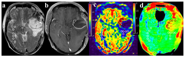
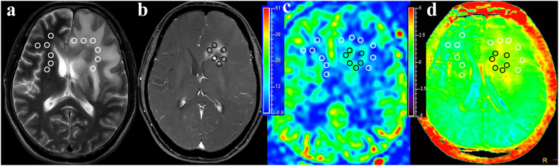
Rationale and objectives: The management of tumor recurrence (TR) and radiation-induced brain injury (RIBI) poses significant challenges, necessitating the development of effective differentiation strategies. In this study, we investigated the potential of amide proton transfer-weighted (APTw) and arterial spin labeling (ASL) imaging for discriminating between TR and RIBI in patients with high-grade glioma (HGG).
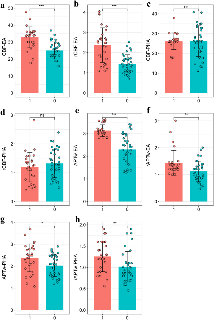
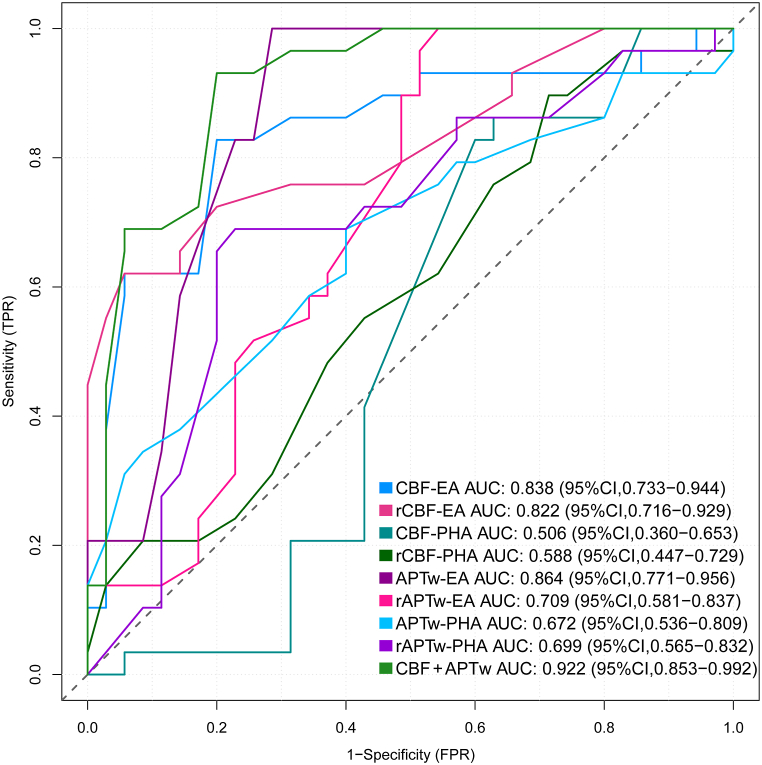
Methods: A total of 64 HGG patients receiving standard treatment were enrolled in this study. The patients were categorized based on secondary pathology or MRI follow-up results, and the demographic characteristics of each group were presented. The APTw, rAPTw, cerebral blood flow (CBF) and rCBF values were quantified. The differences in various parameters between TR and RIBI were assessed using the independent-samples t-test. The discriminative performance of these MRI parameters in distinguishing between the two conditions was assessed using receiver operating characteristic (ROC) curve analysis. Additionally, the Delong test was employed to further evaluate their discriminatory ability.
Results: The APTw and CBF values of TR were significantly higher compared to RIBI (P < 0.05). APTw MRI demonstrated superior diagnostic efficiency in distinguishing TR from RIBI (area under the curve [AUC]: 0.864; sensitivity: 75.0 %; specificity: 81.8 %) when compared to ASL imaging. The combined utilization of APTw and CBF value further enhanced the AUC to 0.922. The Delong test demonstrated that the combination of APTw and ASL exhibited superior performance in the identification of TR and RIBI, compared to ASL alone (P = 0.048).
Conclusion: APTw exhibited superior diagnostic efficacy compared to ASL in the evaluation of TR and RIBI. Furthermore, the combination of APTw and ASL exhibits greater discriminatory capability and diagnostic performance.
Keywords: APTw; ASL; High-grade glioma; Radiation-induced brain injury; Tumor recurrence.
© 2024 The Authors.
Conflict of interest statement
The authors declare that they have no known competing financial interests or personal relationships that could have appeared to influence the work reported in this paper.
Figures
Similar articles
-
Differentiation of true progression from treatment response in high-grade glioma treated with chemoradiation: a comparison study of 3D-APTW and 3D-PcASL imaging and DWI.NMR Biomed. 2023 Jan;36(1):e4821. doi: 10.1002/nbm.4821. Epub 2022 Sep 17. NMR Biomed. 2023. PMID: 36031734
-
Combining amide proton transfer-weighted and arterial spin labeling imaging to differentiate solitary brain metastases from glioblastomas.Magn Reson Imaging. 2023 Oct;102:96-102. doi: 10.1016/j.mri.2023.05.004. Epub 2023 May 10. Magn Reson Imaging. 2023. PMID: 37172748
-
3D Amide Proton Transfer-Weighted Imaging for Grading Glioma and Correlating IDH Mutation Status: Added Value to 3D Pseudocontinuous Arterial Spin Labelling Perfusion.Mol Imaging Biol. 2023 Apr;25(2):343-352. doi: 10.1007/s11307-022-01762-w. Epub 2022 Aug 12. Mol Imaging Biol. 2023. PMID: 35962302
-
Diagnostic performance of multiparametric nonenhanced magnetic resonance imaging (MRI) in grading glioma and correlating IDH mutation status.Clin Radiol. 2025 Mar;82:106791. doi: 10.1016/j.crad.2024.106791. Epub 2024 Dec 26. Clin Radiol. 2025. PMID: 39837107
-
Amide proton transfer-weighted MRI in distinguishing high- and low-grade gliomas: a systematic review and meta-analysis.Neuroradiology. 2019 May;61(5):525-534. doi: 10.1007/s00234-018-02152-2. Epub 2019 Jan 21. Neuroradiology. 2019. PMID: 30666352
References
-
- Li Y., Liu Y., Liang Y., et al. Radiomics can differentiate high-grade glioma from brain metastasis: a systematic review and meta-analysis. Eur. Radiol. 2022;32(11):8039–8051. - PubMed
-
- Zhou Q., Xue C., Ke X., et al. Treatment response and prognosis evaluation in high-grade glioma: an imaging review based on MRI. J. Magn. Reson. Imag. 2022;56(2):325–340. - PubMed
-
- Li C., Gan Y., Chen H., et al. Advanced multimodal imaging in differentiating glioma recurrence from post-radiotherapy changes. Int. Rev. Neurobiol. 2020;151:281–297. - PubMed
LinkOut - more resources
Full Text Sources
