

A case of dedifferentiated liposarcoma discovered due to an intrascrotal calcified ossification
- PMID: 38962045
- PMCID: PMC11217216
- DOI: 10.1007/s13691-024-00682-6
A case of dedifferentiated liposarcoma discovered due to an intrascrotal calcified ossification
Abstract
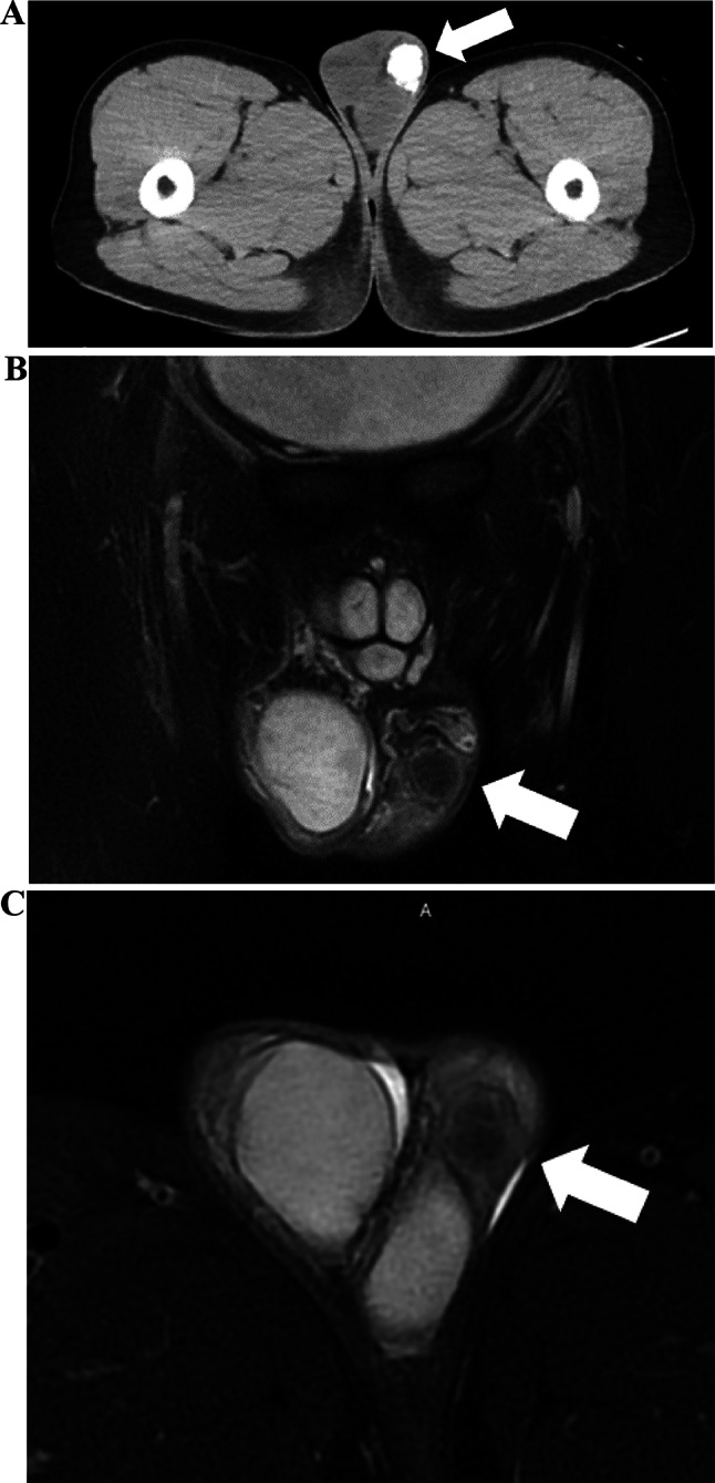
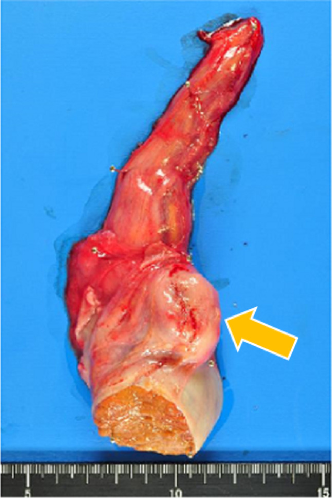
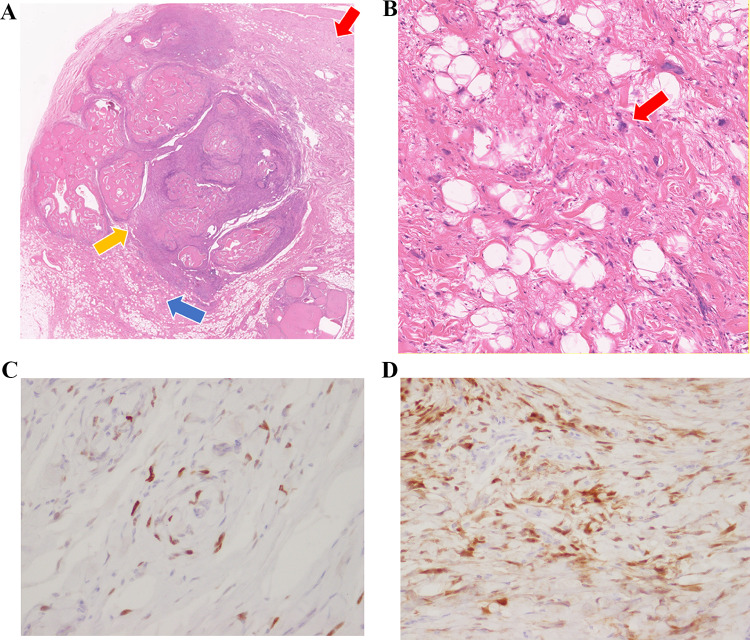
Dedifferentiated liposarcoma is a rare cancer with a poor prognosis. A 52-year-old man presented with a chief complaint of a mass in his left scrotum. He came with suspected testicular tumor, but all the measured tumor markers were negative. Imaging test showed approximately 2 cm diameter mass accompanied by calcification with some substantial components between the testis and epididymis. Left high testicular resection was performed. The tumor had no continuity between the testis and epididymis, and the spermatic cord transection was negative. Pathological findings showed well differentiated fatty component and a dedifferentiated component around the trabecular bone-like tissue. We observed dedifferentiated dysmorphic cells mixed with fatty droplets of unequal size. Immunostaining led to the diagnosis of dedifferentiated liposarcoma. No additional postoperative therapy was performed. The possibility of dedifferentiated liposarcoma should be kept in mind even if mass is confined to the scrotum and consisted of calcification. In the case of an intrascrotal calcified mass with malignant perspective, radical surgery is highly recommended.
Keywords: Calcification; Dedifferentiated liposarcoma; Scrotum.
© The Author(s) under exclusive licence to The Japan Society of Clinical Oncology 2024. Springer Nature or its licensor (e.g. a society or other partner) holds exclusive rights to this article under a publishing agreement with the author(s) or other rightsholder(s); author self-archiving of the accepted manuscript version of this article is solely governed by the terms of such publishing agreement and applicable law.
Conflict of interest statement
Conflict of interestThe authors declare that have no conflict of interest.
Figures
References
-
- Ishibashi Y, et al. A case of dedifferentiated liposarcoma of the spermatic cord. Acta Urol. 2022;68:17–21. - PubMed
-
- Kumada N, et al. A case of liposarcoma of the spermatic cord. Toyota J Med. 2020;29:62–66.
-
- Takaoka N, et al. A case of dedifferentiated liposarcoma of the spermatic cord. Acta Urol. 2019;65:529–532. - PubMed
LinkOut - more resources
Full Text Sources