. 2024 Jul 2;8(7):e619.
doi: 10.1002/pld3.619. eCollection 2024 Jul.

Exploring the diversity of galls on Artemisia indica induced by Rhopalomyia species through morphological and transcriptome analyses

Affiliations

Exploring the diversity of galls on Artemisia indica induced by Rhopalomyia species through morphological and transcriptome analyses

Seiji Takeda et al. Plant Direct. .

Abstract

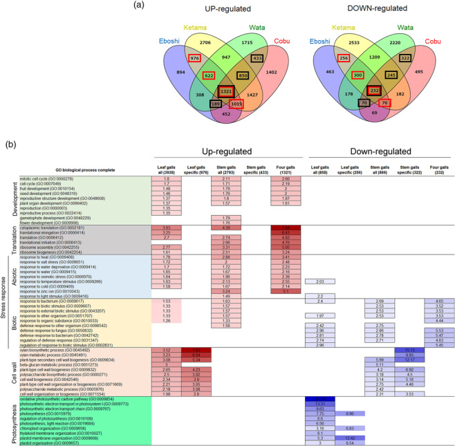
Plant galls generated by insects have highly organized structures, providing nutrients and shelter to the insects living within them. Most research on the physiological and molecular mechanisms of gall development has focused on single galls. To understand the diversity of gall development, we examined five galls with different morphologies generated by distinct species of Rhopalomyia (gall midge; Diptera: Cecidomyiidae) on a single host plant of Artemisia indica var. maximowiczii (Asteraceae). Vasculature developed de novo within the galls, indicating active transport of nutrients between galls and the host plant. Each gall had a different pattern of vasculature and lignification, probably due to differences in the site of gall generation and the gall midge species. Transcriptome analysis indicated that photosynthetic and cell wall-related genes were down-regulated in leaf and stem galls, respectively, compared with control leaf and stem tissues, whereas genes involved in floral organ development were up-regulated in all types of galls, indicating that transformation from source to sink organs occurs during gall development. Our results help to understand the diversity of galls on a single herbaceous host plant.

Keywords: Artemisia indica; RNA sequencing; Rhopalomyia; gall; microCT.


Conflict of interest statement

The authors declare no competing or financial interests.

Figures

FIGURE 1
FIGURE 1
Morphology of galls on leaves or stems. (a–e) Yomogi‐ha‐eboshi‐fushi (Eboshi). (f–j) Yomogi‐ha‐shiro‐ketama‐fushi (Ketama). (k–o) Yomogi‐kuki‐cobu‐fushi (Cobu). Arrowhead in (l) indicates an eclosed midge emerging from the gall. (p–t) Yomogi‐kuki‐wata‐fushi (Wata). (c, h, m, r) Longitudinal sections stained with phloroglucinol, showing lignin staining with purple color. Note that two layers of lignification surround the insect chamber in the Wata gall (arrowheads in r). (d, i, n, s) Longitudinal sections stained with toluidine blue. (e, j, o, t) Schematic illustrations of galls. Yellow, gall tissue; green, leaf or stem of the host plant; pink, lignified tissue; red, phloem; blue, xylem; orange, small cells; l, leaf; st, stem; ic, insect chamber. Bars: q, 1 cm; b–d, g–i, k–p, r, s, 1 mm.
FIGURE 2
FIGURE 2
X‐ray microCT images of galls. (a–d) Eboshi, (e–h) Ketama, (i–l) Cobu, and (m–p) Wata. (a, e, i, j, m, n) Virtual sections showing inside of the galls. Hard tissues are shown in black. (b, f, k) Maximum intensity projection (3D‐MIP) images of galls with grayscale. Hard tissue is shown in white. (c, p) Pseudo‐colored 3D surface model (3D‐SM) images of galls. Purple, lignified vascular tissue; blue, inset chamber; yellow, insects. (d, g, h, l, o) Three‐dimensional volume rendering (3D‐VR) images of galls with pseudo‐color. (a) Longitudinal section of Eboshi gall showing insects and lignified cells in black. Note that the nutritive tissue below the insect chamber is connected to the host vascular bundles. (b, c) lignified hard cells surround the insect chamber. (d) Transverse image of Eboshi gall showing vascular bundles connecting the outer wall and the insect chamber (arrowheads). (e) Longitudinal section of Ketama gall showing insect and several vascular bundles running in the gall tissue. (f, g) Vasculature running throughout the gall reminiscent of tree branching. (h) De novo vasculature emerges from the leaf vasculature of the host plant and runs through the round hole at the bottom of the gall (red arrowheads in f and h). (i, j) transverse (i) and longitudinal (j) sections of a Cobu gall on a stem showing insects and vascular bundles in the gall tissue. (k, l) Vascular bundles connect the gall tissue and host vasculature of the stem. (m, n) Transverse (m) and longitudinal (n) sections of a Wata gall. (o) Two Wata galls on stem. Note that Wata galls are surrounded by many hairs, but the main galls are much smaller than Cobu galls (compare Figure 2o with Figure 2k). Red arrowheads in (n) show the lignified layer surrounding the outside of the insect chamber, as shown in Figure 1r. (p) Hard lignified vascular bundles running around the insect chamber. Images are representative of multiple galls of the same type. g, gall; st, stem. Bars, 1 mm.
FIGURE 3
FIGURE 3
Larva, pupa, and adult of gall midges and parasitic wasps. (a–f) Larvae or pupae in Eboshi galls. (g) Larvae in Ketama galls. (h) Pupa and larvae in Cobu galls. Note that at least three insects were found in a single Cobu gall in this case. (i, j) Pupa and larvae in Wata galls. (k) Female adult midge from an Eboshi gall. (l) Male adult midge from a Ketama gall. (m) Phylogenetic tree of midge species emerging from each gall. Note that they are all Rhopalomyia species, and three of the four species were classified into monophyletic groups, although one Wata midge was classified into the Cobu clade. For comparison, sequences of the mitochondrial cytochrome oxidase subunit I (COI) gene from R. struma (MW817929.1), R. protrahenda (accession number: PP476284.1), and Myetiola destructor (KM936220.1) are shown. Bootstrap values are indicated for nodes (1000 replicates). (n) Parasitic wasp that emerged from a Ketama gall (Aprostocetus sp., male). (o) Parasitic wasp from a Cobu gall (Aprostocetus sp., female). (p, q) parasitic wasps that emerged from Wata galls (p: Aprostecetus sp., female; q, Torymus sp., female). Bars: a–c, g–k, n–q, .5 mm; d–f, .2 mm; l, 1 mm.
FIGURE 4
FIGURE 4
Transcriptome analysis of four galls on A. indica . (a) Venn diagrams showing the number of up‐ and down‐regulated genes in the four types of galls. Numbers in red and black squares represent genes included in the “all” genes category in leaves and stems, respectively, shown in (b). (b) GO analysis of genes expressed in galls. The first column shows the category and number of genes. Numbers in the table indicate fold enrichment of the GO enrichment analysis, and up‐ and down‐regulated genes are colored in red and blue, respectively. GO terms on the left are colored according to function: development, pale green; translation, gray; abiotic stress response, pale blue; biotic stress response, pale orange; cell wall, pale red; photosynthesis, green.
FIGURE 5
FIGURE 5
Morphology of yomogi‐metsubo‐fushi (Metsubo). (a) Axillary buds of A. indica . (b) Metsubo gall generated at the axil. (c) Metsubo gall in (b) cut in half to show a pupa inside. (d–f) Longitudinal section of a Metsubo gall. (e) and (f) are higher magnification images of the bottom and top parts of (d), respectively. Note that Metsubo galls are open at the distal end, which is covered with many hairs (f). (g–j) X‐ray microCT images. (g) Maximum intensity projection (3D‐MIP) image of a Metsubo gall showing the stem vasculature, insect chamber, and insect inside in white. (h) 3D volume rendering (3D‐VR) image showing the development of vasculature from that of the stem. (i, j) Larva inside the insect chamber. (j) Inside the larva body showing a network of fibers and many particles. Bars: a–d, g, h, 2 mm; e, f, 1 mm; i, j, .5 mm.

Similar articles

Cited by

References

    1. Bae, J. , & Jung, S. (2020). First record of the genus Aprostocetus (Hymenoptera: Eulophidae) from Korea with the description of a new species: An inquiline of Rhopalomyia giraldii (Diptera: Cecidomyiidae) including galls on Artemisia princeps (Asterales: Asteraceae). Journal of Asia‐Pacific Entomology, 23, 923–929. 10.1016/j.aspen.2020.07.003 - DOI
    1. Bailey, S. , Percy, D. M. , Hefer, C. A. , & Cronk, Q. C. B. (2015). The transcriptional landscape of insect galls: Phyllid (Hemiptera) gall formation in Hawaiian Metrosideros polymorpha (Myrtaceae). BMC Genomics, 15, 943. 10.1186/s12864-015-2109-9 - DOI - PMC - PubMed
    1. Barnewall, E. C. , & De Clearck‐Floate, R. A. (2012). A preliminary histological investigation of gall induction in an unconventional galling system. Arthropod‐Plant Interactions, 6, 449–459. 10.1007/s11829-012-9193-4 - DOI
    1. Brunner, A. M. , Yakovlex, I. A. , & Strauss, S. H. (2004). Validating internal controls for quantitative plant gene expression studies. BMC Plant Biology, 4, 14. 10.1186/1471-2229-4-14 - DOI - PMC - PubMed
    1. Buchfink, B. , Xie, C. , & Hunson, D. H. (2015). Fast and sensitive protein alignment using DIAMOND. Nature Methods, 12, 59–60. 10.1038/nmeth.3176 - DOI - PubMed

LinkOut - more resources