Tissue Engineering of Outer Blood Retina Barrier for Therapeutic Development
- PMID: 38962280
- PMCID: PMC11218818
- DOI: 10.1016/j.cobme.2024.100538
Tissue Engineering of Outer Blood Retina Barrier for Therapeutic Development
Abstract
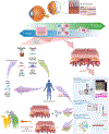
Age related macular degeneration and other retinal degenerative disorders are characterized by disruption of the outer blood retinal barrier (oBRB) with subsequent ischemia, neovascularization, and atrophy. Despite the treatment advances, there remains no curative therapy, and no treatment targeted at regenerating native-like tissue for patients with late stages of the disease. Here we present advances in tissue engineering, focusing on bioprinting methods of generating tissue allowing for safe and reliable production of oBRB as well as tissue reprogramming with induced pluripotent stem cells for transplantation. We compare these approaches to organ-on-a-chip models for studying the dynamic nature of physiologic conditions. Highlighted within this review are studies that employ good manufacturing practices and use clinical grade methods that minimize potential risk to patients. Lastly, we illustrate recent clinical applications demonstrating both safety and efficacy for direct patient use. These advances provide an avenue for drug discovery and ultimately transplantation.
Figures
References
-
- Joussen AM, et al. , Autologous translocation of the choroid and retinal pigment epithelium in patients with geographic atrophy. Ophthalmology, 2007. 114(3): p. 551–60. - PubMed
-
- Schwartz SD, et al. , Subretinal Transplantation of Embryonic Stem Cell-Derived Retinal Pigment Epithelium for the Treatment of Macular Degeneration: An Assessment at 4 Years. Invest Ophthalmol Vis Sci, 2016. 57(5): p. ORSFc1–9. - PubMed
-
-
Liu Z, et al. , Surgical Transplantation of Human RPE Stem Cell-Derived RPE Monolayers into Non-Human Primates with Immunosuppression. Stem Cell Reports, 2021. 16(2): p. 237–251.
*Using RPE-stem cells (RPESCs) as a subpopulation of PRE dissected from the native tissue, the author demonstrated 3 months of recovery progress after transplanting RPESC-derived monolayer into non-human primates. This is one of a few applications for transplanting RPE monolayer, and the first study using RPESCs on a PET membrane.
-
-
-
Yang JM, et al. , Long-term effects of human induced pluripotent stem cell-derived retinal cell transplantation in Pde6b knockout rats. Exp Mol Med, 2021. 53(4): p. 631–642.
*Using iPSC derived reintal cells with the capability of the differntiation into RPE cells and photoreceptors, the authors describe the preserved visions and the expressions of RPE and photoreceptor chracteristics during 10 months after transplanting iPSC-retinal cell suspension into the subretinal region of rat eye. This study shows the promising outcomes of long-term post-tranplanation studies using iPSCs.
-
-
- Chan WH, Hussain AA, and Marshall J, Youngs Modulus of Bruchs Membrane: Implications for AMD. Investigative Ophthalmology & Visual Science, 2007. 48(13): p. 2187–2187.
Grants and funding
LinkOut - more resources
Full Text Sources


