A Case of Neglected Posterior Fracture Dislocation of the Shoulder Treated With Greater Tuberosity Osteotomy
- PMID: 38962284
- PMCID: PMC11221983
- DOI: 10.1155/2024/6486750
A Case of Neglected Posterior Fracture Dislocation of the Shoulder Treated With Greater Tuberosity Osteotomy
Abstract
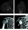
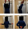
Posterior dislocation of the shoulder joint is a rare condition. It is often misdiagnosed owing to a lack of evident clinical features compared with anterior shoulder dislocation, and inappropriate radiological examination. We present a case of chronic posterior fracture dislocation treated with greater tuberosity osteotomy. A 66-year-old man was injured in a fall while carrying a drone. He was referred to our hospital following 3 months of conservative treatment at a nearby clinic, without reduction of the posterior dislocation. Physical examination revealed a prominent reduction in shoulder joint range of motion and shoulder pain. Radiological examination revealed posterior shoulder dislocation associated with greater tuberosity malunion and a small bone fracture of the posterior portion of the glenoid. Open reduction and internal fixation, including greater tuberosity osteotomy, were performed. Although subluxation of the posterior dislocation persisted postoperatively, the humeral head gradually returned to its centric shoulder joint position owing to rotator cuff force coupling. At 24-month follow-up, the patient showed excellent shoulder results.
Copyright © 2024 Masashi Koide et al.
Conflict of interest statement
The authors declare no conflicts of interest.
Figures


Similar articles
-
Irreducible Posterior Shoulder Dislocation With Concomitant Fracture of Both the Greater and Lesser Tuberosity: An Extremely Rare Shoulder Injury.Cureus. 2024 Jan 15;16(1):e52312. doi: 10.7759/cureus.52312. eCollection 2024 Jan. Cureus. 2024. PMID: 38357043 Free PMC article.
-
Neglected and locked anterior shoulder dislocation: functional outcomes and complications after open reduction and preservation of humeral head.JSES Int. 2023 Oct 5;8(1):11-20. doi: 10.1016/j.jseint.2023.09.003. eCollection 2024 Jan. JSES Int. 2023. PMID: 38312286 Free PMC article.
-
Reconstruction of a Chronic Posterior Dislocation of the Shoulder using a Limited Posterior Deltoid-splitting Approach: A Case Report.J Orthop Case Rep. 2020 Nov;10(8):88-92. doi: 10.13107/jocr.2020.v10.i08.1874. J Orthop Case Rep. 2020. PMID: 33708720 Free PMC article.
-
[Treatment strategy in first traumatic anterior dislocation of the shoulder. Plea for a multi-stage concept of preventive initial management].Unfallchirurg. 1998 May;101(5):328-41; discussion 327. doi: 10.1007/s001130050278. Unfallchirurg. 1998. PMID: 9629045 Review. German.
-
Posterior shoulder fracture-dislocation: A systematic review of the literature and current aspects of management.Chin J Traumatol. 2021 Feb;24(1):18-24. doi: 10.1016/j.cjtee.2020.09.001. Epub 2020 Sep 6. Chin J Traumatol. 2021. PMID: 32980216 Free PMC article.
References
-
- Neer C. S., II Displaced proximal humeral Fractures. The Journal of Bone & Joint Surgery . 1970;52(6):1090–1103. - PubMed
Publication types
LinkOut - more resources
Full Text Sources

