

Morphological characteristics of cerebellum, pons and thalamus in Reccurent isolated sleep paralysis - A pilot study
- PMID: 38962392
- PMCID: PMC11219576
- DOI: 10.3389/fnana.2024.1396829
Morphological characteristics of cerebellum, pons and thalamus in Reccurent isolated sleep paralysis - A pilot study
Abstract
Introduction: Recurrent isolated sleep paralysis (RISP) is a rapid eye movement sleep (REM) parasomnia, characterized by the loss of voluntary movements upon sleep onset and/or awakening with preserved consciousness. Evidence suggests microstructural changes of sleep in RISP, although the mechanism of this difference has not been clarified yet. Our research aims to identify potential morphological changes in the brain that can reflect these regulations.
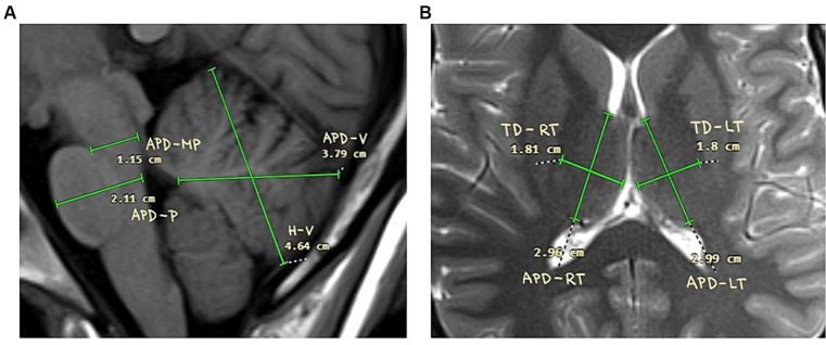
Materials and methods: We recruited 10 participants with RISP (8 women; mean age 24.7 years; SD 2.4) and 10 healthy control subjects (w/o RISP; 3 women; mean age 26.3 years; SD 3.7). They underwent video-polysomnography (vPSG) and sleep macrostructure was analyzed. After that participants underwent magnetic resonance imaging (MRI) of the brain. We focused on 2-dimensional measurements of cerebellum, pons and thalamus. Statistical analysis was done in SPSS program. After analysis for normality we performed Mann-Whitney U test to compare our data.
Results: We did not find any statistically significant difference in sleep macrostructure between patients with and w/o RISP. No evidence of other sleep disturbances was found. 2-dimensional MRI measurements revealed statistically significant increase in cerebellar vermis height (p = 0.044) and antero-posterior diameter of midbrain-pons junction (p = 0.018) in RISP compared to w/o RISP.
Discussion: Our results suggest increase in size of cerebellum and midbrain-pons junction in RISP. This enlargement could be a sign of an over-compensatory mechanism to otherwise dysfunctional regulatory pathways. Further research should be done to measure these differences in time and with closer respect to the frequency of RISP episodes.
Keywords: cerebellum; midbrain; pons; recurrent isolated sleep paralysis; sleep; thalamus.
Copyright © 2024 Miletínová, Kliková, Dostalíková and Bušková.
Conflict of interest statement
The authors declare that the research was conducted in the absence of any commercial or financial relationships that could be construed as a potential conflict of interest.
Figures


Similar articles
-
Objective rapid eye movement sleep characteristics of recurrent isolated sleep paralysis: a case-control study.Sleep. 2021 Nov 12;44(11):zsab153. doi: 10.1093/sleep/zsab153. Sleep. 2021. PMID: 34145456
-
All-night spectral and microstate EEG analysis in patients with recurrent isolated sleep paralysis.Front Neurosci. 2024 Feb 8;18:1321001. doi: 10.3389/fnins.2024.1321001. eCollection 2024. Front Neurosci. 2024. PMID: 38389790 Free PMC article.
-
Rapid eye movement sleep deprivation induces an increase in acetylcholinesterase activity in discrete rat brain regions.Braz J Med Biol Res. 2001 Jan;34(1):103-9. doi: 10.1590/s0100-879x2001000100012. Braz J Med Biol Res. 2001. PMID: 11151034
-
Recurrent Isolated Sleep Paralysis.Sleep Med Clin. 2024 Mar;19(1):101-109. doi: 10.1016/j.jsmc.2023.10.006. Epub 2023 Nov 29. Sleep Med Clin. 2024. PMID: 38368058 Review.
-
[The control of gaze (3). Neurological defects].Med Sci (Paris). 2004 Mar;20(3):357-62. doi: 10.1051/medsci/2004203357. Med Sci (Paris). 2004. PMID: 15067583 Review. French.
References
-
- American Academy of Sleep Medicine (2014). International classification of sleep disorders–third edition (Icsd-3). Darien, IL: American Academy of Sleep Medicine.
LinkOut - more resources
Full Text Sources