

Cerebral air embolism: neurologic manifestations, prognosis, and outcome
- PMID: 38962484
- PMCID: PMC11220112
- DOI: 10.3389/fneur.2024.1417006
Cerebral air embolism: neurologic manifestations, prognosis, and outcome
Abstract
Background: Cerebral air embolism (CAE) is an uncommon medical emergency with a potentially fatal course. We have retrospectively analyzed a set of patients treated with CAE at our comprehensive stroke center and a hyperbaric medicine center. An overview of the pathophysiology, causes, diagnosis, and treatment of CAE is provided.
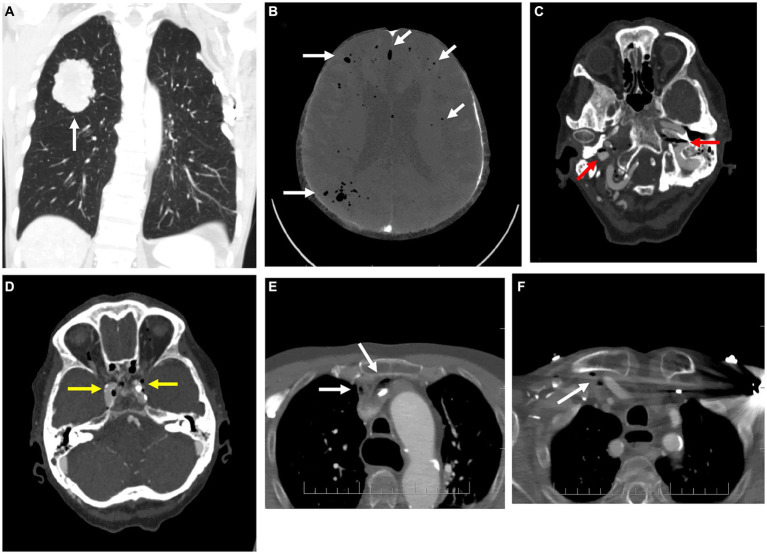
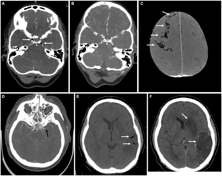
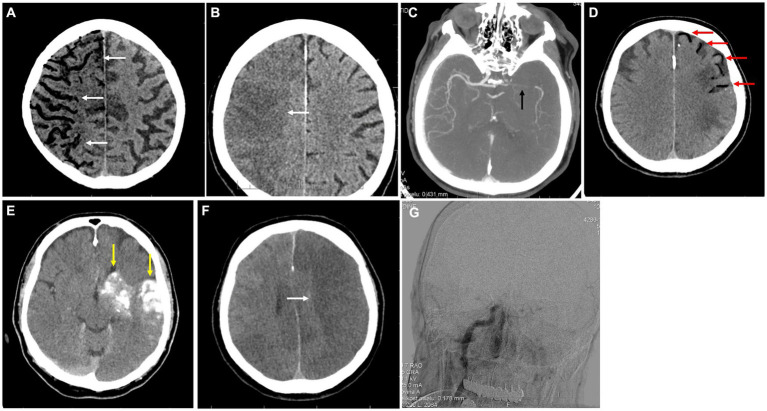
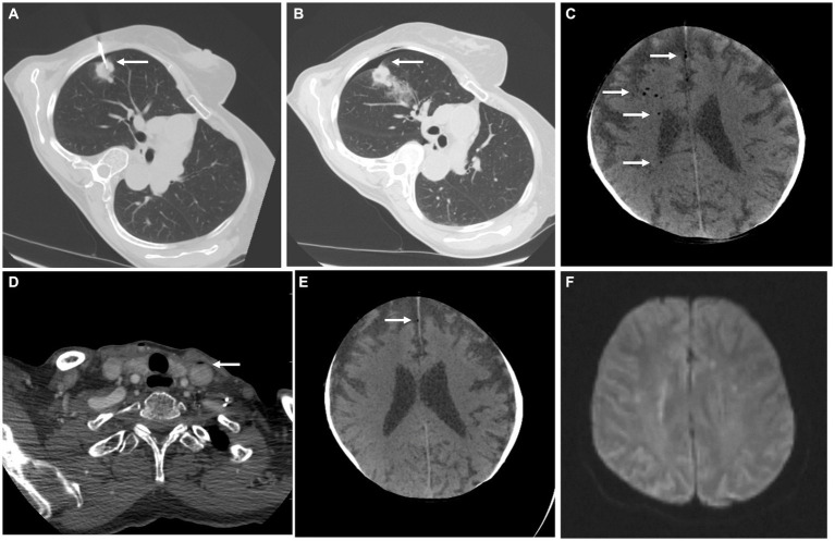
Results: We retrospectively identified 11 patients with cerebral venous and arterial air emboli that highlight the diversity in etiologies, manifestations, and disease courses encountered clinically. Acute-onset stroke syndrome and a progressive impairment of consciousness were the two most common presentations in four patients each (36%). Two patients (18%) suffered from an acute-onset coma, and one (9%) was asymptomatic. Four patients (36%) were treated with hyperbaric oxygen therapy (HBTO), high-flow oxygen therapy without HBOT was started in two patients (18%), two patients (18%) were in critical care at the time of diagnosis and three (27%) received no additional treatment. CAE was fatal in five cases (46%), caused severe disability in two (18%), mild disability in three (27%), and a single patient had no lasting deficit (9%).
Conclusion: Cerebral air embolism is a dangerous condition that necessitates high clinical vigilance. Due to its diverse presentation, the diagnosis can be missed or delayed in critically ill patients and result in long-lasting or fatal neurological complications. Preventative measures and a proper diagnostic and treatment approach reduce CAE's incidence and impact.
Keywords: air embolism; cerebral embolism; cerebral stroke; hyperbaric oxygen therapy; neurological emergency.
Copyright © 2024 Červeňák, Všianský, Cviková, Brichta, Vinklárek, Štefela, Haršány, Hájek, Herzig, Kouřil, Bárková, Filip, Aulický and Weiss.
Conflict of interest statement
The authors declare that the research was conducted in the absence of any commercial or financial relationships that could be construed as a potential conflict of interest.
Figures
Similar articles
-
Clinical Characteristics, Imaging Findings, and Outcomes of Cerebral Air Embolism.Neurocrit Care. 2023 Feb;38(1):158-164. doi: 10.1007/s12028-022-01664-3. Epub 2023 Jan 10. Neurocrit Care. 2023. PMID: 36627433 Review.
-
Cerebral air embolism following a hemodialysis session successfully treated with hyperbaric oxygen: a case report.Ther Adv Neurol Disord. 2024 Oct 9;17:17562864241287457. doi: 10.1177/17562864241287457. eCollection 2024. Ther Adv Neurol Disord. 2024. PMID: 39399101 Free PMC article.
-
Subclinical to catastrophic: a range of outcomes in cerebral air embolism.Emerg Radiol. 2023 Dec;30(6):823-827. doi: 10.1007/s10140-023-02181-2. Epub 2023 Nov 13. Emerg Radiol. 2023. PMID: 37953444
-
Iatrogenic cerebral gas embolism: analysis of the presentation, management and outcomes of patients referred to The Alfred Hospital Hyperbaric Unit.Diving Hyperb Med. 2016 Mar;46(1):15-21. Diving Hyperb Med. 2016. PMID: 27044457
-
Cerebral Air Embolism after Esophagogastroduodenoscopy: Insight on Pathophysiology, Epidemiology, Prevention and Treatment.J Stroke Cerebrovasc Dis. 2019 Dec;28(12):104403. doi: 10.1016/j.jstrokecerebrovasdis.2019.104403. Epub 2019 Sep 26. J Stroke Cerebrovasc Dis. 2019. PMID: 31563566
Cited by
-
Symptomatic air embolism after computed tomography-guided four-hook needle localization of a pulmonary nodule: a case description.Quant Imaging Med Surg. 2024 Dec 5;14(12):9750-9755. doi: 10.21037/qims-24-949. Epub 2024 Nov 18. Quant Imaging Med Surg. 2024. PMID: 39698648 Free PMC article. No abstract available.
-
Point-of-Care Ultrasonography Detects Vanishing Air Embolism Following Central Venous Catheter Removal in a Patient With Chest Tube Drainage: A Case Report.Clin Case Rep. 2025 Jun 30;13(7):e70578. doi: 10.1002/ccr3.70578. eCollection 2025 Jul. Clin Case Rep. 2025. PMID: 40589666 Free PMC article.
-
Cerebral and Portal Venous Air Embolism: A Complication of PICC Line Placement.Case Rep Radiol. 2025 Jul 18;2025:5236025. doi: 10.1155/crra/5236025. eCollection 2025. Case Rep Radiol. 2025. PMID: 40718641 Free PMC article.
References
LinkOut - more resources
Full Text Sources