

Diagnostic Algorithm for Intracranial Lesions in the Emergency Department: Effectiveness of the Relative Brain Volume and Hounsfield Unit Value Measured by Perfusion Tomography
- PMID: 38962639
- PMCID: PMC11221499
- DOI: 10.7759/cureus.61591
Diagnostic Algorithm for Intracranial Lesions in the Emergency Department: Effectiveness of the Relative Brain Volume and Hounsfield Unit Value Measured by Perfusion Tomography
Abstract
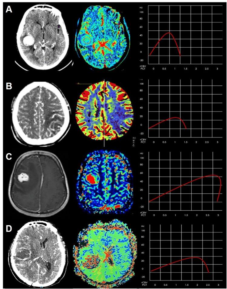
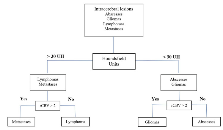
Background Early treatment of intracranial lesions in the emergency department is crucial, but it can be challenging to differentiate between them. This differentiation is essential because the treatment of each type of lesion is different. Cerebral computed tomography perfusion (CTP) imaging can help visualize the vascularity of brain lesions and provide absolute quantification of physiological parameters. Compared to magnetic resonance imaging, CTP has several advantages, such as simplicity, wide availability, and reproducibility. Purpose This study aimed to assess the effectiveness of Hounsfield units (HU) in measuring the density of hypercellular lesions and the ability of CTP to quantify hemodynamics in distinguishing intracranial space-occupying lesions. Methods A retrospective study was conducted from March 2016 to March 2022. All patients underwent CTP and CT scans, and relative cerebral blood volume (rCBV) and HU were obtained for intracranial lesions. Results We included a total of 244 patients in our study. This group consisted of 87 (35.7%) individuals with glioblastomas (GBs), 48 (19.7%) with primary central nervous system lymphoma (PCNSL), 45 (18.4%) with metastases (METs), and 64 (26.2) with abscesses. Our study showed that the HUs for METs were higher than those for GB (S 57.4% and E 88.5%). In addition, rCBV values for PCNSL and abscesses were lower than those for GB and METs. The HU in PCNSL was higher than those in abscesses (S 94.1% and E 96.6%). Conclusion PCT parameters provide valuable information for diagnosing brain lesions. A comprehensive assessment improves accuracy. Combining rCBV and HU enhances diagnostic accuracy, making it a valuable tool for distinguishing between lesions. PCT's widespread availability allows for the use of both anatomical and functional information with high spatial resolution for diagnosing and managing brain tumor patients.
Keywords: abscesses; ct perfusion; glioblastoma; intracranial brain tumors; lymphoma; metastasis; perfusion parameters.
Copyright © 2024, Alvaro-Heredia et al.
Conflict of interest statement
Human subjects: Consent was obtained or waived by all participants in this study. National Institute of Neurology and Neurosurgery issued approval 43-22. Animal subjects: All authors have confirmed that this study did not involve animal subjects or tissue. Conflicts of interest: In compliance with the ICMJE uniform disclosure form, all authors declare the following: Payment/services info: All authors have declared that no financial support was received from any organization for the submitted work. Financial relationships: All authors have declared that they have no financial relationships at present or within the previous three years with any organizations that might have an interest in the submitted work. Other relationships: All authors have declared that there are no other relationships or activities that could appear to have influenced the submitted work.
Figures
Similar articles
-
Role of dynamic CT perfusion study in evaluating various intracranial space-occupying lesions.Indian J Radiol Imaging. 2015 Apr-Jun;25(2):162-6. doi: 10.4103/0971-3026.155866. Indian J Radiol Imaging. 2015. PMID: 25969639 Free PMC article.
-
Differentiation between glioblastoma and primary CNS lymphoma: application of DCE-MRI parameters based on arterial input function obtained from DSC-MRI.Eur Radiol. 2021 Dec;31(12):9098-9109. doi: 10.1007/s00330-021-08044-z. Epub 2021 May 18. Eur Radiol. 2021. PMID: 34003350
-
Improved performance of non-preloaded and high flip-angle dynamic susceptibility contrast perfusion-weighted imaging sequences in the presurgical differentiation of brain lymphoma and glioblastoma.Eur Radiol. 2023 Dec;33(12):8800-8808. doi: 10.1007/s00330-023-09917-1. Epub 2023 Jul 13. Eur Radiol. 2023. PMID: 37439934
-
Perfusion Computed Tomography Parameters Are Useful for Differentiating Glioblastoma, Lymphoma, and Metastasis.World Neurosurg. 2018 Nov;119:e890-e897. doi: 10.1016/j.wneu.2018.07.291. Epub 2018 Aug 9. World Neurosurg. 2018. PMID: 30099179
-
Diagnostic accuracy of flat-panel computed tomography in assessing cerebral perfusion in comparison with perfusion computed tomography and perfusion magnetic resonance: a systematic review.Neuroradiology. 2019 Dec;61(12):1457-1468. doi: 10.1007/s00234-019-02285-y. Epub 2019 Sep 16. Neuroradiology. 2019. PMID: 31523757 Free PMC article.
References
-
- Advanced MR imaging techniques in the diagnosis of intraaxial brain tumors in adults. Al-Okaili RN, Krejza J, Wang S, Woo JH, Melhem ER. Radiographics. 2006;26 Suppl 1:0–89. - PubMed
-
- Contrast nephrotoxicity. Barrett BJ. J Am Soc Nephrol. 1994;5:125–137. - PubMed
-
- Prediction of malignancy grading using computed tomography perfusion imaging in nonenhancing supratentorial gliomas. Beppu T, Sasaki M, Kudo K, et al. J Neurooncol. 2011;103:619–627. - PubMed
-
- Diagnostic imaging for the emergency physician. Broder J. https://books.google.com.ph/books?hl=en&lr=&id=q3_EthzU3dcC&oi=fnd&pg=PP... Elsevier Health Sciences. 2011
-
- A quantitative theory of the Hounsfield unit and its application to dual energy scanning. Brooks RA. J Comput Assist Tomogr. 1977;1:487–493. - PubMed
LinkOut - more resources
Full Text Sources