. 2024 Jun 8:40:148-167.
doi: 10.1016/j.bioactmat.2024.05.040. eCollection 2024 Oct.

Initial therapeutic evidence of a borosilicate bioactive glass (BSG) and Fe3O4 magnetic nanoparticle scaffold on implant-associated Staphylococcal aureus bone infection

Affiliations

Initial therapeutic evidence of a borosilicate bioactive glass (BSG) and Fe3O4 magnetic nanoparticle scaffold on implant-associated Staphylococcal aureus bone infection

Ying Jin et al. Bioact Mater. .

Abstract

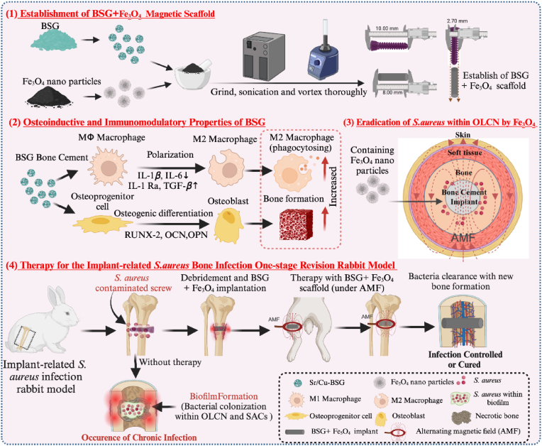
Implant-associated Staphylococcus aureus (S. aureus) osteomyelitis is a severe challenge in orthopedics. While antibiotic-loaded bone cement is a standardized therapeutic approach for S. aureus osteomyelitis, it falls short in eradicating Staphylococcus abscess communities (SACs) and bacteria within osteocyte-lacuna canalicular network (OLCN) and repairing bone defects. To address limitations, we developed a borosilicate bioactive glass (BSG) combined with ferroferric oxide (Fe3O4) magnetic scaffold to enhance antibacterial efficacy and bone repair capabilities. We conducted comprehensive assessments of the osteoinductive, immunomodulatory, antibacterial properties, and thermal response of this scaffold, with or without an alternating magnetic field (AMF). Utilizing a well-established implant-related S. aureus tibial infection rabbit model, we evaluated its antibacterial performance in vivo. RNA transcriptome sequencing demonstrated that BSG + 5%Fe3O4 enhanced the immune response to bacteria and promoted osteogenic differentiation and mineralization of MSCs. Notably, BSG + 5%Fe3O4 upregulated gene expression of NOD-like receptor and TNF pathway in MSCs, alongside increased the expression of osteogenic factors (RUNX2, ALP and OCN) in vitro. Flow cytometry on macrophage exhibited a polarization effect towards M2, accompanied by upregulation of anti-inflammatory genes (TGF-β1 and IL-1Ra) and downregulation of pro-inflammatory genes (IL-6 and IL-1β) among macrophages. In vivo CT imaging revealed the absence of osteolysis and periosteal response in rabbits treated with BSG + 5%Fe3O4 + AMF at 42 days. Histological analysis indicated complete controls of SACs and bacteria within OLCN by day 42, along with new bone formation, signifying effective control of S. aureus osteomyelitis. Further investigations will focus on the in vivo biosafety and biological mechanism of this scaffold within infectious microenvironment.

Keywords: Biofilm; Borosilicate bioactive glass; Magnetic nanoparticles; Osteomyelitis; Staphylococcus aureus.


Conflict of interest statement

Haobo Pan is an editorial board member for Biomedical Technology and was not involved in the editorial review or the decision to publish this article. All authors declare that they have no known competing financial interests or personal relationships that could have appeared to influence the work reported in this paper.

Figures

Image 1
Graphical abstract
Fig. 1
Fig. 1
A Schematic diagram of a model for the treatment of S. aureus related bone infection model with BSG + Fe3O4 magnetic scaffold. (1) To match the size of titanlnium alloy screws used for the establishment of S. aureus bone infection model, the size of BSG + Fe3O4 scaffold was prepared under the premise of full mixing. (2) BSG can promote the autoimmune regulation of M2 macrophage polarization and the bone-inducing properties of promoting osteogenesis. (3) Fe3O4 magnetic particles can eradicate S. aureus within OLCN when exposed to an AMF. (4) Establishment of an implant-related S. aureus bone infection model of rabbit and treatment process. In the untreated group, the infected screws were removed after challenged with S. aureus contaminated screw by day 7, only debridement was performed without scaffolds, as bacteria invasion and biofilm formation, the acute bone infections turned into chronic bone infections eventually. While, the treatment group suffered from debridement and implanted with BSG + Fe3O4 scaffolds at the original infection site, then the treatment last until 42 days under the AMF.
Fig. 2
Fig. 2
Morphological characterization and biomechanical properties of BSG + Fe3O4magnetic scaffold. A) BSG + Fe3O4 scaffolds were compounded to match the size of the titanium screws use in the in vivo model. B) SEM images of the microarchitecture of BSG + Fe3O4 magnetic scaffolds, at 5000X and 20000X magnifications. C-D) FTIR spectra and XRD patterns of 3B3Sr, BSG, Fe3O4, and BSG with 4%, 5%, or 6%Fe3O4. E) Compressive strength (MPa) of each scaffold group (n = 3), presented as mean ± SD. F) Release of ions including BO33−, SiO22−, Ca2+, Sr2+ and Fe3+ from scaffolds incubated in PBS were quantified and plotted across multiple time points. Each data point represents the total released over two days. G) pH of PBS solution containing BSG + Fe3O4 scaffolds, measured to 28 days. H–I) FTIR spectra and XRD patterns of BSG + Fe3O4 scaffolds after immersion in PBS for 7, 14 and 28 days.
Fig. 3
Fig. 3
Cytotoxicity, proliferation, viability, and cytoskeleton of MSCs when cultured with a 128-fold dilution of BSG + Fe3O4magnetic scaffold extracts. A-I) CCK8 results were obtained after culturing cells in extracts of different concentrations of BSG + Fe3O4 for 1 day. Cells exhibited lower survival ratio until the extract was diluted 128 times. J-M) MSCs proliferation was assessed using CCK8 after being culturing in 32-, 64-, 128-, and 256-fold dilutions of BSG + 5%Fe3O4 extract for 1 day. The 128-fold dilution of BSG + 5%Fe3O4 extract had no impact on cell proliferation. N) EdU cell proliferation staining, O) live/dead staining, and P) cytoskeleton staining of MSCs cultured with 128-fold dilutions of the scaffold extracts for 2 days. These results indicated that the extracts of each polymer had no significant effects on cell viability, skeleton and proliferation when diluted 128-fold. Results were shown as Mean ± SD; One-way ANOVA; n = 6; * indicates statistical difference (P < 0.05), ** means P < 0.01, *** means P < 0.001.
Fig. 4
Fig. 4
Osteogenic effects of BSG extracts at 128-fold dilution on MSCs and macrophages in culture. A) ALP staining of MSCs (C3H10T1/2) cultured with scaffold extracts DMEM diluted 32-, 64-, 128-, and 256-fold in OIM for 7 days, and B) alizarin red S staining after 21 days of culture, C-D) with corresponding quantitative analysis (n = 3). Specifically, the 128-fold dilution of BSG + 5%Fe3O4 extract exerted the most substantial impact on ALP activity and late matrix mineralization. E-F) The expression of osteogenesis related mRNA (RUNX2, ALP, OCN) (n = 4) and G-H) proteins (RUNX2 and OCN) were assessed via RT-qPCR and Western blot, with quantification of the blot results displayed in I and J (n = 3). The results suggested that the 128-fold dilution of BSG + 5%Fe3O4 scaffold extracts exhibited the most significant upregulation of osteogenic genes. Results were shown as Mean ± SD; Two-way ANOVA; * indicates statistical difference (P < 0.05), ** means P < 0.01, *** means P < 0.001.
Fig. 5
Fig. 5
BSG+5%Fe3O4enhances immune response to bacteria, osteogenic differentiation, and chemokine receptor gene expression in MSCs. A-B) MSCs (C3H10T1/2) were cultured in 128-fold dilution of BSG + 5%Fe3O4 extract for 7 days, and RNA transcriptome sequencing was performed on three separate samples, along with three unexposed MSCs as negative controls. Genes with significant changes in transcript numbers and high p-values were selected using a thresholding method. C-D) GO enrichment bubble map revealed enrichment of genes associated with the immune response of MSCs to viruses and bacteria (red boxes), osteoblast differentiation, extracellular matrix secretion, bone tissue mineralization (blue boxes), as well as chemokine receptor related pathways (such as CCR2) involved in mediating inflammation response (green boxes). E-F) The molecular pathways of differentially expressed genes were further investigated using KEGG enrichment, these findings revealed that BSG + 5%Fe3O4magnetic scaffold extract significantly upregulated the expression of genes associated with the NOD-like receptor (NLR) and TNF signaling pathways in MSCs (red boxes). This confirmation underscores the potential of the magnetic scaffold to modulate bone immunity and regulate bone homeostasis.
Fig. 6
Fig. 6
BSG scaffolds regulate inflammation in macrophages in vitro. A-B) RAW 264.7 cells were cultured with 128-fold dilution of scaffold extract for 3 days, followed by flow cytometry for CD80 and CD206, the BSG+5%Fe3O4group showed the lowest expression (70.97 %) of CD80 and the highest expression (58.65 %) of CD206. C-D) The expression of anti-inflammatory (TGF-β1 and IL-1Ra) and pro-inflammatory (IL-6 and IL-1β) gene in macrophages were assessed by RT-qPCR. Compared to the control group, RAW 264.7 macrophages cultured with the128-fold dilution of BSG+5%Fe3O4had the significantly promotion effect on expression of TGF-β1and IL-1Ra, and the most obvious inhibition effect on IL-6 and IL-1β. Results were shown as Mean ± SD; Two-way ANOVA; * indicates statistical difference (P < 0.05), ** means P < 0.01, *** means P < 0.001.
Fig. 7
Fig. 7
Magnetic-thermal-induced thermal efficiency and antibacterial properties of BSG + Fe3O4in vitro. A, D) Thermal images of each scaffold were captured over time-course using a far-infrared thermometer. B) When subjected to AMF for 180 s, the temperatures of BSG + 4%Fe3O4, BSG + 5%Fe3O4, and BSG + 6%Fe3O4 reached 46.1 ± 0.7 °C, 54.7 ± 1.1 °C, and 65.3 ± 1.2 °C, respectively. C) White light image of the tissue sample under AMF. E) Linear regression analysis of temperature change from the center of the interface of BSG + 5%Fe3O4 magnetic scaffold radially outward into the air when exposed to AMF for 180 s. F) In the fresh rabbit tibia, linear regression is drawn according to the attenuation of BSG + 5%Fe3O4 interface center temperature with distance. when exposed to AMF for 180 s. G) According to the linear regression of temperature attenuation from the center of BSG + 5%Fe3O4 interface, the effective sterilization temperature of 45 °C was achieved at an average radial distance of 0.1155 mm. H–I) Antibacterial properties of each scaffold in vitro and statistical analysis of diameter of bacteriostatic zone. inhibition zone of BSG + 5%Fe3O4 + AMF exhibited the largest zone of inhibition against S. aureus on TSA plate. Results are shown as Mean± SD; One-way ANOVA; *indicates statistical difference (P<0.05), *** means P<0.001.
Fig. 8
Fig. 8
Representative gross images and CT images of infected tibiae with untreated group, BSG+5%Fe3O4or BSG+5%Fe3O4+AMF as far as 42 days. A) The design of 1-stage revision bone infection model. B) Gross images of the superior tibial metaphysis on days 7. All specimens exhibited well-maintained scaffold positioning without displacement or prolapse, while soft tissue swelling and pus moss formation were observed in all groups. C) Gross images of the superior tibial metaphysis on days 42. By day 42, pus (yellow arrow) and soft tissue infection (orange arrow) were present at the screw tract in the untreated and BSG + 5%Fe3O4 groups, and with a noticeable reduction in the diameter of the screw tract compared to 7 days. In contrast, pus was absent in the BSG + 5%Fe3O4 + AMF group, and the screw tract had nearly disappeared (red arrow) by day 42. D) CT images of rabbit tibiae on days 7, 14, 28, and 42 post revision. On days 28 and 42, the untreated group displayed evident periosteal response (green arrow) and osteolytic destruction or formation of dead bone (orange arrow) in the medullary cavity. Periosteal response was also observed in cortical bone on days 7, 14 and 28 in the BSG + 5%Fe3O4 group (green arrows), while a noticeable periosteal reaction was absent in the BSG + 5%Fe3O4 + AMF group. On day 42, the BSG + 5%Fe3O4 and BSG + 5%Fe3O4 + AMF groups exhibited cortical bone healing (red arrows) at the implant site.
Fig. 9
Fig. 9
Histological evidence of the antibacterial effects of BSG+5%Fe3O4in vivo. A) H&E and Gram staining after infection surgery for 7 days, histological evidence of bone infection could be observed in both the untreated and the BSG + 5%Fe3O4 groups (red arrows). SACs were observed around the scaffolds of BSG + 5%Fe3O4 group, and undegraded iron was also seen in BSG + 5%Fe3O4 group (black arrows). C-E) H&E and Gram staining at 28 days after infection. On day 28, the infection sites had progressed to diffuse inflammatory lesions in both the untreated and BSG + 5%Fe3O4 groups (green arrow), and osteolysis occurred in the two groups (blue arrow). Substantial SACs formation was observed in the pulp cavity of the untreated group (red arrows). In the BSG + 5%Fe3O4 group, S. aureus was detected around and within the scaffold (red arrow). In contrast, the scaffold and pulp cavity of the BSG + 5%Fe3O4 + AMF group exhibited significantly reduced signs of bone infection and early bone formation. F-G) At 42 days post-treatment, H&E and Gram staining revealed partial degradation of the BSG + 5%Fe3O4 scaffold and a reduction in the diffuse medullary lesions observed on day 28 (green arrows). However, some bacteria persisted within the scaffolds (red arrows). Remarkably, the BSG + 5%Fe3O4 + AMF group exhibited significant new bone deposition (yellow arrows) and pronounced scaffold degradation.
Fig. 9
Fig. 9
Histological evidence of the antibacterial effects of BSG+5%Fe3O4in vivo. A) H&E and Gram staining after infection surgery for 7 days, histological evidence of bone infection could be observed in both the untreated and the BSG + 5%Fe3O4 groups (red arrows). SACs were observed around the scaffolds of BSG + 5%Fe3O4 group, and undegraded iron was also seen in BSG + 5%Fe3O4 group (black arrows). C-E) H&E and Gram staining at 28 days after infection. On day 28, the infection sites had progressed to diffuse inflammatory lesions in both the untreated and BSG + 5%Fe3O4 groups (green arrow), and osteolysis occurred in the two groups (blue arrow). Substantial SACs formation was observed in the pulp cavity of the untreated group (red arrows). In the BSG + 5%Fe3O4 group, S. aureus was detected around and within the scaffold (red arrow). In contrast, the scaffold and pulp cavity of the BSG + 5%Fe3O4 + AMF group exhibited significantly reduced signs of bone infection and early bone formation. F-G) At 42 days post-treatment, H&E and Gram staining revealed partial degradation of the BSG + 5%Fe3O4 scaffold and a reduction in the diffuse medullary lesions observed on day 28 (green arrows). However, some bacteria persisted within the scaffolds (red arrows). Remarkably, the BSG + 5%Fe3O4 + AMF group exhibited significant new bone deposition (yellow arrows) and pronounced scaffold degradation.

References

    1. Schwarz E.M., Parvizi J., Gehrke T., et al. International consensus meeting on musculoskeletal infection: research priorities from the general assembly questions. J. Orthop. Res. : Off. Publ. Orthop. Res. Soc. 2018;37(5) doi: 10.1002/jor.24293. 2019. - DOI - PubMed
    1. Ren Y., Liu L., Sun D., et al. Epidemiological updates of post-traumatic related limb osteomyelitis in China: a 10 years multicentre cohort study. Int. J. Surg. 2023;109(9):2721–2731. doi: 10.1097/JS9.0000000000000502. - DOI - PMC - PubMed
    1. Kong P., Ren Y., Yang J., et al. Relapsed boyhood tibia polymicrobial osteomyelitis linked to dermatophytosis: a case report. BMC Surg. 2022;22(1):156. doi: 10.1186/s12893-022-01600-4. - DOI - PMC - PubMed
    1. Zoller S.D., Hegde V., Burke Z.D.C., et al. Evading the host response: Staphylococcus "hiding" in cortical bone canalicular system causes increased bacterial burden. Bone Res. 2020;8(1):43. doi: 10.1038/s41413-020-00118-w. - DOI - PMC - PubMed
    1. Ren Y., Weeks J., Xue T., et al. Evidence of bisphosphonate-conjugated sitafloxacin eradication of established methicillin-resistant S. aureus infection with osseointegration in murine models of implant-associated osteomyelitis. Bone Res. 2023;11(1):51. doi: 10.1038/s41413-023-00287-4. - DOI - PMC - PubMed

LinkOut - more resources