. 2025 Feb;60(2):177-188.
doi: 10.1111/jre.13317. Epub 2024 Jul 4.

Cytokine responses of CD4+ T cells and NKT cells to periodontitis-associated bacteria in individuals with or without periodontitis

Affiliations

Cytokine responses of CD4+ T cells and NKT cells to periodontitis-associated bacteria in individuals with or without periodontitis

Anne Katrine Danielsen et al. J Periodontal Res. 2025 Feb.

Abstract

Aim: Periodontitis is an inflammatory disease driven by opportunistic bacteria including Porphyromonas gingivalis and Fusobacterium nucleatum, where T-cell and NKT-cell responses to these bacteria in patients with periodontitis grade B or C are not fully elucidated. The objective is to determine if exaggerated proinflammatory Th-cell responses to periodontitis-associated bacteria, but not commensal bacteria, is a characteristic of increased periodontitis grade.

Methods: Mononuclear cells from patients with periodontitis grade C (n = 26) or grade B (n = 33) and healthy controls (HCs; n = 26) were stimulated with P. gingivalis, F. nucleatum or the commensal bacteria, Staphylococcus epidermidis and Cutibacterium acnes. Cytokine production by different T-cell populations and FOXP3-expression by regulatory T cells were assessed by flow cytometry.

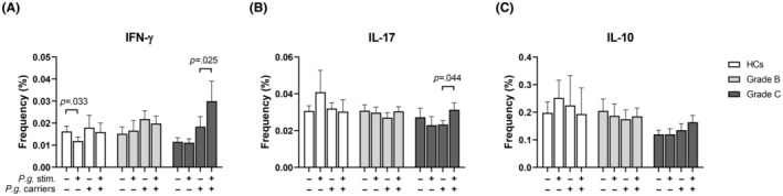
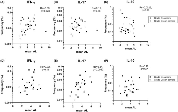
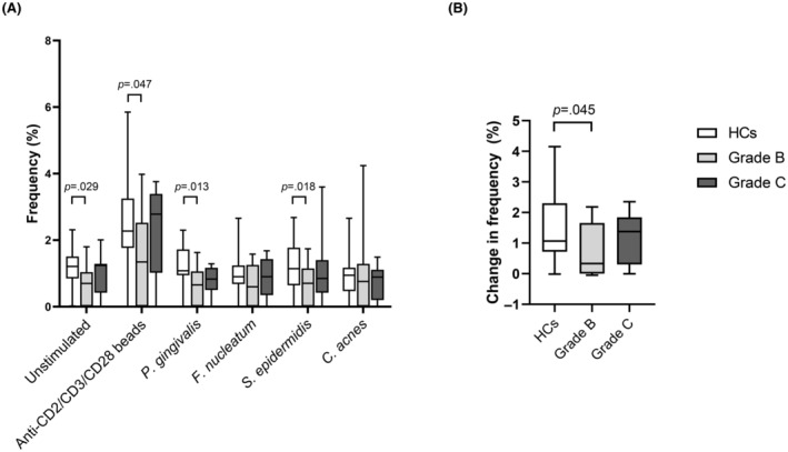
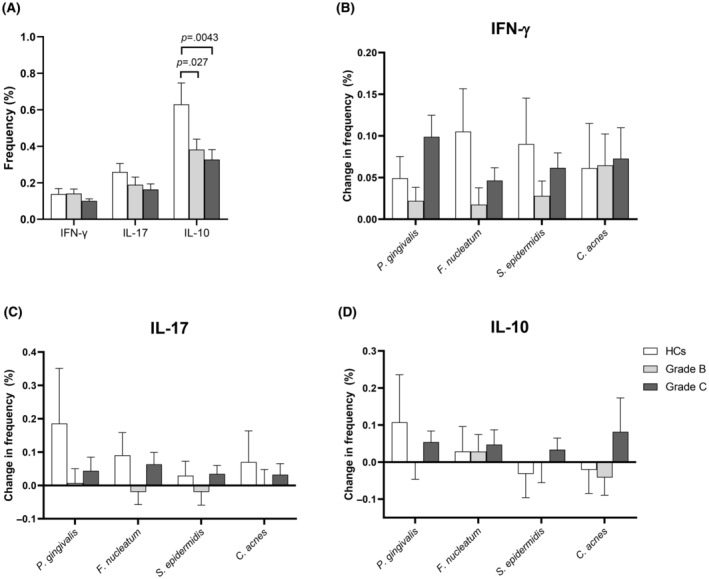
Results: Compared to HCs, grade C patients had decreased frequencies of interleukin (IL)-10-producing CD4+ T cells before stimulation (p = .02) and increased frequencies of IFN-y-producing CD4+ T cells after stimulation with P. gingivalis (p = .0019). Grade B patients had decreased frequencies of FOXP3+ CD4+ T cells before (p = .030) before and after stimulation with anti-CD2/anti-CD3/anti-CD28-loaded beads (p = .047), P. gingivalis (p = .013) and S. epidermidis (p = .018). Clinical attachment loss correlated with the frequencies of IFN-y-producing Th1 cells in P. gingivalis- and F. nucleatum-stimulated cultures in grade B patients (p = .023 and p = .048, respectively) and with the frequencies of Th17 cells in P. gingivalis-stimulated cultures (p = .0062) in grade C patients. Patients with periodontitis grade C or grade B showed lower frequencies of IL-10-producing NKT cells than HCs in unstimulated cultures (p = .0043 and p = .027 respectively).

Conclusions: Both periodontitis groups showed decreased frequencies of immunoregulatory T-cell and NKT cell subsets at baseline. Clinical attachment loss correlated with P. gingivalis-induced Th17-responses in grade C patients and with Th1-responses in grade B patients when cells were stimulated with P. gingivalis, supporting that dysregulated pro-inflammatory T-cell responses to periodontitis-associated bacteria contribute to the pathogenesis of periodontitis.

Keywords: Fusobacterium Nucleatum; Porphyromonas gingivalis; NKT cells; T‐helper cells; cytokines; periodontitis.


Conflict of interest statement

All authors declare no financial conflicts of interest.

Figures

FIGURE 1
FIGURE 1
Induction of cytokine‐producing CD4+ T cells by periodontitis‐associated bacteria and commensal bacteria. Peripheral blood mononuclear cells from 26 periodontally healthy controls (HCs; white bars), 33 periodontitis grade B patients (light grey bars) and 26 periodontitis grade C patients (dark grey bars) were stimulated with P. gingivalis, F. nucleatum, S. epidermidis, C. acnes or left unstimulated for 19 h and assessed for intracellular cytokines by flow cytometry. In addition, T cells from 16 HCs, 18 grade B patients, and 16 grade C patients were stimulated with anti‐CD2/CD3/CD28 beads. (A) Gates defining IFN‐γ‐ and IL‐17‐producing and IL‐10‐producing CD4+ T cells from a representative patient with periodontitis grade C in unstimulated cultures and after stimulation with P. gingivalis. (B) Frequencies of CD4+ T cells producing IFN‐γ, IL‐17A, or IL‐10 in unstimulated samples.1 (C–E) Frequencies of CD4+ T cells producing IFN‐γ, IL‐17A, or IL‐10 in peripheral blood mononuclear cell cultures stimulated with bacteria. The corresponding frequencies observed in unstimulated cultures have been subtracted. Statistical analysis was performed on log‐transformed data. Mean baseline frequencies of cytokine‐producing cells were compared between groups with repeated measures of one‐way ANOVA (B). Comparisons between groups after stimulation with bacteria were carried out with mixed‐effects analysis with uncorrected Fishers LSD test (C–E). Bars and error bars represent means and SEMs. 1Data on Th17 cells from two HCs, five periodontitis grade B patients and two grade C patients and on S. epidermidis‐stimulated cells from one grade B and two grade C patients were missing.
FIGURE 2
FIGURE 2
Frequencies of cytokine‐producing CD4+ T‐cell in response to P. gingivalis according to carriage of the bacterium. Peripheral blood mononuclear cells from healthy controls (HCs; 3 carriers of P. gingivalis and 23 non‐carriers) (white bars), patients with periodontitis grade B (18 carriers and 15 non‐carriers) (light grey bars) and patients with periodontitis grade C (18 carriers and 8 non‐carriers) (dark grey bars) were stimulated with P. gingivalis for 19 h or left unstimulated and assessed by flow cytometry for the frequencies of CD4+ T cells producing (A) IFN‐γ, (B) IL‐17A or (C) IL‐10.1 Mixed‐effects model with uncorrected Fisher LSD test was used for statistical analysis. Bars and error bars indicate mean and SEM. 1Data on IL‐17‐producing cells were missing for two HCs, five grade B patients and two grade C patients.
FIGURE 3
FIGURE 3
Correlation between tooth attachment loss and frequencies of CD4+ T cells producing IFN‐γ, IL‐17, or IL‐10 after stimulation with P. gingivalis. (A–C) Association within the periodontitis grade B group between mean attachment loss (AL) and the frequencies of IFN‐γ‐, IL‐17‐ or IL‐10‐producing CD4+ T cells in cultures of peripheral blood mononuclear cells stimulated with P. gingivalis. (D–F) The corresponding associations within the grade C group. Closed and open symbols indicate patients with and without P. gingivalis in saliva, respectively; i.e. carriers vs. non‐carriers. Correlation coefficients were calculated using Spearman's rank correlation test.
FIGURE 4
FIGURE 4
FOXP3+ expression by CD4+ T cells. (A) Peripheral blood mononuclear cells from 16 periodontally healthy controls (HCs; white bars), 18 patients with periodontitis grade B (light grey bars), and 15 patients with periodontitis grade C (dark grey bars) were stimulated with P. gingivalis, F. nucleatum, S. epidermidis, or C. acnes or left unstimulated for 19 h and assessed for FOXP3 content. Stimulation with anti‐CD2/anti‐CD3/anti‐CD28‐loaded MACSiBeads was used as positive control. Frequencies of FOXP3 + CD4+ T cells are shown as mean and SEM. (B) Net increase in the frequency of FOXP3 + CD4+ T cells after stimulation with the beads. Kruskal–Wallis with uncorrected Dunn's test was used to compare the three groups.
FIGURE 5
FIGURE 5
Induction of cytokine‐producing NKT cell in response to periodontitis‐associated bacteria and commensal bacteria. Peripheral blood mononuclear cells from 26 periodontally healthy controls (HCs), 33 patients with periodontitis grade B, and 26 patients with periodontitis grade C were stimulated with P. gingivalis, F. nucleatum, S. epidermidis, or C. acnes or left unstimulated for 19 h at 37°C in the presence of 10% normal human serum. The intracellular content of cytokines in CD3 + CD56+ NKT cells was assessed by flow cytometry. (A) Frequencies of CD3+ CD56+ NKT cells producing IFN‐γ, IL‐17A or IL‐10 in unstimulated samples, shown as mean and SEM. Statistical analysis was performed on log‐transformed data using repeated measures one‐way ANOVA. (B–D) Frequencies of CD3+ CD56+ T cells producing IFN‐γ, IL‐17A, or IL‐10 in cultures stimulated with bacteria after subtraction of the corresponding frequencies in unstimulated cell cultures. Comparisons between groups were carried out using mixed‐effects analysis with uncorrected Fishers LSD test (B–D). 1Data from S. epidermidis‐stimulated samples from one grade B and two grade C patients were excluded due to insufficient CD3 staining, and data on IL‐17 were missing for two HCs, five grade B patients, and two grade C patients.

Similar articles

References

    1. Page RC, Schroeder HE. Pathogenesis of inflammatory periodontal disease. A summary of current work. Lab Investig. 1976;34(3):235‐249. - PubMed
    1. Papapanou PN, Sanz M, Buduneli N, et al. Periodontitis: consensus report of workgroup 2 of the 2017 world workshop on the classification of periodontal and Peri‐implant diseases and conditions. J Clin Periodontol. 2018;45(Suppl 20):S162‐S170. - PubMed
    1. Baker PJ, Dixon M, Evans RT, Dufour L, Johnson E, Roopenian DC. CD4(+) T cells and the proinflammatory cytokines gamma interferon and interleukin‐6 contribute to alveolar bone loss in mice. Infect Immun. 1999;67(6):2804‐2809. - PMC - PubMed
    1. Alvarez C, Suliman S, Almarhoumi R, et al. Regulatory T cell phenotype and anti‐osteoclastogenic function in experimental periodontitis. Sci Rep. 2020;10(1):19018. - PMC - PubMed
    1. Sato K, Suematsu A, Okamoto K, et al. Th17 functions as an osteoclastogenic helper T cell subset that links T cell activation and bone destruction. J Exp Med. 2006;203(12):2673‐2682. - PMC - PubMed

MeSH terms