. 2024 Jul 15;137(14):jcs261469.
doi: 10.1242/jcs.261469. Epub 2024 Jul 25.

The intracellular domain of Sema6A is essential for development of the zebrafish retina

Affiliations

The intracellular domain of Sema6A is essential for development of the zebrafish retina

Caroline M Dumas et al. J Cell Sci. .

Abstract

Semaphorin6A (Sema6A) is a repulsive guidance molecule that plays many roles in central nervous system, heart and bone development, as well as immune system responses and cell signaling in cancer. Loss of Sema6A or its receptor PlexinA2 in zebrafish leads to smaller eyes and improper retinal patterning. Here, we investigate a potential role for the Sema6A intracellular domain in zebrafish eye development and dissect which phenotypes rely on forward signaling and which rely on reverse signaling. We performed rescue experiments on zebrafish Sema6A morphants with either full-length Sema6A (Sema6A-FL) or Sema6A lacking its intracellular domain (Sema6A-ΔC). We identified that the intracellular domain is not required for eye size and retinal patterning, however it is required for retinal integrity, the number and end feet strength of Müller glia and protecting against retinal cell death. This novel function for the intracellular domain suggests a role for Sema6A reverse signaling in zebrafish eye development.

Keywords: Eye development; Retina; Reverse signaling; Semaphorin; Zebrafish.


Conflict of interest statement

Competing interests The authors declare no competing or financial interests.

Figures
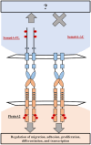
Fig. 1.
Fig. 1.
Schematic of Sema6A constructs. Schematic of (left) Sema6A full-length (FL) with both forward and reverse signaling, and (right) Sema6A-ΔC with only forward signaling. Areas shaded blue and peach represent intracellular areas.
Fig. 2.
Fig. 2.
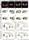
The intracellular domain of Sema6A is not required for eye field proliferation and eye size. (A–L) Transgenic embryos of (A,E,I) control (UIC, uninjected control), (B,F,J) Sema6A morphant (MO), (C,G,K) Sema6A-FL rescue and (D,H,L) Sema6A-ΔC rescue. (A–D) Immunohistochemistry for pHH3 (red) on 18 hpf rx3:GFP (gray) embryos; (E–H) lateral images of 48 hpf embryos; (I–L) Lateral images of 72 hpf embryos. Scale bars: 100 μm (A); 500 μm (E). (M) Quantification of 18 hpf eye area, n=48–67. (N) Quantification of 18 hpf pHH3-positive cells per eye field, n=18–33. (O) Quantification of 48 hpf eye diameter, n=33–43. (P) Quantification of 72 hpf eye diameter, n=30. (M–P) N=3 experiments. P-values: ns (not significant) P>0.05, *P≤0.05, **P≤0.01, ***P≤0.001, **** P≤0.0001 (Tukey's multiple comparison). All box and whiskers plots show the first and third quartiles represented by the box, with the median being the middle line. The whiskers extend from the quartile ranges to 1.5 times the interquartile range from the Q1 and Q3 boundaries. The box and whisker plots are overlaid by individual points representing one fish per point.
Fig. 3.
Fig. 3.
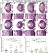
The intracellular domain of Sema6A is not required for retinal lamination but is required for retinal integrity. Transverse retinal sections stained with H&E at (A–D) 48 hpf and (E–H) 72 hpf of (A,A′,E,E′) control (UIC, uninjected control), (B,B′,F,F′) Sema6A morphant (MO), (C,C′,G,G′) Sema6A-FL rescue, and (D,D′,H,H′) Sema6A-ΔC rescue. Scale bar: 20 μm. Arrows indicate retinal lens detachment and acellular regions, and white boxes indicate the regions shown in the magnifications. (I) Quantification of acellular regions per retina and (J) area of acellular regions within the retina at 72 hpf. (I,J) N=3 experiments, n=19–22, P-values: ns (not significant) P>0.05, ***P≤0.001, ****P≤0.0001 (Tukey's multiple comparison). Box plots are presented as described in Fig. 2.
Fig. 4.
Fig. 4.
The intracellular domain of Sema6A is not required for retinal patterning. Transverse retinal sections of Sofa1 transgenic embryos at (A–D) 48 hpf and (E–H) 72 hpf of (A,E) control (UIC, uninjected control), (B,F) Sema6A morphant (MO), (C,G) Sema6A-FL rescue, and (D,H) Sema6A-ΔC rescue. Representative of N=3 experiments, n=16. Scale bar: 50 μm.
Fig. 5.
Fig. 5.
Retinal cell number is unaffected with loss of the intracellular domain of Sema6A. Quantification of retinal cell numbers in (A) whole retina, (B) the retinal ganglion cell layer (RGC), (C) the inner nuclear layer (INL), and (D) the outer nuclear layer (ONL). N=3 experiments, n=19–22. P-values: ns (not significant) P>0.05, * P≤0.05, *** P≤0.001, **** P≤0.0001 (Tukey's multiple comparison). Box plots are presented as described in Fig. 2.
Fig. 6.
Fig. 6.
The intracellular domain of Sema6A is contributes to Müller glia number and end feet strength. (A–D) Transverse retinal sections at 72 hpf gfap:GFP transgenic embryos of (A,A′) control (UIC, uninjected control), (B,B′) Sema6A morphant (MO), (C,C′) Sema6A-FL rescue, and (D,D′) Sema6A-ΔC rescue. (A′–D′) Magnification of the area highlighted by the white box in above image. Arrows indicate glial end feet. Scale bars: 20 μm (A); 10 µm (A′). (E) Quantification of GFAP-positive cells per eye, and (F) Müller glia end feet fluorescence intensity divided by nuclei fluorescence intensity. N=3 experiments, n=19–21. P-values: ns (not significant) P>0.05, *P≤0.05, ***P≤0.001, ****P≤0.0001 (Tukey's multiple comparison). Box plots are presented as described in Fig. 2.
Fig. 7.
Fig. 7.
The intracellular domain of Sema6A protects against retinal cell death. (A–D) Transverse retinal sections of immunohistochemistry labeling for cleaved-caspase-3 of (A) control, (B) Sema6A morphant, (C) Sema6A-FL rescue, and (D) Sema6A-ΔC rescue. Scale bar: 10 μm. (E) Quantification of immuno-positive puncta per retinal area. N=2 experiments, n=18–21. P-values: ns (not significant) P>0.05, ***P≤0.001, ****P≤0.0001 (Tukey's multiple comparison). Box plots are presented as described in Fig. 2.
Fig. 8.
Fig. 8.
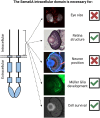
Roles of the Sema6A intracellular domain in zebrafish eye development. The intracellular domain of Sema6A is required for retinal integrity, Müller glia development and cell survival. It is not required for eye size and retinal neuronal position. Created using BioRender.com.

References

    1. Almeida, A. D., Boije, H., Chow, R. W., He, J., Tham, J., Suzuki, S. C. and Harris, W. A. (2014). Spectrum of Fates: a new approach to the study of the developing zebrafish retina. Development 141, 1971-1980. 10.1242/dev.104760 - DOI - PMC - PubMed
    1. Alto, L. T. and Terman, J. R. (2017). Semaphorins and their signaling mechanisms. Methods Mol. Biol. 1493, 1-25. 10.1007/978-1-4939-6448-2_1 - DOI - PMC - PubMed
    1. Amini, R., Rocha-Martins, M. and Norden, C. (2018). Neuronal migration and lamination in the vertebrate retina. Front. Neurosci. 11, 742. 10.3389/FNINS.2017.00742 - DOI - PMC - PubMed
    1. Andermatt, I., Wilson, N. H., Bergmann, T., Mauti, O., Gesemann, M., Sockanathan, S. and Stoeckli, E. T. (2014). Semaphorin 6B acts as a receptor in post-crossing commissural axon guidance. Development 141, 3709-3720. 10.1242/dev.112185 - DOI - PMC - PubMed
    1. Barberis, D., Casazza, A., Sordella, R., Corso, S., Artigiani, S., Settleman, J., Comoglio, P. M. and Tamagnone, L. (2005). p190 Rho-GTPase activating protein associates with plexins and it is required for semaphorin signalling. J. Cell Sci. 118, 4689-4700. 10.1242/jcs.02590 - DOI - PubMed

LinkOut - more resources