Review
. 2024 Aug;46(8):1165-1175.
doi: 10.1007/s00276-024-03432-8. Epub 2024 Jul 4.

Retroperitoneum revisited: a review of radiological literature and updated concept of retroperitoneal fascial anatomy with imaging features and correlating anatomy

Affiliations
Review

Retroperitoneum revisited: a review of radiological literature and updated concept of retroperitoneal fascial anatomy with imaging features and correlating anatomy

B Boekestijn et al. Surg Radiol Anat. 2024 Aug.

Abstract

Purpose: Spread of disease in the retroperitoneum is dictated by the complex anatomy of retroperitoneal fasciae and is still incompletely understood. Conflicting reports have led to insufficient and incorrect anatomical concepts in radiological literature.

Methods: This review will discuss previous concepts prevalent in radiological literature and their shortcomings will be highlighted. New insights from recent anatomical and embryological research, together with imaging examples, will be used to clarify patterns of disease spread in the retroperitoneum that remain unexplained by these concepts.

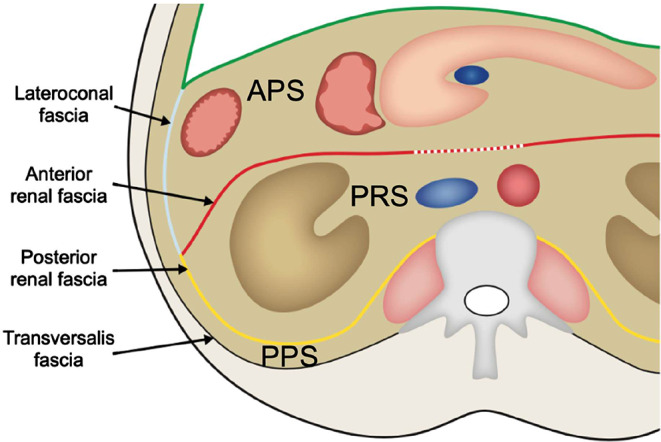
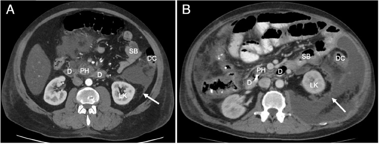
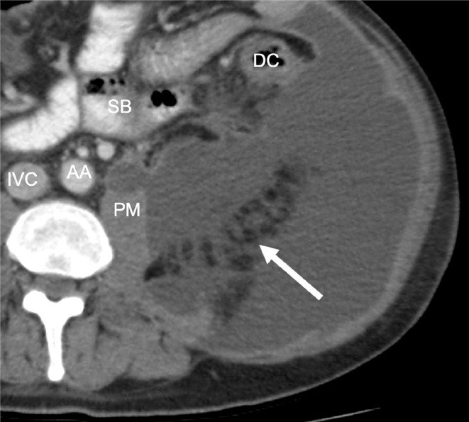
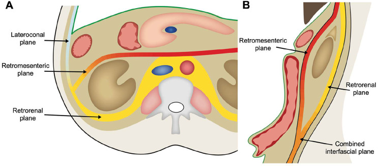
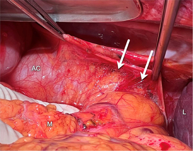
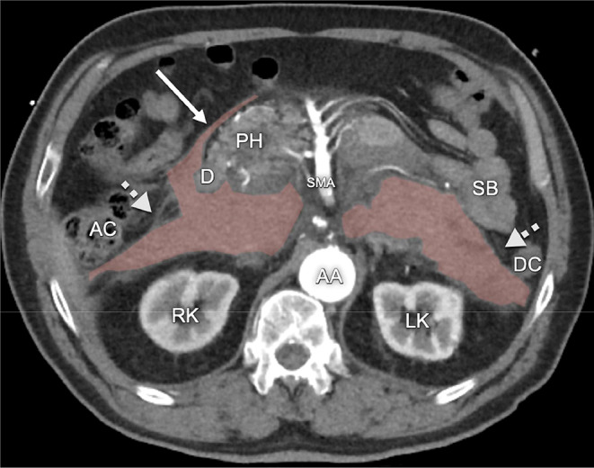
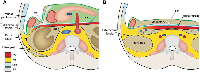
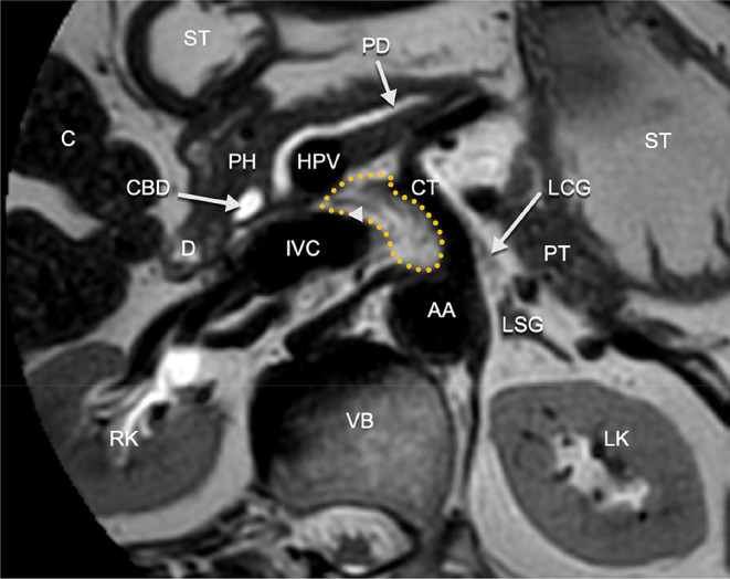
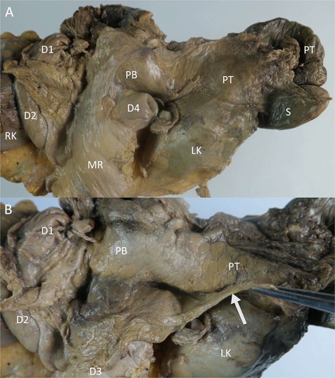
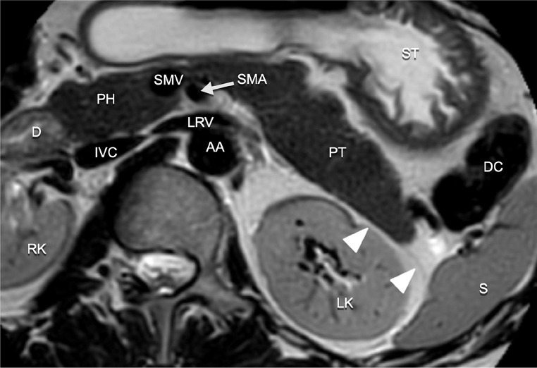
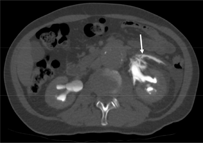
Results: The fusion fascia and the renal fascia in particular give rise to planes and spaces that act as vectors for spread of disease in the retroperitoneum. Some of these planes and structures, such as the caudal extension of the renal fascia, have previously not been described in radiological literature.

Conclusion: New insights, including the various fasciae, potential spaces and planes, are incorporated into an updated combined retroperitoneal fascial concept.

Keywords: Anatomy; Fascia; Imaging; Radiology; Retroperitoneum.


Conflict of interest statement

The authors declare no competing interests.

Figures

Fig. 1
Fig. 1
Schematic illustration of the tricompartmental concept. APS: anterior pararenal space, PRS: perirenal space, PPS: posterior pararenal space. The green line represents the peritoneum. The dotted line at the medial aspect of the anterior lamina of the renal fascia represents the supposed fusion of the fascia with connective tissue in the root of the mesentery and the dorsal aspect of the pancreas and duodenum
Fig. 2
Fig. 2
Imaging examples in CT of fluid tracking posteriorly along the renal fascia in two patients with pancreatitis. A) Fluid starting to spread posterior along the posterior lamina of the renal fascia (arrow). B) Massive amount of fluid spreading posteriorly around the renal fascia (arrow) extending medially all the way to the psoas major muscle D: duodenum, PH: pancreatic head, SB: small bowel, DC: descending colon, LK: left kidney
Fig. 3
Fig. 3
CT image of the left retroperitoneum at the level of the L4 vertebra below the kidneys. A large amount of fluid in the retroperitoneum tracks around and also dorsal to the flank pad (arrow pointing to the flank pad itself). AA: abdominal aorta, DC: descending colon, IVC: inferior vena cava, PM: psoas major muscle, SB: small bowel
Fig. 4
Fig. 4
A) Schematic illustration of the concept of interfascial spread at the level of the pancreas in the transverse plane, showing the retromesenteric, retrorenal and lateroconal planes. These planes are continuous with each other lateral to the kidney, which is illustrated by the color gradient. The green line represents the peritoneum. B) Schematic illustration in the sagittal plane. The retromesenteric and retrorenal planes fuse inferior to the perirenal space to form the combined interfascial plane, illustrated by the color gradient. The peritoneum is represented in green
Fig. 5
Fig. 5
Retroperitoneal fluid in a patient with pancreatitis demonstrated on CT imaging. A) At the level of the pelvis fluid tracks around the caudal extension of the renal fascia which is visible as a bilateral sheath of adipose tissue. The gonadal vessels (arrows), mark the approximate lateral border of the caudal extension of the renal fascia. B) Sagittal reconstructions demonstrate the caudal extension of the renal fascia (arrow heads), containing the ureter and testicular vessels coursing to the inguinal canal. AC: ascending colon, CIA: common iliac artery, CIV: common iliac vein, DC: descending colon, LK: left kidney, PM: psoas major muscle, SB: small bowel, SC: sigmoid colon
Fig. 6
Fig. 6
Loose connective tissue around the ascending colon visible during surgery as thin irregular strands (arrows). AC: ascending colon, L: liver, M: mesentery
Fig. 7
Fig. 7
Patient with a retroperitoneal hematoma due to a ruptured abdominal aortic aneurysm. The hematoma (highlighted in red) spreads in a plane dorsal to the mesocolon (dotted arrow bilaterally marks the position of adipose tissue in the mesocolon) and also extends in a plane anterior to the pancreatic head within the fusion plane of Fredet (arrow). AA: abdominal aorta, AC: ascending colon, D: duodenum (descending part), DC: descending colon, LK: left kidney, RK: right kidney, SB: small bowel, SMA: superior mesenteric artery, PH: pancreatic head
Fig. 8
Fig. 8
(A) Combined retroperitoneal fascial concept at the level of the pancreas. PS: prerenal space; RS: retrorenal space; LES: lateral extraperitoneal space; FP: fusion plane; PRS: perirenal space; PPS: peripancreatic space. The prerenal and retrorenal spaces, as well as the lateral extraperitoneal space, are continuous with each other indicated by the color gradient. The fusion plane (light gray) is present in between remnants of the peritoneum (light blue). The retropancreatic peritoneal remnant is illustrated as a dotted line (also marked with a dotted light blue arrow) to the left side of the superior mesenteric artery because of its close relationship and potential fusion with the anterior lamina of the renal fascia. (B) Combined retroperitoneal fascial concept at a level inferior to the kidneys. PS: prerenal space; RS: retrorenal space; LES: lateral extraperitoneal space; FP: fusion plane; PRS: perirenal space (caudal extension containing the ureters and gonadal vessels). The ‘flank pad’ is the major body of retroperitoneal adipose tissue at this level. The lateroconal fascia along the anteromedial border of the flank pad is represented by the thickened black line
Fig. 9
Fig. 9
T2-weighted magnetic resonance image of the pancreatic head. The neural plexus contained by the retropancreatic fascia is visible (dotted yellow area in which several linear structures (arrow head) are visible corresponding to nerve fibers of the plexus). The left celiac ganglion (LCG) is visible to the left side of the celiac trunk (CT). AA: abdominal aorta, C: colon, CBD: common bile duct, D: duodenum (descending part), HPV: hepatic portal vein, IVC: inferior vena cava, LK: left kidney, RK: right kidney, LSG: left suprarenal gland, PD: pancreatic duct, PH: pancreatic head, PT: pancreatic tail, ST: stomach, VB: vertebral body
Fig. 10
Fig. 10
Photographs of the pancreas during dissection on a specimen from a Fix for Life © embalmed human cadaver. A) The ventral side of the pancreas is still covered by its peritoneal covering.B) The peritoneal covering is detached from the pancreatic body and tail and pulled caudally (arrow), revealing the pancreas itself and the peripancreatic space. D1: duodenal bulb, D2: descending part of the duodenum, D3: horizontal part of the duodenum, D4: ascending part of the duodenum, LK: left kidney, MR: mesenteric root, PB: pancreatic body, PT: pancreatic tail, S: spleen
Fig. 11
Fig. 11
T2-weighted MR image of the pancreatic tail. The anterior renal fascia (arrow heads) is in close proximity to the pancreatic tail. AA: abdominal aorta, D: duodenum (descending part), DC: descending colon, IVC: inferior vena cava, LK: left kidney, LRV: left renal vein, PH: pancreatic head, PT: pancreatic tail, RK: right kidney, S: spleen, SMA: superior mesenteric artery, SMV: superior mesenteric vein, ST: stomach
Fig. 12
Fig. 12
A patient with blow-out of the renal pelvis due to retroperitoneal fibrosis. An excretory phase at CT shows leakage of contrast from the renal pelvis into the perirenal space and also extending through the ARF into the prerenal space (arrow). The focal discontinuation of the fascia is most likely pathological due to the presence of the retroperitoneal fibrosis

Similar articles

Cited by

References

    1. Meyers MA, Whalen JP, Peelle K, Berne AS. Radiologic features of extraperitoneal effusions. An anatomic approach. Radiology. 1972;104:249–257. doi: 10.1148/104.2.249. - DOI - PubMed
    1. Tirkes T, Sandrasegaran K, Patel AA, Hollar MA, Tejada JG, Tann M, Akisik FM, Lappas JC. Peritoneal and retroperitoneal anatomy and its relevance for cross-sectional imaging. Radiogr Rev Publ Radiol Soc N Am Inc. 2012;32:437–451. doi: 10.1148/rg.322115032. - DOI - PubMed
    1. Hayes MA. Abdominopelvic fasciae. Am J Anat. 1950;87:119–161. doi: 10.1002/aja.1000870105. - DOI - PubMed
    1. Cho BH, Kimura W, Song CH, Fujimiya M, Murakami G. An investigation of the embryologic development of the fascia used as the basis for pancreaticoduodenal mobilization. J Hepatobiliary Pancreat Surg. 2009;16:824–831. doi: 10.1007/s00534-009-0126-2. - DOI - PubMed
    1. Rajiah P, Sinha R, Cuevas C, Dubinsky TJ, Bush WH, Kolokythas O. Imaging of uncommon retroperitoneal masses. Radiogr Rev Publ Radiol Soc N Am Inc. 2011;31:949–976. doi: 10.1148/rg.314095132. - DOI - PubMed

MeSH terms

LinkOut - more resources