

Classification of variant portal vein anatomy based on three-dimensional CT: surgical implications
- PMID: 38963433
- PMCID: PMC11246292
- DOI: 10.1007/s00276-024-03427-5
Classification of variant portal vein anatomy based on three-dimensional CT: surgical implications
Abstract
Purposes: The purpose of this study was to develop a new and more comprehensive classification system for portal vein (PV) variations using three-dimensional visualization and evaluation (3DVE) and to discuss the prevalence rates and clinical implications of the variants.
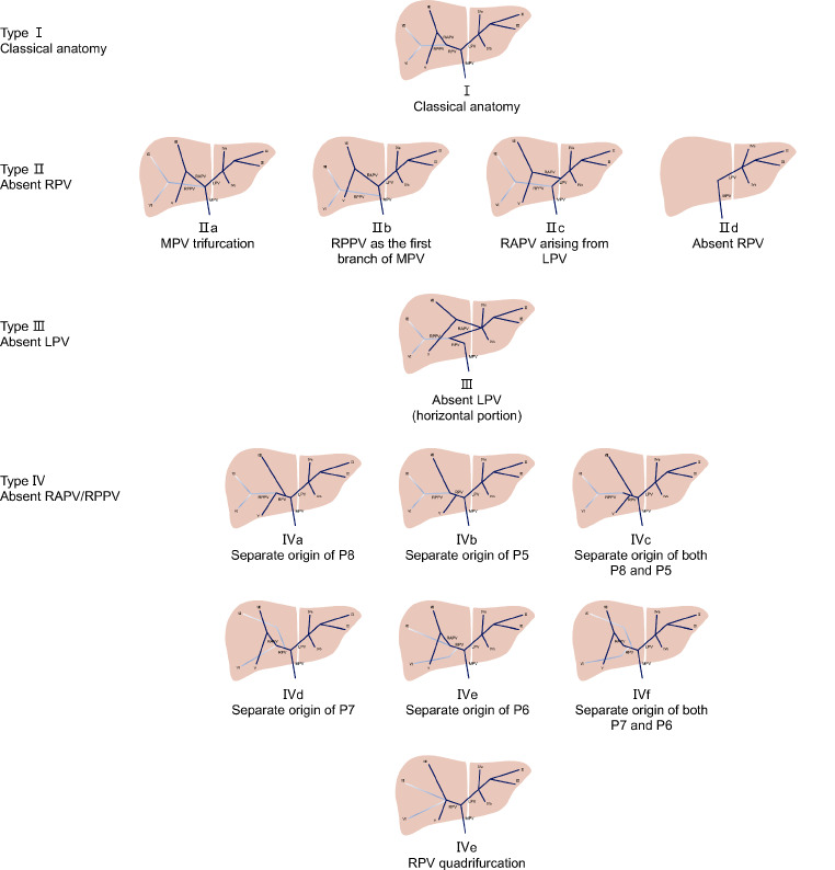
Methods: The anatomies of PVs were tracked and analyzed by using three-dimensional visualization of CT images acquired between 2013 and 2022. Scans from 200 adults were evaluated and a total of 178 patients (N = 178) were included in the study. The new classification system, named BLB classification, was developed based on the level of the absent PV branch in each variant anatomy.
Results: Using the BLB classification system, PVs were divided into thirteen subtypes. Only 82.6-84.8% of the portal veins of the 178 patients were depicted in Atri's, Cheng's or Covey's classification, compared with 100% identified by the BLB classification. The BLB classification was validated against external data sets from previous studies, with 97.0-98.9% of patients classified by the BLB system.
Conclusion: Variant PV anatomies are more commonly seen based on 3DVE than in previous reports. The BLB classification covers almost all portal vein variants and may be used for planning liver surgery.
Keywords: Anatomic variation; Clinical practice; Portal vein; Three-dimensional imaging.
© 2024. The Author(s).
Conflict of interest statement
The authors declare no competing interests.
Figures

Similar articles
-
CT imaging-based determination and classification of anatomic variations of left gastric vein.Surg Radiol Anat. 2017 Mar;39(3):249-255. doi: 10.1007/s00276-016-1722-x. Epub 2016 Jul 8. Surg Radiol Anat. 2017. PMID: 27393662
-
Variations of the right branch of hepatic portal vein in children based on three-dimensional simulation technology.Surg Radiol Anat. 2020 Dec;42(12):1467-1473. doi: 10.1007/s00276-020-02499-3. Epub 2020 May 18. Surg Radiol Anat. 2020. PMID: 32424682
-
[Efficacy of three-dimensional visualization technology in the precision diagnosis and treatment for primary liver cancer: a retrospective multicenter study of 1 665 cases in China].Zhonghua Wai Ke Za Zhi. 2020 May 1;58(5):375-382. doi: 10.3760/cma.j.cn112139-20200220-00105. Zhonghua Wai Ke Za Zhi. 2020. PMID: 32393005 Chinese.
-
Hepatic portal vein branching patterns according to different liver assessment methods and classifications of branching type.Ann Anat. 2024 Feb;252:152204. doi: 10.1016/j.aanat.2023.152204. Epub 2023 Dec 23. Ann Anat. 2024. PMID: 38142799 Review.
-
Cross-sectional imaging of congenital and acquired abnormalities of the portal venous system.Diagn Interv Radiol. 2016 Nov-Dec;22(6):501-507. doi: 10.5152/dir.2016.16012. Diagn Interv Radiol. 2016. PMID: 27731302 Free PMC article. Review.
Cited by
-
[3D visualization-based classification of left intrahepatic vessels and its application in precision hepatectomy].Nan Fang Yi Ke Da Xue Xue Bao. 2025 May 20;45(5):1047-1055. doi: 10.12122/j.issn.1673-4254.2025.05.18. Nan Fang Yi Ke Da Xue Xue Bao. 2025. PMID: 40415437 Free PMC article. Chinese.
-
Examining the Branching Patterns of the Hepatis Portae Vena with Computed Tomography Images.J Clin Med. 2025 Jul 8;14(14):4835. doi: 10.3390/jcm14144835. J Clin Med. 2025. PMID: 40725529 Free PMC article.
References
MeSH terms
LinkOut - more resources
Full Text Sources
Medical
Research Materials
