Review
. 2024 Dec;43(4):1165-1183.
doi: 10.1007/s10555-024-10196-5. Epub 2024 Jul 4.

Cancer treatments as paradoxical catalysts of tumor awakening in the lung

Affiliations
Review

Cancer treatments as paradoxical catalysts of tumor awakening in the lung

Emmanuelle Nicolas et al. Cancer Metastasis Rev. 2024 Dec.

Abstract

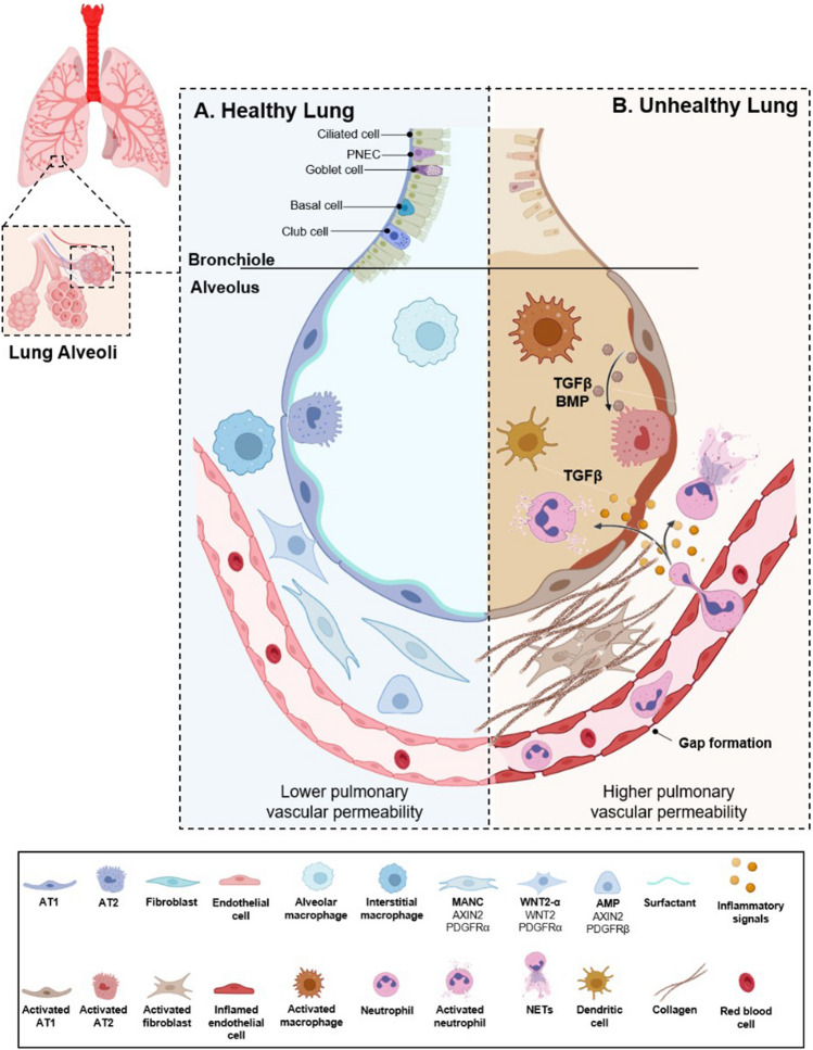
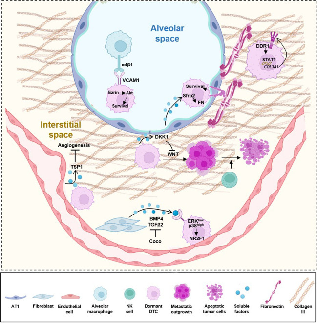
Much of the fatality of tumors is linked to the growth of metastases, which can emerge months to years after apparently successful treatment of primary tumors. Metastases arise from disseminated tumor cells (DTCs), which disperse through the body in a dormant state to seed distant sites. While some DTCs lodge in pre-metastatic niches (PMNs) and rapidly develop into metastases, other DTCs settle in distinct microenvironments that maintain them in a dormant state. Subsequent awakening, induced by changes in the microenvironment of the DTC, causes outgrowth of metastases. Hence, there has been extensive investigation of the factors causing survival and subsequent awakening of DTCs, with the goal of disrupting these processes to decrease cancer lethality. We here provide a detailed overview of recent developments in understanding of the factors controlling dormancy and awakening in the lung, a common site of metastasis for many solid tumors. These factors include dynamic interactions between DTCs and diverse epithelial, mesenchymal, and immune cell populations resident in the lung. Paradoxically, among key triggers for metastatic outgrowth, lung tissue remodeling arising from damage induced by the treatment of primary tumors play a significant role. In addition, growing evidence emphasizes roles for inflammation and aging in opposing the factors that maintain dormancy. Finally, we discuss strategies being developed or employed to reduce the risk of metastatic recurrence.

Keywords: Chemoradiation; Dormancy; Extracellular matrix; Fibrosis; Inflammation; Neutrophil extracellular trap.


Conflict of interest statement

Declarations Ethical approval N/A. Informed consent N/A. Competing interests The authors declare no competing interests.

Figures

Fig. 1
Fig. 1
Lung alveoli in health and disease. A Air enters and exits the lung through large and small branching airways (the bronchi and bronchioles) that terminate in alveoli, which participate in gas exchange and are surrounded by capillary plexus. The cellular lining of bronchi and bronchioles is complex, including a respiratory epithelium composed of basal cells, multi-ciliated cells, secretory (club) cells, goblet cells, brush (tuft) cells, and others, supported by an underlying layer of stromal cells. The alveoli are lined by two epithelial cell types: alveolar type-1 (AT1) and alveolar type-2 (AT2) cells. AT2s are cuboidal, sparsely distributed among the AT1s, and comprise 5% of the total surface area. AT2s have a unique multi-apical polarity and multi-lumen enfacement organization [47] and secrete surfactant, a lipid-protein complex which decreases alveolar surface tension at the air–liquid interface. AT2s both self-renew and differentiate into AT1s, thereby serving as stem cells. At homeostasis, turn-over is infrequent. However, in settings of lung injury and tissue repair, changes in cell signaling including activation of WNT [48, 49] and reprogramming of the transforming growth factor (TGF) β and bone morphogenic protein (BMP) support AT2 replication and differentiation to AT1 cells. AT1s are very thin, terminally differentiated and have a large surface area covering 95% of the alveolar luminal. AT1s share a basement membrane and mediate gas exchange with the endothelial cells of the pulmonary capillaries. The capillary endothelium is covered with pericytes, which help regulate blood flow [50]. In addition, several types of interstitial fibroblasts comprise a supportive mesenchyme encompassing the alveoli. The pulmonary extracellular matrix (ECM) provides mechanical stability and elastic recoil, which are essential for physiological lung function. Tissue homeostasis is in part maintained by biomechanical functional units made up of resident fibroblasts and self-generated interstitial ECM. These units provide the lung (and other organs) with the needed tensile strength (e.g., collagen) and elasticity (e.g., elastin). Of note, any disturbance or aggravation to the organ is repaired with the participation of these units [35]: through secretion and activation of proteases [51], release or storage of ECM stored factors such as TGFβ that trigger local and systemic changes, as well as the activation of ECM enzymes such as lysyl hydroxylases (LOX), which catalyze crosslinking of collagen and/or elastin fibrils to remodel the fibrous ECM bundles [, –54]. In cases of transient lung injury, ECM proteases and LOX [55] play an important role in the degradation and turnover of all matrix components needed for repair and regeneration, preventing fibrosis [56, 57]. Besides systems required for air exchange, the lymphatic network penetrates the lung, allowing movement of lymphocytes and other immune cells through the tissue, and performing other functions including drainage of interstitial fluid, and removal of cellular debris [58]. The narrow lymphatics drain into larger collecting vessels that are components of bronchovascular bundles and also reside in interlobular septa; these collecting vessels in turn drain to collecting lymph nodes. Immune homeostasis in the healthy lung is maintained through interactions of several cell types, including fibroblast units, resident alveolar macrophages, dendritic cells, alveolar type-1 (AT1), and alveolar type-2 (AT2) cells. Surfactant is composed of surfactant proteins which bind and neutralize viruses. Vascular capillaries line the alveolar walls to facilitate gaseous exchange and infiltration of circulating immune cells. In the interstitial compartment, interstitial fibroblast units, including mesenchymal alveolar niche cells (MANCs), Axin2-positive myogenic precursors (AMPs), and Wnt2-expressing platelet-derived growth factor-α (PDGFRα)-positive cells (WNT2–Pα), comprise a supportive mesenchyme encompassing the alveoli. B In pathological conditions, many tissue-resident alveolar macrophages are lost and replaced by bone marrow-derived macrophages. This occurs in parallel with infiltration of inflammatory cells such as neutrophils, recruitment of which is facilitated by chemokines and disrupted barrier integrity. The activated epithelium secretes a plethora of mediators that induce proliferation and activation of fibroblast units, which in turn dynamically modify the interstitial ECM, promoting fibrosis [35]. During lung injury and subsequent tissue repair, activation of WNT signaling and secretion of TGFβ-related factors such as BMP and others support AT2 replication and differentiation to AT1 cells. This figure was created in part using BioRender (BioRender.com)
Fig. 2
Fig. 2
Signaling supporting DTC dormancy in the lung. Cues from the lung microenvironment are critical in maintaining dormancy of DTCs. In the alveolar space, DTCs expressing VCAM-1 receive survival signaling from resident macrophages through juxtacrine activation of a VCAM-1-Ezrin-PI3K/Akt survival pathway. In an epithelial niche, induction of SFRP2 in DTCs causes them to conduct de novo fibrillogenesis, resulting in increases in fibrillar fibronectin as a component of the ECM being altered, and thus promoting survival signaling. Enrichment of type III collagen in the interstitial ECM niche also sustains DTC dormancy, signaling through the DDR1 receptor to activate STAT1. In the perivascular niche, deposited basement membrane components like TSP-1 suppress angiogenesis to also support dormancy. Local resident fibroblast/ECM units secrete TGFβ2 and BMP4 to inhibit ERK1/2 signaling, promoting dormancy. Additionally, DTCs can enter dormancy by secreting DKK1, an inhibitor of the WNT signaling pathway, and evading NK-cell-mediated immunity within the dormant/metastatic niche. See text for details. This figure was created in part using BioRender (BioRender.com)
Fig. 3
Fig. 3
Signaling activated by cancer treatments that promote awakening and metastatic outgrowth. Chemotherapy promotes infiltration of LOX-secreting CD8+ T cells in the lung, which modulates the ECM, promoting metastasis. Chemotherapy also stimulates tumor cells to release cytokines including IL1β, IL8/CXCL8, TGFβ1, and EGF, that caused fibroblast recruitment. Fibroblasts then secrete collagen-1 and COX2, triggering integrin/SRC and prostaglandin E signaling in the cancer cells, stimulating awakening of dormant DTCs. Chemotherapy action on lung DTCs stimulates them to secrete IL-1β, which in turn triggers NET formation, promoting chemoresistance and promoting metastasis. Furthermore, chemotherapy promotes complement signaling in lung fibroblasts, leading to recruitment of MDSCs and the formation of an immunosuppressive niche, favorable to metastatic relapse. Other pro-metastatic mechanisms linked to cancer treatment include the release of pro-inflammatory cytokines and growth factors such as IL-6, IL-8, and VEGF, which awaken DTCs and micrometastases in the lung. Additionally, radiation in healthy lung tissue leads to infiltration and activation of neutrophils and induces Notch activation within epithelial cells, fueling the subsequent growth of arriving DTCs. Additional details are provided in the main text. This figure was created in part using BioRender (BioRender.com)

Similar articles

Cited by

References

    1. Siegel, R. L., Giaquinto, A. N., & Jemal, A. (2024). Cancer statistics, 2024. CA: A Cancer Journal for Clinicians,74(1), 12–49. - PubMed
    1. Massagué, J., & Ganesh, K. (2021). Metastasis-initiating cells and ecosystems. Cancer Discovery,11(4), 971–994. - PMC - PubMed
    1. Borriello, L., et al. (2022). Primary tumor associated macrophages activate programs of invasion and dormancy in disseminating tumor cells. Nature Communications,13(1), 626. - PMC - PubMed
    1. Goddard, E. T., et al. (2018). Dormant tumour cells, their niches and the influence of immunity. Nature Cell Biology,20(11), 1240–1249. - PubMed
    1. Pu, Y., et al. (2023). Drug-tolerant persister cells in cancer: The cutting edges and future directions. Nature Reviews. Clinical Oncology,20(11), 799–813. - PubMed