

Exploring the Function of Epicardial Cells Beyond the Surface
- PMID: 38963865
- PMCID: PMC11225799
- DOI: 10.1161/CIRCRESAHA.124.321567
Exploring the Function of Epicardial Cells Beyond the Surface
Abstract
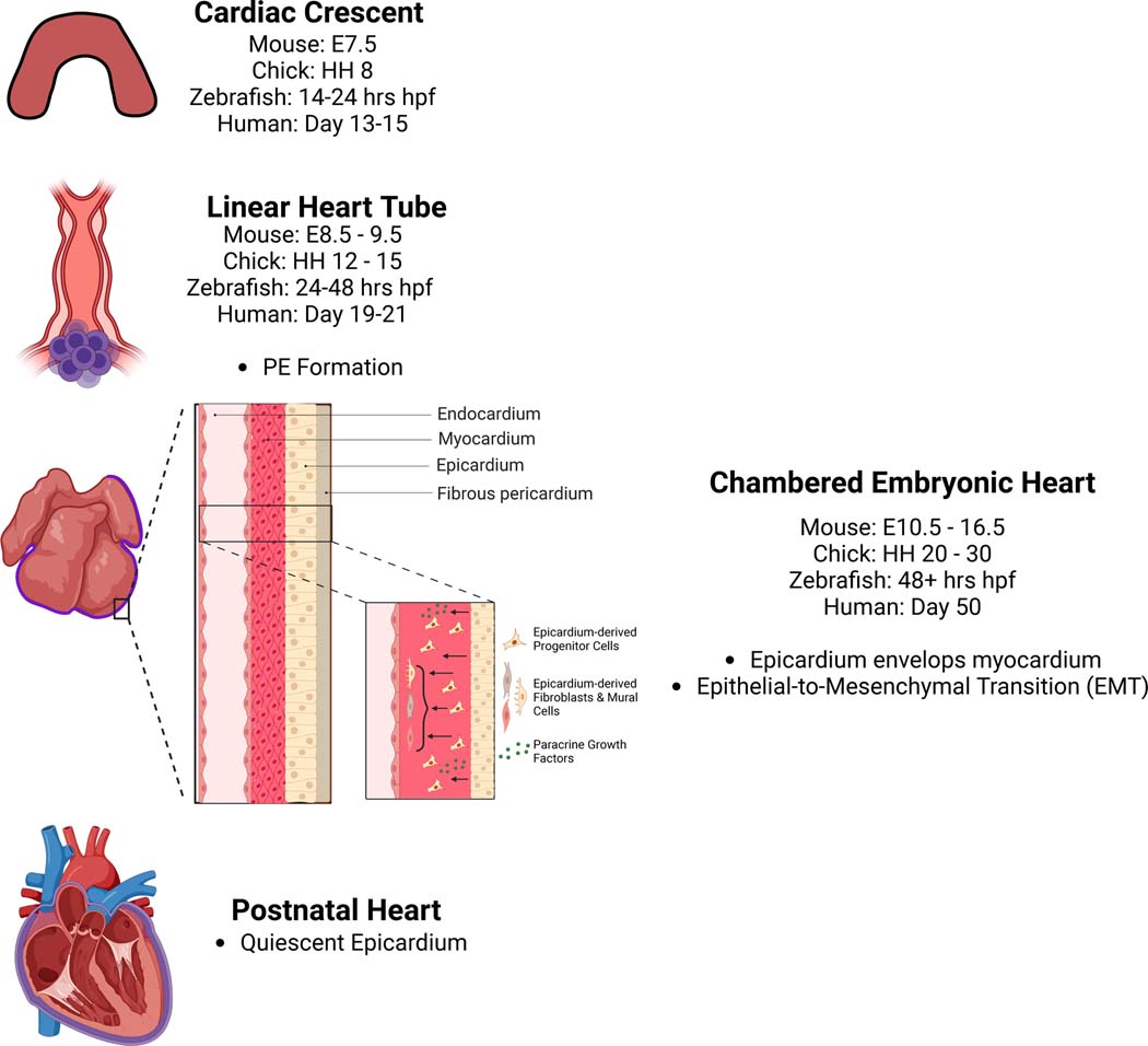
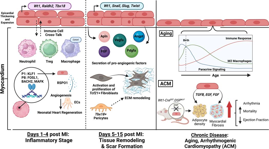
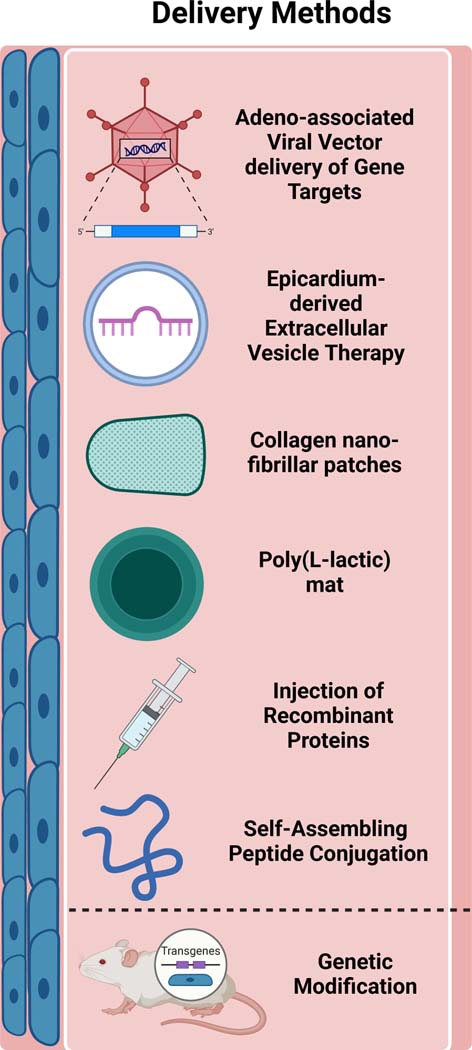
The epicardium, previously viewed as a passive outer layer around the heart, is now recognized as an essential component in development, regeneration, and repair. In this review, we explore the cellular and molecular makeup of the epicardium, highlighting its roles in heart regeneration and repair in zebrafish and salamanders, as well as its activation in young and adult postnatal mammals. We also examine the latest technologies used to study the function of epicardial cells for therapeutic interventions. Analysis of highly regenerative animal models shows that the epicardium is essential in regulating cardiomyocyte proliferation, transient fibrosis, and neovascularization. However, despite the epicardium's unique cellular programs to resolve cardiac damage, it remains unclear how to replicate these processes in nonregenerative mammalian organisms. During myocardial infarction, epicardial cells secrete signaling factors that modulate fibrotic, vascular, and inflammatory remodeling, which differentially enhance or inhibit cardiac repair. Recent transcriptomic studies have validated the cellular and molecular heterogeneity of the epicardium across various species and developmental stages, shedding further light on its function under pathological conditions. These studies have also provided insights into the function of regulatory epicardial-derived signaling molecules in various diseases, which could lead to new therapies and advances in reparative cardiovascular medicine. Moreover, insights gained from investigating epicardial cell function have initiated the development of novel techniques, including using human pluripotent stem cells and cardiac organoids to model reparative processes within the cardiovascular system. This growing understanding of epicardial function holds the potential for developing innovative therapeutic strategies aimed at addressing developmental heart disorders, enhancing regenerative therapies, and mitigating cardiovascular disease progression.
Keywords: angiogenesis; cardiovascular diseases; fibrosis; inflammation; myocardial infarction.
Conflict of interest statement
None.
Figures
Similar articles
-
Relationship between Pituitary Gland and Stem Cell in the Aspect of Hormone Production and Disease Prevention: A Narrative Review.Endocr Metab Immune Disord Drug Targets. 2025;25(7):509-526. doi: 10.2174/0118715303314551241031093717. Endocr Metab Immune Disord Drug Targets. 2025. PMID: 39812047 Review.
-
Systemic pharmacological treatments for chronic plaque psoriasis: a network meta-analysis.Cochrane Database Syst Rev. 2021 Apr 19;4(4):CD011535. doi: 10.1002/14651858.CD011535.pub4. Cochrane Database Syst Rev. 2021. Update in: Cochrane Database Syst Rev. 2022 May 23;5:CD011535. doi: 10.1002/14651858.CD011535.pub5. PMID: 33871055 Free PMC article. Updated.
-
Systemic pharmacological treatments for chronic plaque psoriasis: a network meta-analysis.Cochrane Database Syst Rev. 2017 Dec 22;12(12):CD011535. doi: 10.1002/14651858.CD011535.pub2. Cochrane Database Syst Rev. 2017. Update in: Cochrane Database Syst Rev. 2020 Jan 9;1:CD011535. doi: 10.1002/14651858.CD011535.pub3. PMID: 29271481 Free PMC article. Updated.
-
Generation of mature epicardium derived from human-induced pluripotent stem cells via inhibition of mTOR signaling.Nat Commun. 2025 Jul 1;16(1):5902. doi: 10.1038/s41467-025-60934-8. Nat Commun. 2025. PMID: 40593704 Free PMC article.
-
Foxf1-mediated co-regulation of miR-495 and let-7c modulates epicardial cell migration and myocardial specification.Cell Mol Life Sci. 2025 Jun 25;82(1):254. doi: 10.1007/s00018-025-05735-4. Cell Mol Life Sci. 2025. PMID: 40555850 Free PMC article.
Cited by
-
HGF Overexpression in Mesenchymal Stromal Cell-Based Cell Sheets Enhances Autophagy-Dependent Cytoprotection and Proliferation to Guard the Epicardial Mesothelium.Int J Mol Sci. 2025 Jul 28;26(15):7298. doi: 10.3390/ijms26157298. Int J Mol Sci. 2025. PMID: 40806435 Free PMC article.
-
Beyond the Heartbeat: Single-Cell Omics Redefining Cardiovascular Research.Curr Cardiol Rep. 2024 Nov;26(11):1183-1196. doi: 10.1007/s11886-024-02117-3. Epub 2024 Aug 19. Curr Cardiol Rep. 2024. PMID: 39158785 Review.
-
Molecular convergence of risk variants for congenital heart defects leveraging a regulatory map of the human fetal heart.medRxiv [Preprint]. 2024 Nov 22:2024.11.20.24317557. doi: 10.1101/2024.11.20.24317557. medRxiv. 2024. PMID: 39606363 Free PMC article. Preprint.
References
-
- Tsao CW, Aday AW, Almarzooq ZI, Anderson CAM, Arora P, Avery CL, Baker-Smith CM, Beaton AZ, Boehme AK, Buxton AE, et al. Heart Disease and Stroke Statistics-2023 Update: A Report From the American Heart Association. Circulation. 2023;147:e93–e621. doi: 10.1161/CIR.0000000000001123 - DOI - PMC - PubMed
-
- Scalise RFM, De Sarro R, Caracciolo A, Lauro R, Squadrito F, Carerj S, Bitto A, Micari A, Bella GD, Costa F, et al. Fibrosis after Myocardial Infarction: An Overview on Cellular Processes, Molecular Pathways, Clinical Evaluation and Prognostic Value. Med Sci (Basel). 2021;9. doi: 10.3390/medsci9010016 - DOI - PMC - PubMed
Publication types
MeSH terms
Grants and funding
LinkOut - more resources
Full Text Sources
