

Identification of heparin-binding amino acid residues in antibody HS4C3 with the potential to design antibodies against heparan sulfate domains
- PMID: 38963938
- PMCID: PMC11231949
- DOI: 10.1093/glycob/cwae046
Identification of heparin-binding amino acid residues in antibody HS4C3 with the potential to design antibodies against heparan sulfate domains
Abstract
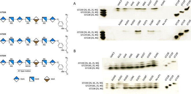
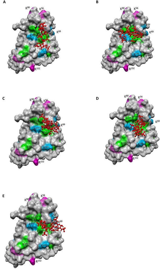
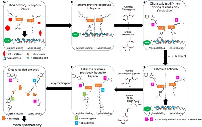
Heparan sulfate (HS) is a linear polysaccharide with high structural and functional diversity. Detection and localization of HS in tissues can be performed using single chain variable fragment (scFv) antibodies. Although several anti-HS antibodies recognizing different sulfation motifs have been identified, little is known about their interaction with HS. In this study the interaction between the scFv antibody HS4C3 and heparin was investigated. Heparin-binding lysine and arginine residues were identified using a protect and label methodology. Site-directed mutagenesis was applied to further identify critical heparin-binding lysine/arginine residues using immunohistochemical and biochemical assays. In addition, computational docking of a heparin tetrasaccharide towards a 3-D homology model of HS4C3 was applied to identify potential heparin-binding sites. Of the 12 lysine and 15 arginine residues within the HS4C3 antibody, 6 and 9, respectively, were identified as heparin-binding. Most of these residues are located within one of the complementarity determining regions (CDR) or in their proximity. All basic amino acid residues in the CDR3 region of the heavy chain were involved in binding. Computational docking showed a heparin tetrasaccharide close to these regions. Mutagenesis of heparin-binding residues reduced or altered reactivity towards HS and heparin. Identification of heparin-binding arginine and lysine residues in HS4C3 allows for better understanding of the interaction with HS and creates a framework to rationally design antibodies targeting specific HS motifs.
Keywords: antibodies; heparan sulfate; heparin-binding residues; protect & label; site-specific mutations.
© The Author(s) 2024. Published by Oxford University Press.
Conflict of interest statement
None declared.
Figures


Similar articles
-
Construction and evaluation of an antibody phage display library targeting heparan sulfate.Glycoconj J. 2020 Aug;37(4):445-455. doi: 10.1007/s10719-020-09925-z. Epub 2020 May 28. Glycoconj J. 2020. PMID: 32468289 Free PMC article.
-
Generation and application of type-specific anti-heparan sulfate antibodies using phage display technology. Further evidence for heparan sulfate heterogeneity in the kidney.J Biol Chem. 1998 May 22;273(21):12960-6. doi: 10.1074/jbc.273.21.12960. J Biol Chem. 1998. PMID: 9582329
-
3-O-sulfated oligosaccharide structures are recognized by anti-heparan sulfate antibody HS4C3.J Biol Chem. 2006 Feb 24;281(8):4654-62. doi: 10.1074/jbc.M506357200. Epub 2005 Dec 22. J Biol Chem. 2006. PMID: 16373349
-
The role of heparin/heparan sulphate in the IFN-γ-led Arena.Biochimie. 2020 Mar;170:1-9. doi: 10.1016/j.biochi.2019.11.018. Epub 2019 Nov 30. Biochimie. 2020. PMID: 31794784 Review.
-
Molecular Aspects of Heparanase Interaction with Heparan Sulfate, Heparin and Glycol Split Heparin.Adv Exp Med Biol. 2020;1221:169-188. doi: 10.1007/978-3-030-34521-1_6. Adv Exp Med Biol. 2020. PMID: 32274710 Review.
References
-
- Ausekar MV, Mawale RM, Pazdera P, Havel J. Matrix assisted and/or laser desorption ionization quadrupole ion trap time-of-flight mass spectrometry of WO3 clusters formation in gas phase. Nanodiamonds, fullerene, and graphene oxide matrices. J Am Soc Mass Spectrom. 2018:29(3):581–587. - PubMed
-
- Becker S, Boch J. TALE and TALEN genome editing technologies. Gene Genome Ed. 2021:2:100007.
-
- Brummell DA, Sharma VP, Anand NN, Bilous D, Dubuc G, Michniewicz J, MacKenzie CR, Sadowska J, Sigurskjold BW, Sinnott B. Probing the combining site of an anti-carbohydrate antibody by saturation-mutagenesis: role of the heavy-chain CDR3 residues. Biochemistry. 1993:32(4):1180–1187. - PubMed
Publication types
MeSH terms
Substances
Grants and funding
LinkOut - more resources
Full Text Sources
Medical
