

Histopathological, morphological, and molecular characterization of fish-borne zoonotic parasite Eustrongylides Excisus infecting Northern pike (Esox lucius) in Iran
- PMID: 38965518
- PMCID: PMC11223293
- DOI: 10.1186/s12917-024-04146-0
Histopathological, morphological, and molecular characterization of fish-borne zoonotic parasite Eustrongylides Excisus infecting Northern pike (Esox lucius) in Iran
Abstract
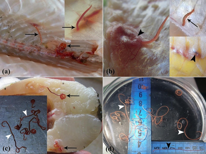
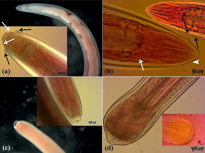
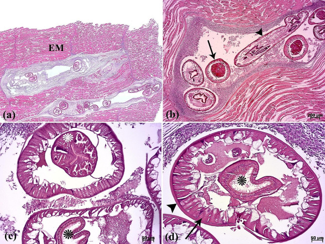
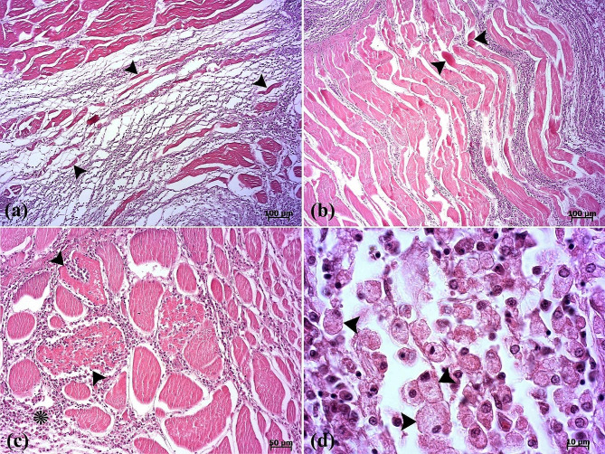
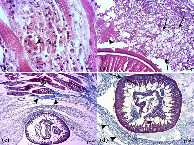
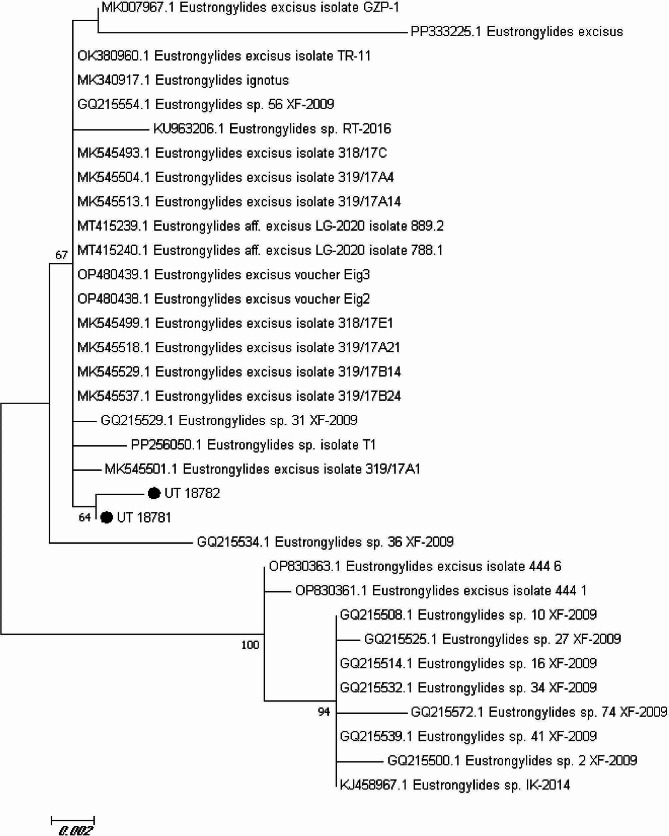
Eustrongylides excisus is a fish-borne zoonotic parasite known to infect various fish species, including Northern pike (Esox Lucius). This nematode, belonging to the family Dioctophymatidae, has a complex life cycle involving multiple hosts. This study aimed to investigate the occurrence of Eustrongylides nematodes in Northern pike (E. Lucius) collected from Mijran Dam (Ramsar, Iran). Between June and October 2023, an investigation was conducted on Northern pike from Mijran Dam in Ramsar, Iran, following reports of reddish parasites in their muscle tissues. Sixty fish were examined at the University of Tehran, revealing live parasites in the muscles, which were then analyzed microscopically and preserved for a multidisciplinary study. The skeletal muscle tissues of 85% (51/60) of fish specimens were infected by grossly visible larvae which were microscopically identified as Eustrongylides spp. In histopathological examination, the lesion was composed of encapsulated parasitic granulomatous myositis. Microscopically, the cystic parasitic granulomas compressed the adjacent muscle fibers, leading to their atrophy and Zenker's necrosis. Moreover, epithelioid macrophages, giant cells and mononuclear inflammatory cells were present around the larvae and between the muscle fibers. Finally, a molecular analysis by examining the ITS gene region, revealed that they belong to the species E. excisus. Eustrongylidiasis in northern Iran necessitates further research into the biology, epidemiology, and control of Eustrongylides nematodes, focusing on various hosts. This study is the first to comprehensively characterize E. excisus in Northern pike in Ramsar, Iran, raising concerns about possible zoonotic transmission.
Keywords: Eustrongylides excisus; Freshwater ecosystem; Granulomatous myositis; Iran; Northern pike.
© 2024. The Author(s).
Conflict of interest statement
The authors declare no competing interests.
Figures
Similar articles
-
Morphological and molecular characterization of Eustrongylides excisus larvae (Nematoda: Dioctophymatidae) in Sander lucioperca (L.) from Northern Turkey.Parasitol Res. 2021 Jun;120(6):2269-2274. doi: 10.1007/s00436-021-07187-8. Epub 2021 May 18. Parasitol Res. 2021. PMID: 34002260
-
Morphological and Molecular Characterization of Larval and Adult Stages of Eustrongylides excisus (Nematoda: Dioctophymatoidea) with Histopathological Observations.J Parasitol. 2019 Dec;105(6):882-889. J Parasitol. 2019. PMID: 31738125
-
Histopathology and the inflammatory response of European perch, Perca fluviatilis muscle infected with Eustrongylides sp. (Nematoda).Parasit Vectors. 2015 Apr 15;8:227. doi: 10.1186/s13071-015-0838-x. Parasit Vectors. 2015. PMID: 25889096 Free PMC article.
-
A review on fish-borne zoonotic parasites in Iran.Vet Med Sci. 2023 Mar;9(2):748-777. doi: 10.1002/vms3.981. Epub 2022 Oct 21. Vet Med Sci. 2023. PMID: 36271486 Free PMC article. Review.
-
The Occurrence of Freshwater Fish-Borne Zoonotic Helminths in Italy and Neighbouring Countries: A Systematic Review.Animals (Basel). 2023 Dec 8;13(24):3793. doi: 10.3390/ani13243793. Animals (Basel). 2023. PMID: 38136832 Free PMC article. Review.
References
-
- Anderson JL, Asche F, Garlock T, Chu J, Aquaculture. Its role in the future of food. In: Schmitz A, Kennedy PL, Schmitz TG, editors. World agricultural resources and food security. Emerald Publishing Limited; 2017. pp. 159–73. 10.1108/S1574-871520170000017011
-
- Rahmati Holasoo H, Marandi A, Ebrahimzadeh Mousavi H, Azizi A. Study of the losses of siberian sturgeon (Acipenser baerii) due to gill infection with Diclybothrium armatum in sturgeon farms of Qom and Mazandaran provinces. J Anim Environ. 2021;13:193–200. doi: 10.22034/aej.2021.165929. - DOI
-
- Ziafati Kafi Z, Ghalyanchilangeroudi A, Nikaein D, Marandi A, Rahmati-Holasoo H, Sadri N, Erfanmanesh A, Enayati A. Phylogenetic analysis and genotyping of Iranian infectious haematopoietic necrosis virus (IHNV) of rainbow trout (Oncorhynchus mykiss) based on the glycoprotein gene. Veterinary Med Sci. 2022;8:2411–7. doi: 10.1002/vms3.931. - DOI - PMC - PubMed
-
- Arlinghaus R, Alós J, Pieterek T, Klefoth T. Determinants of angling catch of northern pike (Esox lucius) as revealed by a controlled whole-lake catch-and-release angling experiment—the role of abiotic and biotic factors, spatial encounters and lure type. Fish Res. 2017;1:648–57. doi: 10.1016/j.fishres.2016.09.009. - DOI
MeSH terms
LinkOut - more resources
Full Text Sources
Miscellaneous
