Review
. 2024 Jul 4;22(1):395.
doi: 10.1186/s12951-024-02634-x.

Exosome for mRNA delivery: strategies and therapeutic applications

Affiliations
Review

Exosome for mRNA delivery: strategies and therapeutic applications

Zoya Iqbal et al. J Nanobiotechnology. .

Abstract

Messenger RNA (mRNA) has emerged as a promising therapeutic molecule with numerous clinical applications in treating central nervous system disorders, tumors, COVID-19, and other diseases. mRNA therapies must be encapsulated into safe, stable, and effective delivery vehicles to preserve the cargo from degradation and prevent immunogenicity. Exosomes have gained growing attention in mRNA delivery because of their good biocompatibility, low immunogenicity, small size, unique capacity to traverse physiological barriers, and cell-specific tropism. Moreover, these exosomes can be engineered to utilize the natural carriers to target specific cells or tissues. This targeted approach will enhance the efficacy and reduce the side effects of mRNAs. However, difficulties such as a lack of consistent and reliable methods for exosome purification and the efficient encapsulation of large mRNAs into exosomes must be addressed. This article outlines current breakthroughs in cell-derived vesicle-mediated mRNA delivery and its biomedical applications.

Keywords: Exosomes; Targeted delivery; mRNA.


Conflict of interest statement

The authors declare that the work was conducted in the absence of any commercial or financial relationships that could be construed as a potential competing interests.

Figures
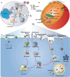
Fig. 1
Fig. 1
Biogenesis of exosoems and the mechanisms of uptake of exosomes involved by the recipient cell. A Early endosomes are produced from the invegination of the cell membrane, which mature into MVBs with ILVs inside. The inward budding of the endosomal membrane leads to ILVs formation. The fusion of the MVBs to the plasma membrane results in the secretion of exosomes extracellularly [71]. B Typical structure of exosomes containing functional DNA, proteins and RNA biomolecules surrounded by a lipid bilayer. C Several mechanisms have been reported for the uptake of exosomes, e.g., include micropinocytosis, phagocytosis, caveolae/raft-dependent endocytosis, receptor-mediated endocytosis, and direct fusion [72]. Adhesion molecules, including integrin, ICAM-1, LFA-1, CD81, and CD9 on the vesicle membranes, also play important roles in the binding and uptake of exosomes. Moreover, several receptor-ligand interactions, such as heparin sulfate proteoglycans-fibronectin, TIM receptors-phosphatidylserine, and epidermal growth factor receptor (EGFR)-epidermal growth factor (EGF), mediate the exosome endocytosis [73]
Fig. 2
Fig. 2
Schematic representation of programmable exosomes for mRNAs loading
Fig. 3
Fig. 3
Different strategies for loading cargoes into exosomes
Fig. 4
Fig. 4
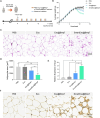
The efficiency of the US triggered a smart exosome-based system for Bmp7 delivery for OAT browning. A Schematic representation of the experimental design. B Each group's average weight was observed from the high-fat diet till the end of the treatment period. C HE staining images of OAT from mice after i.v treatment. D Adipocyte area for HE staining images. E Ucp1 expression level in OAT from mice after i.v treatment. F Images of Ucp1 staining of a section of OAT depots from mice after i.v treatments. Reprinted with permission from Ref. [121]. Copyright © 2023 BioMed Central Ltd
Fig. 5
Fig. 5
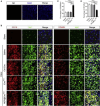
Role of NGF@ExoRVG on Microglia Polarization in brain injury. A Immunofluorescence images confirming the specificity of immunostaining via secondary antibody negative control (NC) B Representative immunofluorescence images of different antibodies staining in the ischemic cortex of different treatments. NGF@ExoRVG showed a significant decrease in CD16 expression and a remarkable increase in the CD206 expression compared with other groups C, D Cell quantification of the % age of CD206 and CD16 in the ischemic region. Reprinted with permission from Ref. [124], Copyright © 2023 Elsevier B.V
Fig. 6
Fig. 6
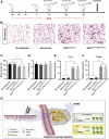
Efficient delivery of mIRES-PGC1α mRNA promotes browning in mouse adipose tissues. A Schematic illustration of the experimental procedure. Mice were fed with a high-fat diet for 3 weeks, followed by exosome delivery with the aid of UTMD. The exosome delivery was performed once a week for 3 continuous weeks. Mice were sacrificed at the end of experiments for histology and gene expression. B H&E staining of the adipose tissue in mice treated as indicated. Smaller browning adipocytes are indicated by arrows. Scale bar, 50 μm. Data shown are representative of five to seven mice in each group. C Body weight in mice treated as indicated. Data are expressed as mean ± SEM of seven mice in each group. ∗ p < 0.05 by one-way ANOVA. D Food intake in mice treated as indicated. Data are expressed as mean ± SEM of seven mice in each group. ∗ p < 0.05 by one-way ANOVA. E and F qPCR analysis of Ucp1 (E) and Cidea (F) expression in adipose tissue. Gapdh served as internal control. Data are expressed as mean ± SEM of three mice in each group. ∗ p < 0.05 by one-way ANOVA. G Schematic illustration of the study. Reprinted with permission from Ref. [97]. Copyright © 2023 BioMed Central Ltd

Similar articles

Cited by

References

    1. Hombach S, Kretz M. Non-coding RNAs: classification, biology and functioning. Adv Exp Med Biol. 2016;937:3–17. doi: 10.1007/978-3-319-42059-2_1. - DOI - PubMed
    1. Kaczmarek JC, Kowalski PS, Anderson DG. Advances in the delivery of RNA therapeutics: from concept to clinical reality. Genome Med. 2017;9:60. doi: 10.1186/s13073-017-0450-0. - DOI - PMC - PubMed
    1. Fang E, Liu X, Li M, Zhang Z, Song L, Zhu B, Wu X, Liu J, Zhao D, Li Y. Advances in COVID-19 mRNA vaccine development. Signal Transduct Target Ther. 2022;7:94. doi: 10.1038/s41392-022-00950-y. - DOI - PMC - PubMed
    1. Baden LR, El Sahly HM, Essink B, Kotloff K, Frey S, Novak R, Diemert D, Spector SA, Rouphael N, Creech CB, et al. Efficacy and safety of the mRNA-1273 SARS-CoV-2 vaccine. N Engl J Med. 2021;384:403–416. doi: 10.1056/NEJMoa2035389. - DOI - PMC - PubMed
    1. Szabó GT, Mahiny AJ, Vlatkovic I. COVID-19 mRNA vaccines: platforms and current developments. Mol Ther. 2022;30:1850–1868. doi: 10.1016/j.ymthe.2022.02.016. - DOI - PMC - PubMed

LinkOut - more resources