. 2024 Jul 5;43(1):188.
doi: 10.1186/s13046-024-03103-x.

lncRNA FGD5-AS1 is required for gastric cancer proliferation by inhibiting cell senescence and ROS production via stabilizing YBX1

Affiliations

lncRNA FGD5-AS1 is required for gastric cancer proliferation by inhibiting cell senescence and ROS production via stabilizing YBX1

Shanshan Qin et al. J Exp Clin Cancer Res. .

Abstract

Background: The vast majority of lncRNAs have low expression abundance, which greatly limits their functional range and impact. As a high expression abundance lncRNA, FGD5-AS1's non-ceRNA biological function in cancer is unclear.

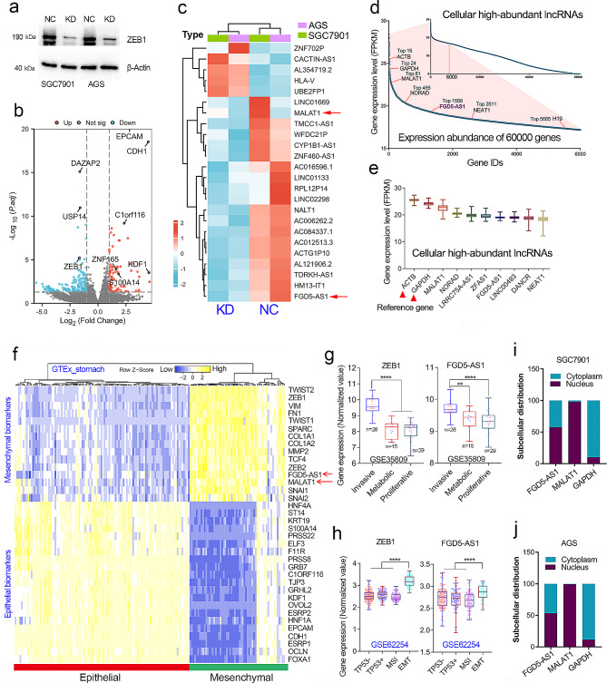
Methods: RNA-seq studies and chromatin immunoprecipitation (Chip) assays were performed to identify ZEB1-regulated lncRNAs. RNA sequencing, RNA pulldown, RNA Immunoprecipitation assays, and rescue assays were conducted to explore the molecular mechanisms of FGD5-AS1 in GC.

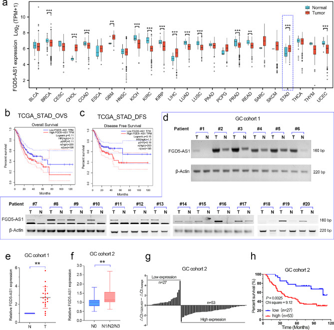
Results: As one of the most abundant lncRNAs in cells, FGD5-AS1 has been shown to be transcriptionally activated by ZEB1, thus closely related to epithelial-mesenchymal transition (EMT) signaling. Clinical analysis showed that FGD5-AS1 overexpression was clinically associated with lymph node metastasis, and predicted poor survival in GC. Loss-of-function studies confirmed that FGD5-AS1 knockdown inhibited GC proliferation and induced cisplatin chemosensibility, cell senescence, and DNA damage in GC cells. Mechanismically, FGD5-AS1 is a YBX1-binding lncRNA due to its mRNA contains three adjacent structural motifs (UAAUCCCA, ACCAGCCU, and CAGUGAGC) that can be recognized and bound by YBX1. And this RNA-protein interaction prolonged the half-life of the YBX1 protein in GC. Additionally, a rescue assay showed that FGD5-AS1 promotes GC by repressing cell senescence and ROS production via YBX1.

Conclusion: FGD5-AS1 is a cellular high-abundant lncRNA that is transcriptionally regulated by ZEB1. FGD5-AS1 overexpression promoted GC progression by inhibiting cell senescence and ROS production through binding and stabilizing the YBX1 protein.

Keywords: Cell senescence; Gastric cancer; RNA-protein interaction; ROS; Transcriptional regulation.


Conflict of interest statement

The authors declare that they have no competing interests.

Figures

Fig. 1
Fig. 1
FGD5-AS1 is an EMT-related, highly abundant lncRNA in GC. (a) The knockdown efficiency of ZEB1 was examined by western blotting assay in the ZEB1-depleted GC cell lines. (b) RNA sequencing studies were conducted in the ZEB1-depleted GC cell lines. As expected, the expression of the classic epithelial biomarkers was greatly increased. (c) The top 24 differentially expressed lncRNAs after ZEB knockdown were shown in the heat map. (d) According to the normalized expression level (FPKM value) of each gene (including protein-coding genes and lncRNAs), FGD5-AS1 was a highly abundant lncRNA in GC. (e) FGD5-AS1 is one of 10 lncRNAs with the highest expression in GC. (f) MALAT1 and FGD5-AS1 were both EMT-related lncRNAs that are highly co-expressed with mesenchymal biomarkers in GC. (g) Both ZEB1 and FGD5-AS1 were highly expressed in the invasive subtype (also known as the mesenchymal phenotype) in the GSE35809 cohort. (h) Both ZEB1 and FGD5-AS1 were highly expressed in the EMT subtype GC patients of the GSE62254 cohort. (i, j) The subcellular distribution of lncRNA FGD5-AS1 was examined by nuclear-cytoplasmic separation assay in the SGC7901 and AGS cell lines
Fig. 2
Fig. 2
LncRNA FGD5-AS1 was transcriptionally induced by ZEB1 in GC. (a) The knockdown efficiency of ZEB1 in the GC cell lines was verified by the qRT-PCR analysis. (b) The FGD5-AS1 expression was detected in the GC cell lines with or without ZEB1 knockdown. (c) The overexpression efficiency of ZEB1 in GC cell lines was verified by qRT-PCR assay. (d) Overexpression of ZEB1 significantly increased the expression level of FGD5-AS1 in the GC cell lines. (e) The chip-seq analysis using the Cistrome web tool confirmed that ZEB1 can bind to the promoter of FGD5-AS1 by recognizing the “CCCA.CCTGCTG” motif in the HepG2 cell line. (f) Promoter analysis showed that the promoter DNA of the FGD5-AS1 gene contains a conserved ZEB1 binding motif. (g) The chip-qPCR analysis confirmed that ZEB1 can directly bind to the promoter of FGD5-AS1 in the AGS cells transfected with ZEB1 overexpression plasmids. (h, i) Transcription factor ZEB1 was highly co-expressed with FGD5-AS1 in pan-tissue (GTEx cohort) and pan-cancer (TCGA cohort), indicating the transcriptional regulation of FGD5-AS1 by ZEB1 may be conserved in human cells and tissues. **, P < 0.01
Fig. 3
Fig. 3
FGD5-AS1 overexpression predicted a poor prognosis in GC. (a) Pan-cancer analysis showed that FGD5-AS1 is frequently dysregulated in cancers and is overexpressed in stomach cancer. (b, c) Survival analysis using the GEPIA web tool showed that FGD5-AS1 overexpression predicted poor overall survival and disease-free survival in GC. (d, e) The expression level of FGD5-AS1 was evaluated in GC cohort 1 (n = 20) using RT-PCR analysis. The results showed that FGD5-AS1 expression was significantly increased in the GC tissues, compared to the normal adjacent tissues. (f) The relative expression level of FGD5-AS1 was evaluated in GC cohort 2 (n = 80). Clinical analysis showed that FGD5-AS1 was positively correlated with lymph node metastasis in GC cohort 2. (g) According to the relative FGD5-AS1 expression level, the GC patients in the GC cohort 2 were divided into two groups, FGD5-AS1_high group and the FGD5-AS1_low group. (h) Prognostic analysis showed that GC patients in the FGD5-AS1_high group had a shorter overall survival than the patients in the FGD5-AS1_low group. **, P < 0.01
Fig. 4
Fig. 4
FGD5-AS1 knockdown inhibits cell proliferation and cisplatin chemoresistance in GC. (a) The relative expression level of FGD5-AS1 was examined in the five GC cell lines and the normal gastric cell line GES-1. (b, c) FGD5-AS1 was successfully silenced in the HGC-27 and SGC7901 cell lines. (d) The CCK-8 assays confirmed that FGD5-AS1 depletion significantly inhibited cell growth in the GC cell lines. (e) The cell colony formation assay was performed in the GC cell lines with or without FGD5-AS1 knockdown. (f, g) The cell apoptosis experiments showed that FGD5-AS1 knockdown significantly increased the number of apoptotic and PANoptotic cells in the HGC-27 and SGC7901 cell lines. (h) The CCK-8 assays confirmed that FGD5-AS1 depletion significantly decreased the chemoresistance of cisplatin in the GC cell lines. (i, j) After 30 days of subcutaneous tumor bearing, nude mice were euthanized. The subcutaneous xenograft tumor in nude mice was taken out and weighed. (k) Tumor volumes for the indicated day after injecting shNC and shFGD5-AS1 cells into nude mice. Data represent mean tumor volumes ± SEM. (l) The body weight of nude mice for the indicated day after injecting shNC and shFGD5-AS1 cells into nude mice. The data represent mean tumor volumes ± SEM. **, P < 0.01
Fig. 5
Fig. 5
FGD5-AS1 silencing induces senescence and DNA damage in GC cells. (a) The RNA-seq analysis was performed in the SGC7901 cell line with or without FGD5-AS1 knockdown. (b) The GO/KEGG analysis showed that the differently expressed genes were enriched in cellular senescence signaling. (c) The transcript abundance of the SASP factors according to the RNA-seq data of the FGD5-AS1 knockdown. (d) The expression levels of cellular senescence biomarkers, including IL1A, IL1B, and IL8/CXCL8, were significantly increased in the FGD5-AS1 depleted GC cell lines. (e) The SA-β-gal staining assay showed that FGD5-AS1 knockdown obviously increased the number of senescence cells in the HGC-27 and SGC7901 cell lines. (f) The comet assay confirmed that knockdown of FGD5-AS1 significantly promoted DNA damage in the GC cell lines. **, P < 0.01
Fig. 6
Fig. 6
FGD5-AS1 directly binds and stabilizes the YBX1 protein in GC. (a) The RNA pull-down assay for identification of proteins interacted with FGD5-AS1 was performed. The sense (S) and anti-sense (AS) of FGD5-AS1 RNA were biotinylated, refolded, and incubated with HGC-27 cell lysates. The red arrow represents the differential band containing the potential FGD5-AS1 binding proteins. (b) The differential band was separated for mass spectrum analysis. The FGD5-AS1 binding proteins were finally confirmed according to the workflow. (c, d) According to the MS analysis, YBX1 was the most possible RNA-binding protein that interacted with FGD5-AS1. (e) The western blotting using RNA pulldown samples confirmed the binding of FGD5-AS1 and YBX1. (f) FGD5-AS1 has a similar sub-cellular location to the YBX1 protein in the GC cell lines. (g) The interaction of FGD5-AS1 and YBX1 was further verified using the RIP-qPCR assay in the GC cell lines. (h) The secondary structure of the lncRNA FGD5-AS1 was predicted by the RNAfold web server. According to the predicted structure, we divided FGD5-AS1 into three fragments. Notably, fragment 2 contains three 8-mer linear YBX1 recognized binding motifs. (i) The RNA pull-down experiments using three truncated fragments of FGD5-AS1 were performed. It was found that only fragment 2 and full-length FGD5-AS1 fragments could interact with YBX1. (j, k) The knockdown of FGD5-AS1 significantly prolonged the half-life of the YBX1 protein in the HGC-27 cell line. (l) The gene expression correlation analysis showed that FGD5-AS1 was highly co-expressed with YBX1 in GC. **P < 0.01
Fig. 7
Fig. 7
FGD5-AS1 promoted GC proliferation and repressed senescence in a YBX1-dependent manner. (a) The differentially expressed genes after YBX1 knockdown were shown in the heat map after RNA-seq analysis. (b) YBX1 knockdown significantly increased the expression level of SASP factors, including IL1A, IL1B, IGFBP1/3, CYR61, ANGPTL4, and SERPINE2 in the GC cell line BGC823. (c) The western blotting assays showed that YBX1 was successfully overexpressed in GC cell lines. (d) The cell colony formation assay was performed in the GC cell lines with or without YBX1 overexpression. (e) The SA-β-gal staining assay showed that YBX1 overexpression significantly inhibited cell senescence in the HGC-27 and SGC7901 cell lines. (f) The rescue colony formation assay showed that the inhibitory effects of FGD5-AS1 depletion on cell proliferation could be rescued by additional YBX1 overexpression in GC. (g) The rescue SA-β-gal staining assay showed that the promotion effects of FGD5-AS1 knockdown on cell senescence could be rescued by additional YBX1 overexpression in GC. (h, i) The rescue CCK-8 assay showed that the inhibitory effects of FGD5-AS1 knockdown on GC cell growth could be rescued by additional YBX1 overexpression. (j, k) Evaluate the effect of FGD5-AS1 knockdown on intracellular ROS content using flow cytometry. The 2’,7’-dichlorofluorescein (DCF) is an oxidatively sensitive fluorescent probe used to measure intracellular reactive oxygen species (ROS) production. The inhibitory effects of FGD5-AS1 knockdown on ROS production of GC cell lines could be rescued by additional YBX1 overexpression. **P < 0.01
Fig. 8
Fig. 8
The working model of the role of lncRNA FGD5-AS1 in driving GC progression. The cellular high-abundant lncRNA FGD5-AS1 was transcriptionally activated by the EMT-TF ZEB1 in GC. Due to the upregulation of ZEB1, lncRNA FGD5-AS1 expression was increased, and its overexpression was clinically associated with metastasis and poor survival in GC. Mechanically, FGD5-AS1 directly binds to the YBX1 protein to enhance its protein stability. FGD5-AS1 promotes GC progression by repressing cell senescence and ROS production in a YBX1-dependent manner

References

    1. Siegel RL, Giaquinto AN, Jemal A. Cancer statistics, 2024. CA Cancer J Clin. 2024;74(1):12–49. doi: 10.3322/caac.21820. - DOI - PubMed
    1. Bray F, Laversanne M, Sung H, Ferlay J, Siegel RL, Soerjomataram I, et al. Global cancer statistics 2022: GLOBOCAN estimates of incidence and mortality worldwide for 36 cancers in 185 countries. CA Cancer J Clin. 2024;74(3):229–63. doi: 10.3322/caac.21834. - DOI - PubMed
    1. Joshi SS, Badgwell BD. Current treatment and recent progress in gastric cancer. Ca-Cancer J Clin. 2021;71(3):264–79. doi: 10.3322/caac.21657. - DOI - PMC - PubMed
    1. Li D, Huang P, Xia L, Leng W, Qin S. Cancer-associated fibroblasts promote gastric cancer cell proliferation by paracrine FGF2-driven ribosome biogenesis. Int Immunopharmacol. 2024;131:111836. doi: 10.1016/j.intimp.2024.111836. - DOI - PubMed
    1. Li DD, Wang JJ, Zhang MX, Hu XH, She JJ, Qiu XM, et al. LncRNA MAGI2-AS3 is regulated by BRD4 and promotes gastric Cancer Progression via maintaining ZEB1 overexpression by sponging miR-141/200a. Mol Ther-Nucl Acids. 2020;19:109–23. doi: 10.1016/j.omtn.2019.11.003. - DOI - PMC - PubMed

MeSH terms